Cardiovascular system labeling
 Call Kai
 Learn
 Practice Test
 Spaced Repetition
 Match
 Flashcards
 Knowt Play
1/52

There's no tags or description
Looks like no tags are added yet.
53 Terms
right atrium

left atrium

right ventricle

left ventricle

tricuspid valve
valve between the right atrium and the right ventricle

mitral valve
valve between the left atrium and the left ventricle

pulmonary valve
valve between the right ventricle and pulmonary artery

aortic valve
valve between the left ventricle and the aorta

SA node (sinoatrial node)
pacemaker of the heart

AV node (atrioventricular node)

AV bundle

Purkinje fibers

left bundle branch

interatrial septum
separates atria

interventricular septum
separates ventricles

artery
A blood vessel that carries oxygenated blood away from the heart (1)

Arterioles
small vessels that receive blood from the arteries (3)
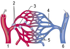
capillary beds
a small blood vessel that allows diffusion of gases, nutrients and wastes between plasma and interstitial fluid (5)

Venules
small vessels that gather blood from the capillaries into the veins (4)
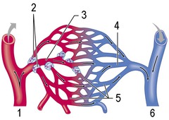
Veins
Blood vessels that carry deoxygenated blood back to the heart (6)

Pericardium
Double-layered membrane surrounding the heart.

epicardium
outer layer of the heart

Myocardium
muscular, middle layer of the heart

endocardium
Inner layer of the heart

tunica intima
the innermost layer of a blood vessel

tunica media
middle smooth muscle layer

tunica adventitia
outer layer of blood vessel

elastic artery
-thick, elastic intima layer
-thicker, most elastic media layer
-relatively thin adventitia layer (contains blood vessels)

muscular artery
-thin, elastic intima layer (squiggly)
-smooth muscle spirally arranged media layer
-connective tissue holding vessel together adventitia layer

epicardium, myocardium, endocardium
three layers of the heart wall

continuous capillaries
most common type of capillary
-relatively impermeable to large molecules
found in muscle, CNS, skin

fenestrated capillaries
type of capillary with fenestrated endothelial cell wall
leaky in terms of fluid flowing
found in kidney

Discontinous capillaries
type of capillary with wide gaps
super leaky
found in liver, spleen, bone marrow
allows passage of cells

muscular venules
-irregular in shape
recieve blood from the postcapillary venules
-1-2 layers of smooth muscle
-thin adventitia

P wave
atrial depolarization

QRS complex
ventricular depolarization (and atrial repolarization)

T wave
ventricular repolarization
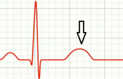
Tachycardia
EKG abnormality showing a rapid heart rate of >100 bpm

Bradycardia
EKG abnormality showing slowed heart rate of < 65 bpm

Atrial Flutter
EKG showing rapid, but regular sequence of atrial depolarization at rates between 200 and 380 bpm

atrial fibrillation
EKG showing rapid, irregular, uncoordinated depolarization of the atria with no definite P waves
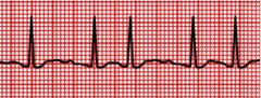
ventricular fibrillation
EKG showing very serious rhythmic abnormality in which the ventricular musculature exhibits uncoordinated, chaotic contractions

ischemia
EKG shows pathology of an inadequate blood supply to an organ or part of the body, especially the heart muscles.
-T wave is symmetrically inverted

Infarct
injury with ST segment elevations and deep Q waves

Arteriolar vasoconstriction
-reduces blood flow to injured area

primary hemostasis
platelet plug formation

secondary hemostasis
fibrin clot formation

Clot stabilization and resorption
platelets and fibrin aggregats contract forming a permanent plug

von Willebrand factor
bridge between collagen and platelets

True aneurysm (saccular)
what type of aneurysm has a bulge

True aneurysm (fusiform)
what type of aneurysm is ballooned on both sides

false aneurysm
break in both the intima layer and media layer

dissection
break in intima layer but not media layer
