Section 3: Central Nervous System
 Call Kai
 Learn
Learn Practice Test
 Spaced Repetition
 Match
Match Flashcards
 Knowt Play
1/31

There's no tags or description
Looks like no tags are added yet.
Name | Mastery | Learn | Test | Matching | Spaced | Call with Kai | Chat |
|---|
No analytics yet
Send a link to your students to track their progress
32 Terms
What is the Cerebrum? what are the two parts?
The cerebrum makes up the bulk of the brain. It is composed of the telencephalon and the diencephalon.
What is Telencephalon and its 6 parts
which makes up the bulk of the cerebrum. responsible for interpreting sensory signals, sending out motor signals, and decision making.
1. Cerebral Hemispheres
2. Cerebral Cortex
3. Sulci and Gyri
4. Lobes of the Cerebrum
5. Grey Matter of the Brain
6. White Matter of the Brain

1. Cerebral Hemispheres
The cerebrum is made up of two paired hemispheres: the right and left cerebral hemisphere. separated by the longitudinal fissure.

2. Cerebral Cortex
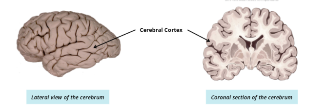
The outer layer of the cerebrum is known as the cerebral cortex. In a coronal section, it can be distinguished from other neural tissue by its dark grey colour.
3. Sulci and Gyri
The cerebrum has many smaller fissures, or grooves, which create folds in the cerebrum. This folding increases the surface area of neural tissue, which increases the functional capacity of the tissue.
The fissures and grooves are known as sulci.
The lumps and bumps between the sulci are known as gyri.

SULCI
The fissures and grooves are known as sulci.
Sulci on the cerebral cortex create the boundaries of the lobes, and divide the brain into regions with different functions.
What are the 5 kinds of sulcus? and where they are
Precentral Sulcus: Creates the anterior border of the motor cortex.
Central Sulcus: Divides the frontal and parietal lobes. It also divides the motor from the sensory cortex.
Postcentral Sulcus: Creates the posterior border of the sensory cortex.
Parieto-occipital Sulcus: Divides the parietal and occipital lobes. This sulcus is more prominent on medial surface on the hemisphere.
Lateral Sulcus: Divides the temporal lobe from the frontal and parietal lobes.
Midsagittal section: Note that the parieto-occipital sulcus is more prominent in the midsagittal section, while the lateral sulcus is absent

What are the two types of GYRI
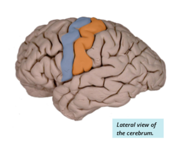
Precentral Gyrus (Motor Cortex): Responsible for sending motor output signals. It is located anteriorly to the central sulcus (Blue).
Postcentral Gyrus (Somatosensory Cortex): Responsible for receiving sensory input related to touch and feel. It is located posteriorly to the central sulcus (Orange).
4. Lobes of the Cerebrum. what are the 4 lobes and where are they?
The telencephalon is divided into four lobes, each with specific functions. The lobes are named after the overlying skull bones.

5. Grey Matter of the Brain
Grey matter consists of nerve cell bodies, short interneurons, and glial cells. It can be found in the cerebral cortex and the inner areas of the brain.

6. White Matter of the Brain
White matter consists of bundles of nerve fibres and axons. It can be found in the inner regions of the cerebrum.
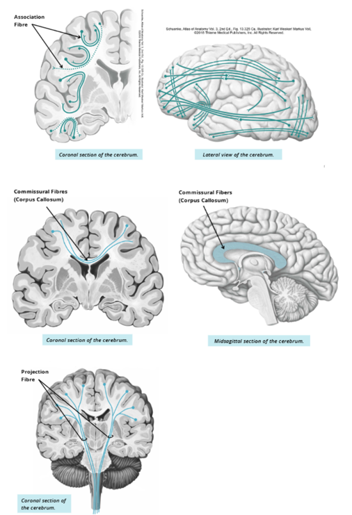
A specialized region of white matter, known as the corpus callosum, connects the two hemispheres of the brain.

what are the three categories of white matter tracts based upon the structures they connect and the type of communication they facilitate
Association fibres: facilitate communication within one hemisphere.
Commissural fibres: facilitate communication between the two hemispheres. The word “commissure” commonly refers to the site where two structures join. An example is a structure known as the corpus callosum, which runs horizontally to connect the two sides of the brain.
Projection fibres: facilitate communication between different levels of the CNS, such as between the brain and spinal cord
What is Diencephalon
The second layer, the diencephalon, is deep to the telencephalon. It is the central core of the cerebrum, surrounded by the cerebral hemispheres. The diencephalon is responsible for sensory and motor relay into and out of the brain, as well as control of the autonomic nervous system.
Encloses a structure known as the third ventricle. The diencephalon is composed of the thalamus and the hypothalamus.
Explain what the thalamus and the hypothalamus of the Diencephalon
Thalamus: The Thalamus is a deep brain structure with two bodies, one per hemisphere. It is responsible for relaying sensory information, except sense of smell, to other areas of the brain.
Hypothalamus: a small structure below the thalamus that is connected to the pituitary gland. The hypothalamus controls the release of hormones through the pituitary gland.

What is the Brainstem and name the three parts?
The brainstem is the area of the brain that connects the brain to the spinal cord. It is situated just below the diencephalon and is comprised of the midbrain, the pons, and the medulla oblongata.
The brainstem is very important as it contains many autonomic reflex centers and white matter tracts necessary for human functioning and survival.

1. Midbrain:
The midbrain is the most superior aspect of the brainstem.
Anteriorly, the midbrain consists of large white matter tracts that make up the cerebral peduncles. From a postero-lateral view, the midbrain has two pairs of raised bumps, known as the superior and inferior colliculi.

2. Pons:
The pons is the structure in the middle of the brainstem.
It is located anterior to the 4th ventricle and the cerebellum.
It is important to note that the pons has fibers connecting it to the cerebellum, allowing for communication between these two structures.

3. Medulla:
The medulla oblongata, or simply the medulla, is the most inferior structure of the brainstem. It is continuous with the spinal cord.

What is cerebellum?
The cerebellum is a structure that is appended to the back of the brainstem and sits underneath the occipital lobe of the cerebrum.
The cerebellum functions to coordinate motor activity.

What is the purpose of Support and Protection of the Brain, and the two parts
The brain is a vital organ for life. It has ample support and protection to prevent injury to itself.
Two protective measures: the meninges and the ventricles.
what are Meanings and the three structures?
Connective tissue membranes protecting the structures of the CNS.
Dura Mater:
Arachnoid Mater:
Pia Mater:
What are Dura Mater? + the projections
Dura mater is the most superficial and tough of the meninges. It is composed of a double layer of connective tissue that attaches firmly to the skull.
Projections of the Dura Mater:
The dura mater also invaginates into:
• The longitudinal fissure: creating the falx cerebri between the cerebral hemispheres.
• Tentorium cerebelli: The transverse fissure between the cerebrum and cerebellum
• Falx cerebelli: the space at the midline of the cerebellum.

What is the Arachnoid Mater?
The arachnoid mater loosely covers the brain. It creates an area called the subarachnoid space. This space is filled with cerebrospinal fluid (a protective fluid) and blood vessels.

What is the Pia Mater?
Pia mater is firmly attached to the cerebral cortex and is the innermost layer of the meninges.

What are ventricles? What are the 4 hollow spaces?
Ventricles also provide support and protection to the brain.
They are made up of 4 hollow spaces that are continuous with each other and the spinal cord. These spaces contain cerebrospinal fluid (C S F)
Lateral ventricle
Third ventricle
Fourth ventricle
Lateral Ventricle:
The lateral ventricles are paired ‘C’ shaped spaces deep within the cerebral hemispheres. A portion of the lateral ventricles can be found in each lobe.

Third Ventricle:
The singular third ventricle is located in the middle of the diencephalon between the two halves of the thalamus.
It is connected to the lateral ventricles by the interventricular foramina (of Monro).

Fourth Ventricle:
The fourth ventricle sits between the pons/medulla and the cerebellum. It is connected to the third ventricle by the cerebral aqueduct.
It is continuous with the central canal of the spinal cord and the subarachnoid space.

What is Cerebrospinal fluid (CSF)?
CSF is contained within the ventricles and flows from the lateral ventricle to the bloodstream.
CSF is a specialized fluid that bathes the structures of the CNS (brain and spinal cord). It flows through various spaces within the brain known as ventricles, the central canal of the spinal cord, and the subarachnoid space.
Cerebrospinal fluid is produced inside the ventricles primarily by the choroid plexus, a collection of ependymal cells and blood vessels.
What are the main functions of CSF?
First: Providing a Buoyancy effect to the brain, and spinal cord, as these structures are suspended by the CSF. These structures weigh less when surrounded by CSF.
Second: CSF also provides support and protection to the CNS, acting as shock absorber.
Third: CSF also serves as a medium for the transport of nutrients, waste, dissolved gases and other substances
What are the steps in the CSF flow?
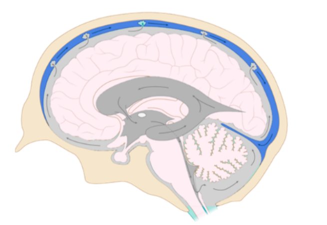
Step 1: Lateral Ventricles → Third Ventricle
CSF is produced in the two lateral ventricles (one in each hemisphere).
It flows through the interventricular foramina (of Monro) into the third ventricle.
Step 2: Third Ventricle → Fourth Ventricle
CSF flows from the third ventricle through the cerebral aqueduct into the fourth ventricle.(which is surrounded by the pons and medulla anteriorly and by the cerebellum posteriorly )
Step 3: Fourth Ventricle → Circulation
CSF leaves the fourth ventricle and enters the subarachnoid space (around the brain and spinal cord) and central canal (inside the spinal cord).
It circulates to protecting and providing buoyancy to the brain and spinal cord.
Finally, CSF is reabsorbed into the bloodstream.

CSF flow sequence
