Hoof structure and Function

0.0(0)
Studied by 0 people
call kaiCall Kai
learnLearn
examPractice Test
spaced repetitionSpaced Repetition
heart puzzleMatch
flashcardsFlashcards
GameKnowt Play
Card Sorting

1/37

encourage image

There's no tags or description

Looks like no tags are added yet.

38 Terms
1
New cards
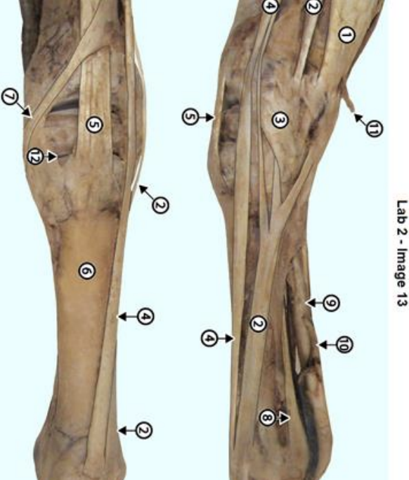
<p><span data-name="star" data-type="emoji">⭐</span> Question: Identify equine extensor structures 1, 2, 4, and 5.</p><p></p><p>mc</p><ul><li><p>Extensor carpi radialis</p></li><li><p>Common digital extensor</p></li><li><p>Ulnaris lateralis</p></li><li><p>Lateral digital extensor</p></li></ul><p></p>

⭐ Question: Identify equine extensor structures 1, 2, 4, and 5.


mc

  • Extensor carpi radialis

  • Common digital extensor

  • Ulnaris lateralis

  • Lateral digital extensor


  • 1 = Ulnaris lateralis

  • 2 = Lateral digital extensor

  • 4 = Common digital extensor

  • 5 = Extensor carpi radialis


2
New cards
<p><span data-name="star" data-type="emoji">⭐</span> Question: Identify structures 4, 5, 6, and 7.</p><p>mc</p><ul><li><p>Suspensory ligament (interosseous)</p></li><li><p>Superficial digital flexor tendon (SDFT)</p></li><li><p>Distal check/accessory ligament</p></li><li><p>Deep digital flexor tendon (DDFT)</p></li></ul><p></p>

⭐ Question: Identify structures 4, 5, 6, and 7.

mc

  • Suspensory ligament (interosseous)

  • Superficial digital flexor tendon (SDFT)

  • Distal check/accessory ligament

  • Deep digital flexor tendon (DDFT)


  • 4 = Superficial digital flexor tendon (SDFT)

  • 5 = Deep digital flexor tendon (DDFT)

  • 6 = Distal check/accessory ligament

  • 7 = Suspensory ligament (interosseous)


3
New cards
<p><span data-name="star" data-type="emoji">⭐</span> Question: Identify 1, 1′, 4, 5, and 6 in this ligament diagram.</p><p></p><p>mc</p><ul><li><p>Straight distal sesamoidean ligament</p></li><li><p>Intersesamoid ligament</p></li><li><p>Interosseous/suspensory ligament</p></li><li><p>Oblique distal sesamoidean ligament</p></li><li><p>Extensor branch</p></li></ul><p></p>

⭐ Question: Identify 1, 1′, 4, 5, and 6 in this ligament diagram.


mc

  • Straight distal sesamoidean ligament

  • Intersesamoid ligament

  • Interosseous/suspensory ligament

  • Oblique distal sesamoidean ligament

  • Extensor branch


  • 1 = Interosseous/suspensory ligament

  • 1′ = Extensor branch

  • 4 = Straight distal sesamoidean ligament

  • 5 = Oblique distal sesamoidean ligament

  • 6 = Intersesamoid ligament


4
New cards
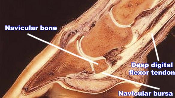
<p><span data-name="star" data-type="emoji">⭐</span> Question: Explain the anatomical relationship among the navicular bone, navicular bursa, and deep digital flexor tendon.</p><p></p>

⭐ Question: Explain the anatomical relationship among the navicular bone, navicular bursa, and deep digital flexor tendon.


Answer: The navicular bone is the distal sesamoid bone behind the coffin joint. The navicular bursa lies between the navicular bone and DDFT, reducing friction as the tendon passes over the bone toward P3.

5
New cards
<p><span data-name="star" data-type="emoji">⭐</span> Question: Identify the external hoof regions labeled A–D.</p><p>mc</p><ul><li><p>Toe</p></li><li><p>Coronet/coronary band</p></li><li><p>Heel</p></li><li><p>Quarter</p></li></ul><p></p>

⭐ Question: Identify the external hoof regions labeled A–D.

mc

  • Toe

  • Coronet/coronary band

  • Heel

  • Quarter


  • A = Coronet/coronary band

  • B = Toe

  • C = Quarter

  • D = Heel


6
New cards
<p><span data-name="star" data-type="emoji">⭐</span> Question: Which animal is (a) and which is (b)? What are the two principal weight-bearing digits in these cloven-hoofed animals?</p><p></p>

⭐ Question: Which animal is (a) and which is (b)? What are the two principal weight-bearing digits in these cloven-hoofed animals?


Answer:

(a) Pig;

(b) Ox. Digits III and IV are the principal weight-bearing digits.

7
New cards
<p><span data-name="star" data-type="emoji">⭐</span> Question: Identify structures 1, 2, and 3.</p><p></p><ul><li><p>Bar</p></li><li><p>Toe</p></li><li><p>Frog</p></li><li><p>Heel Bulb</p></li><li><p>Sole</p></li><li><p>White Line<br></p></li></ul><p></p>

⭐ Question: Identify structures 1, 2, and 3.


  • Bar

  • Toe

  • Frog

  • Heel Bulb

  • Sole

  • White Line


  • 1 = Frog

  • 2 = Bar

  • 3 = Sole


8
New cards
<p><span data-name="star" data-type="emoji">⭐</span> Question: Identify structures 4, 5, and 6.</p><p></p><ul><li><p>Bar</p></li><li><p>Toe</p></li><li><p>Frog</p></li><li><p>Heel Bulb</p></li><li><p>Sole</p></li><li><p>White Line</p></li></ul><p></p>

⭐ Question: Identify structures 4, 5, and 6.


  • Bar

  • Toe

  • Frog

  • Heel Bulb

  • Sole

  • White Line


  • 4 = White line

  • 5 = Toe

  • 6 = Heel bulb


9
New cards
<p><span data-name="star" data-type="emoji">⭐</span> Question: Identify the apex, crura, central sulcus, and paracuneal grooves of the frog.</p><p></p>

⭐ Question: Identify the apex, crura, central sulcus, and paracuneal grooves of the frog.


  • Apex: Pointed tip of the frog, toward the toe.

  • Crura: Two branches extending toward the heels.

  • Central sulcus: Groove between the two crura.

  • Paracuneal grooves: Grooves on either side of the frog.


10
New cards
<p><span data-name="star" data-type="emoji">⭐</span> Question: On the left specimen, identify numbers 1, 2, and 3.</p><p></p><ul><li><p>bar dermis</p></li><li><p>sole dermis</p></li><li><p>perioplic dermis</p></li><li><p>laminar dermis</p></li><li><p>coronary dermis</p></li><li><p>frog dermis</p></li></ul><p></p>

⭐ Question: On the left specimen, identify numbers 1, 2, and 3.


  • bar dermis

  • sole dermis

  • perioplic dermis

  • laminar dermis

  • coronary dermis

  • frog dermis


  • 1 = Perioplic dermis

  • 2 = Coronary dermis

  • 3 = Laminar dermis


11
New cards
<p><span data-name="star" data-type="emoji">⭐</span> Question: On the right specimen, identify numbers 3, 4, and 5.</p><ul><li><p>bar dermis</p></li><li><p>sole dermis</p></li><li><p>perioplic dermis</p></li><li><p>laminar dermis</p></li><li><p>coronary dermis</p></li><li><p>frog dermis</p></li></ul><p></p>

⭐ Question: On the right specimen, identify numbers 3, 4, and 5.

  • bar dermis

  • sole dermis

  • perioplic dermis

  • laminar dermis

  • coronary dermis

  • frog dermis


  • 3 = Bar dermis

  • 4 = Frog dermis

  • 5 = Sole dermis


12
New cards
<p>Identify the white line</p>

Identify the white line

Answer: White line — the visible junction between the hoof wall and sole, associated with the hoof's epidermal laminar region.

13
New cards
<p><span data-name="star" data-type="emoji">⭐</span> Question: What structures have lost their normal attachment in this image, and which tendon can contribute to P3 displacement?</p><p></p>

⭐ Question: What structures have lost their normal attachment in this image, and which tendon can contribute to P3 displacement?


Answer: The dermal and epidermal laminae have lost their normal connection, consistent with laminar failure/laminitis. The deep digital flexor tendon (DDFT) can exert pull that contributes to rotation of P3.

14
New cards
⭐ Which digits bear weight in horses versus cloven-hoofed animals?
Horse: digit III. Ruminants: digits III and IV.
15
New cards
⭐ How does the relationship of P3 to the hoof/nail differ in horses and carnivores?
Horse: entire P3 lies inside hoof capsule. Dog/cat: primarily ungual process enters claw.
16
New cards
⭐ Define fetlock, pastern and coffin joints using bone names.
Fetlock: cannon–P1 (MCP/MTP). Pastern: P1–P2 (PIP). Coffin: P2–P3 (DIP).
17
New cards
⭐ What do equine collateral (ungual) cartilages contribute?
Shock absorption and expansion of the hoof during weight-bearing.
18
New cards
⭐ Where does the equine common digital extensor tendon insert?
Dorsal extensor process of P3; extends the digit.
19
New cards
⭐ Where do common digital extensor tendons insert in dogs?
Distal phalanges (P3) of digits II–V.
20
New cards
⭐ What does extensor carpi radialis do?
Extends the carpus.
21
New cards
⭐ What does lateral digital extensor do?
Helps extend lateral digits; also contributes to carpal extension.
22
New cards
⭐ What does ulnaris lateralis do?
Can flex the carpus and provide lateral support; grouped with forelimb extensors.
23
New cards
⭐ What is the function of the extensor retinaculum?
Holds the extensor tendons near the carpus as they cross the joint.
24
New cards
⭐ Where does the superficial digital flexor tendon (SDFT) insert?
Distal P1 and proximal P2, primarily on the palmar/plantar aspect.
25
New cards
⭐ Where does the deep digital flexor tendon (DDFT) insert?
Flexor surface of P3; acts to flex the digit.
26
New cards
⭐ What is the manica flexoria?
A cuff formed by the SDFT through which the DDFT passes.
27
New cards
⭐ What is the distal check/accessory ligament attached to?
It joins the deep digital flexor tendon (DDFT).
28
New cards
⭐ What does the suspensory/interosseous ligament do?
Supports the fetlock against excessive extension; sends extensor branches toward the digital extensor tendon.
29
New cards
⭐ Name the major distal sesamoidean ligament groups shown.
Straight, oblique and deeper cruciate distal sesamoidean ligaments.
30
New cards
⭐ What is the navicular bone and why is its bursa needed?
Distal sesamoid bone; the bursa reduces friction between it and the DDFT.
31
New cards
⭐ What is the distal navicular (impar) ligament?
A ligament connecting the distal navicular bone to P3.
32
New cards
⭐ What does the digital cushion do?
Supports weight-bearing and absorbs shock within the hoof.
33
New cards
⭐ Define the coronary band, toe, quarter, heel and bar.
Coronet: skin/hoof junction. Toe: dorsal wall. Quarter: sides. Heel: palmar wall. Bar: inward-folded wall beside frog.
34
New cards
⭐ What are the four named parts/regions of the frog?
Apex, crura, central sulcus and paracuneal grooves.
35
New cards
⭐ What is the white line, and why is it clinically important?
Junction of wall and sole associated with laminae; a potential entry site for infection.
36
New cards
⭐ Match stratum externum, medium and internum to their hoof functions.
Externum: periople/moisture barrier. Medium: bulk of wall. Internum: laminar attachment.
37
New cards
⭐ Why do dermal and epidermal laminae interdigitate?
They create a strong attachment suspending P3 within the hoof wall.
38
New cards
⭐ Primary versus secondary hoof laminae: difference?
Primary laminae are larger folds; secondary laminae branch from them, increasing attachment area.