Only Corrections + More: Shoulder, Clavicle, AC Joints, & Scapula Positioning (RAD 1101)
 Call Kai
 Learn
 Practice Test
 Spaced Repetition
 Match
 Flashcards
 Knowt Play
1/61

There's no tags or description
Looks like no tags are added yet.
62 Terms

What is the error in this internal rotation X-ray of the shoulder? How do you correct it?
Humerus is not in alignment. It is angled outward. To correct, the humerus should be placed parallel to the body if possible.

What is the error in this axillary shoulder projection? How do you correct this?
Humerus is not in same plane as shoulder. Humerus is foreshortened. To correct, place entire arm in the same plane to correctly align CR, part, and IR. Arm is foreshortened.

What is the error in this axillary shoulder projection? How do you correct this?
Central ray not directed correctly to axilla. To correct, correctly align CR, part, and IR. Arm is elongated.

What is the error in this axillary shoulder projection? How do you correct this?
Incorrect centering of IR, also quantum mottle and incorrect collimation. To correct, center to IR correctly, collimate, and increase the amount of radiation.
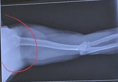
What is the error in this axillary shoulder projection?
Collimation is open too much.
What projections is considered a non-trauma series when looking at X-raying the shoulder?
1. AP projections: external and internal rotations
2. inferosuperior axial projection (axillary view) (Lawrence Method)

What projections are used for trauma patients when looking at X-raying the shoulder?
1. AP projection in neutral rotation
2. transthoracic lateral projection (Lawrence Method)
3. PA oblique projection (scapular Y method)

What is the error in this Grashey projection? How should it be corrected?
Patient under-rotated. Head of humerus overlaps glenoid fossa. To correct, rotate the patient more.

What projection is this X-ray? Is this dislocation anterior or posterior? How can you tell?
neutral rotation, it shows a posterior dislocation as the humeral head is inferior to the acromion process

What projection is this X-ray? Is this dislocation anterior or posterior? How can you tell?
Y-view, it shows an anterior dislocation as the humeral head is inferior to the coracoid process

What is the error in this scapula Y-view error?
Obliqued too little. Humerus is too far from the body.

What is the error in this scapula Y-view error?
Obliqued too much. Humerus is too close to the body.
NOTE: How can a scapula Y-view for severely injured patients be performed?
in RPO or LPO position
NOTE: For an AP projection of the clavicle, what would reduce OID if a patient is able to stand?
erect PA

NOTE: How should the patient be positioned for a bilateral projection of the AC joints (Pearson Method) to reduce OID?
erect PA

NOTE: What should be used or done for a bilateral AP (or PA) projection of the AC joints (Pearson Method) to include both joints?
14x17 cross-wise or do AC joints separate if it will not fit

NOTE: For patients that cannot stand for bilateral AP projection of the acromioclavicular joints, what is the alternative position?
supine

What is the exception for an AP projection of the scapula?
when there should be no arm movement in cases of trauma


This X-ray of the lateral scapula is incorrect due to...
incorrect centering causing quantum mottle
Situation: A radiograph of an AP oblique projection for the glenoid cavity reveals that the anterior and posterior rims of the glenoid process are not superimposed. What is the solution?
To produce a more acceptable image, the rotation
of the body should be increased
Situation: A radiograph of a transthoracic lateral projection reveals that it is difficult to visualize the proximal humerus due to the ribs and lung markings. The exposure was made on suspended respiration. What is the solution?
Using a breathing technique would improve the quality of the image (or shallow breathing)
Situation: A radiograph of an AP clavicle reveals that the sternal extremity is partially collimated off. What should the technologist do?
Repeat with the correct collimation to include both ends and joints

Situation: A radiograph of a scapular Y lateral position reveals that the scapula is slightly rotated (the vertebral and axillary borders are not superimposed). The axillary border of the scapula is determined to be more lateral compared with the vertebral border. What is the solution?
The rotation of the thorax should be increased for the repeat exposure
Situation: A patient comes to radiology for treatment of an arthritic condition of the right shoulder. The radiologist orders AP internal/external rotation projections and an inferosuperior axial projection of the scapulohumeral joint. However, the patient cannot abduct the arm for this projection. Which other projection/method will best demonstrate a profile view of the glenohumeral joint space?
Grashey method

Situation: A patient comes to the ER with a possible right AC joint separation. Right clavicle and AC joint exams are ordered. The clavicle is taken first, and a small linear fracture of the midshaft of the clavicle is discovered. What should the technologist do in this situation?
Consult with the ER physician before AC joint study as they are done with weights and could possibly be broken more with adding weights
Situation: A patient enters the ER with multiple injuries. The physician is concerned about a dislocation of the left shoulder. The patient is unable to stand. Which two projections are advisable to best demonstrate this condition?
AP in neutral rotation and scapular Y-view (AP oblique projection or transthoracic lateral
Situation: A patient enters the ER with a possible AC joint separation. The patient is paraplegic; therefore, the study cannot be done erect. What is the solution?
Nonweight-bearing and weight-bearing type of projections with the patient recumbent and pulling down on the shoulders could be performed to diagnose this condition
Situation: A patient enters the ER with a dislocated shoulder. The technologist attempts to position the patient into the transthoracic lateral projection, but the patient is unable to raise the unaffected arm over his head
completely. What is the solution?
The technologist can angle the CR 10° to 15° cephalad to compensate for the patient's inability to raise his arm completely.
Situation: A radiograph of an AP axial projection of the clavicle demonstrates that the clavicle is within the midportion of the lung apices. What is the solution?
The technologist should angle more cephalad to correct this error

What is the structure in this should X-ray at A?
scapula

What is the structure in this should X-ray at B?
surgical neck

What is the structure in this should X-ray at C?
lesser tuberosity

What is the structure in this should X-ray at D?
coracoid process

What is the structure in this should X-ray at E?
clavicle

What is the structure in this should X-ray at F?
AC joint

What is the structure in this should X-ray at G?
acromion process

What is the structure in this should X-ray at H?
head of humerus

What is the structure in this should X-ray at I?
anatomical neck

What is the structure in this should X-ray at J?
greater tuberosity

What projection is shown?
AP projection - internal rotation

What projection is shown?
AP projection - external rotation

What projection is shown?
AP oblique - Grashey Method

What projection is shown?
transthoracic lateral projection - Lawrence Method

What projection is shown?
Y-view for shoulder

What projection is shown?
lateral projection of scapula

What projection is shown?
axillary view - Lawrence Method

What projection is shown?
AP projection of AC joints

What projection is shown?
AP projection of scapula

What projection is shown?
AP projection of clavicle

What projection is shown in this shoulder X-ray?
neutral rotation

What projection is shown in this shoulder X-ray?
external rotation

What projection is shown in this shoulder X-ray?
internal rotation

What projection is shown in this shoulder X-ray?
axillary view

What projection is shown in this shoulder X-ray?
Grashey Method

What projection is shown in this shoulder X-ray?
transthoracic lateral (Lawrence Method)

What projection is shown in this shoulder X-ray?
Y-view shoulder

What projection is shown in this X-ray?
AP clavicle

What projection is shown in this X-ray?
AP axial clavicle

What projection is shown in this X-ray?
Y view scapula

What projection is shown in this X-ray?
AP scapula

What projection is shown in this X-ray?
AP AC joints

Which of these images would be obtained for a trauma patient?
2, 3, & 4