Ch. 7 - Microscopic Examination of Urine
 Call Kai
 Learn
 Practice Test
 Spaced Repetition
 Match
 Flashcards
 Knowt Play
1/91

There's no tags or description
Looks like no tags are added yet.
92 Terms
what is the least standardized and most-time consuming portion of urinalysis?
microscopic urinalysis
what could be the significance of color in macroscopic examination that could relate to microscopic examination
blood, infections
what could be the significance of clarity in macroscopic examination that could relate to microscopic examination
hematuria vs. hemoglobinuria/myoglobinuria
confirm pathologic or nonpathologic cause of turbidity
what could be the significance of blood in macroscopic examination that could relate to microscopic examination
RBCs/ RBC casts
what could be the significance of protein in macroscopic examination that could relate to microscopic examination
casts/cells
what could be the significance of nitrite in macroscopic examination that could relate to microscopic examination
bacteria/ WBCs
what could be the significance of leukocyte esterase (LE) in macroscopic examination that could relate to microscopic examination
WBCs/ WBC casts/ bacteria
what could be the significance of glucose in macroscopic examination that could relate to microscopic examination
yeast
Clinical and Laboratory Standards Institute (CLSI) recommended reasons to perform microscopic analysis
requested by clinician
laboratory-specified population
pregnant, pediatric, geriatric, diabetic, immunocompromised or renal patients
any abnormal physical or chemical results
reflex criteria
microscopic screening
identification and quantitation of insoluble substances (formed elements)
-ex. RBCs, WBCs, epithelial cells, bacteria, yeast, parasites, mucus, spermatozoa, casts, crystals, artifacts
how should the specimen be prepared for microscopic analysis
midstream clean catch - less contamination
examine when fresh or preserved
rbcs, wbcs, and casts lyse quickly in dilute, alkaline urine
refrigeration precipitates urates, phosphates and crystal
measure and record urine volume
too little volume will yield fewer formed elements
examination of sediment in urine
occurs after centrifugation
low power (10x): casts and general composition
initial focusing on low power w/ reduced light
helpful hint- focus on epithelial cells
high power (40x): everything else
minimum 10 lpfs and 10 hpfs
use fine adjustment continuously for best view
what are common microscopy techniques
brightfield
phase-contrast
polarizing
bright field microscopy
most commonly used microscopic technique
phase-contrast micrscopy
allows more detailed visualization of translucent or low-refractile components and living cells
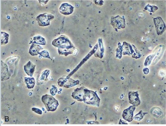
polarizing light microscopy
confirms presence of cholesterol
lipids from a maltese cross pattern w/ polarized light
also helpful for evaluating crystals
cholesterol and uric acid

how are casts reported in microscopy
avg. # seen per lpf
how are RBC/WBCs reported in microscopy
avg. number seen per hpf
number per lpf per facility
semiquantitative terms
rare, few, moderate, or many
1+, 2+, 3+, 4+
how are epithelial cells and crystals reported in microscopy
how are trans/renal cells reported in microscopy
number per hpf per facility
semiquantitative terms
rare, few, moderate, or many
1+, 2+, 3+, 4+
sediment stains
low refractive index elements are often difficult to see under bright-field microscopy
stains improve visibility
Sternheimer-Malbin stain
Lipid stains
Gram stain
Hansel stain
Prussian blue stain
Sternheimer-Malbin stain
sediment stain
crystal violet & safranin O
most commonly used
increases refractive index
absorbed well by WBCs, epithelial cells, and casts
stains nuclei, cytoplasm and inclusions

Lipid stain
sediment stain
Oil Red O and Sudan II
Triglycerides, neutral fats and cholesterol
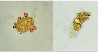
Gram stain
identification of bacterial casts
hansel stain
urinary eosinophils
methylene blue and eosin Y

Prussian blue stain
Hemosiderin granules seen w/ hemoglobinuria
Formed elements in sediment
small amounts can be normal or pathogenic based on the clinical picture
large numbers of abnormal formed elements is diagnostically significant
many urines have just a rare epithelial cell or mucus strand
some cellular elements are easily distorted
RBCs
small, smooth, non-nucleated, biconcave disks
identify and quantitate using high power (40x)
shape or appearance can aid in diagnosis
crenated in concentrated urine (shrink)
ghost cells (no hgb) in dilute urine (lyse)
dysmorphic - usually benign
acanthocytic, fragmented or hypochromic
acanthocytes - glomerular disorder

what can RBCs be mistaken for when identifying elements microscopically
Yeast - look for buds
oil droplets - refractility
air bubbles - refractility and possibly in a different plane
starch - refractile, polarizes

RBCs - reagent strip correlation - positive strip for blood w/ no RBCs seen microscopically
RBCs lysed; released hemoglobin tests positive
false-positive test - detects myoglobin, peroxidases
RBCs - reagent strip correlation - negative strip for blood w/ RBCs seen microscopically
ascorbic acid interference causes false-negative result
RBC look-alikes present, such as yeast or crystals
RBC clinical significance and reference value
ref value= 0-3 or 5/hpf
damage to glomerular membrane or vascular injury to the genitourinary tract - # cells correlate with damage
along w/ RBC casts - renal bleeding
no casts or protein - bleeding below the kidney or caused by contamination (menstruation)
macroscopic versus microscopic hematuria
cloudy, red urine
advanced disease, trauma, acute infection or coagulation disorders
clear urine
early glomerular disease, malignancy, strenuous exercise or renal calculi confirmation
early glomerular disease, malignancy, strenuous exercise or renal calculi confirmation
what can clear red urine indicate
early glomerular disease, malignancy, strenuous exercise or renal calculi confirmation
what can cloudy red urine indicate
WBCs
neutrophil is predominant → visible lobes and granules
glitter cells
hypotonic urine creates cell swelling of neutrophil
Brownian movement of granules → cells appear to sparkle
Eosinophils
Mononuclear cells (lymphocytes, monocytes, macrophages, or histiocytes are rare)
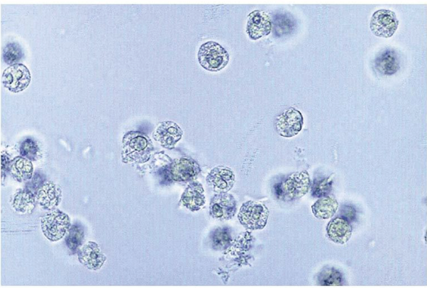
WBCs - Eosinophils
associated with: drug-induced nephritis and renal transplant rejection
evaluation:
concentrate sediment (centrifuge or cytocentrifuge)
Hansel stain
report % eosinophils per 100-500 cells (>1% significant, seen or not seen)
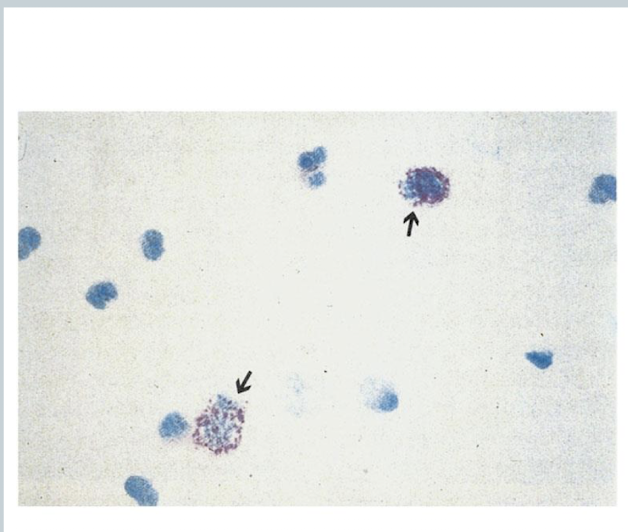
WBCs - Mononuclear cells
lymphocytes, monocytes, macrophages, or histiocytes are rare
lymphocytes may resemble RBCS
seen in early transplant rejection
differentiate from renal tubular epithelial (RTE) cells
may need to refer to cytodiagnostic testing
WBC clinical significance and ref value
ref value= <5 per hpf
more in females
may enter thru glomerulus or trauma but also be amoeboid migration
increased = pyuria
bacterial infections (cystitis, pyelonephritis, prostatitis, or urethritis
nonbacterial disorders (glomerulonephritis, lupus, interstitial nephritis, or tumors
possible presence of bacteria
squamous epithelial cells
vagina, female urethra and lower portion of male urethra
largest cell in urine
good for focusing microscope
rare, few, mod, or many (lpf or hpf per laboratory)
Normal sloughing
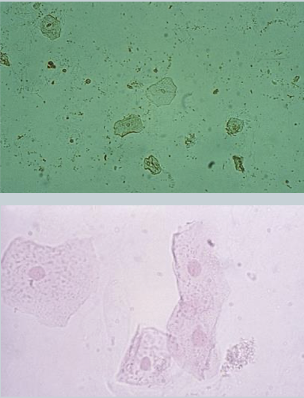
clue cells
squamous cell with pathologic significance
Gardnerella vaginalis - causes bacterial vaginosis infection
Coccobacillus covers most of the cell and extends over the edges of the squamous cell
seen in urine but more common in vaginal wet preparation

transitional epithelial cells
line renal calyces, pelvis, ureters, bladder, part of male urethra
vary in size due to separate layers
provides integrity and flexibility
centrally located nucleus
clinical significance: inflammation, infection, urinary procedure, carcinoma

what are the three forms of the transitional epithelial cells in bladder
spherical → superficial layer
caudate → intermediate layer
polyhedral → deep basal layer
clumps (malignancy) and sheets (instrumentation)

renal tubular epithelial cells
located in the kidney
involved in kindey function
each portion of the tubule is lined with different types of cell. shape varies based on location
maintain passage of water and electrolytes
clinical significance: kidney damage and renal tubular disease

PCT cells
larger than other RTEs
columnar, convoluted and/or rectangular
eccentric nucleus

DCT cells
round or oval-shaped
smaller
may resemble WBCs or spherical transitional cells
eccentrically placed nucleus
differentiates them from spherical transitional

collecting duct RTEs
cuboidal, never round
at least one straight edge
eccentric nucleus
renal fragment
3 or more cuboidal cells in a group
often large sheets
PCT and DCT not seen in clumps

clinical significance of RTE cells
most clinically significant urine epithelial cells
indicate tubular necrosis
Renal fragments indicate basement membrane destruction
at least 3 cells are still intact
heavy metals, drug toxicity, hemoglobin, myoglobin, viral infections, pyelonephritis, transplant rejection, salicylate poisoning
casts usually present
Oval fat bodies
often seen with free-floating refractile droplets
renal tubular cells w/ absorbed fat
confirm w/ sudan III or Oil red O stain
stains tryglycerides and neutral fats
view w/ polarizing light
cholesterol polarizes
“maltese cross”
lipiduria
nephrotic syndrome
acute tubular necrosis
diabetes
crush syndromes

bacteria
urine is usually sterile
contaminates during voiding
contaminants multiply fast
rods and cocci may be seen
rods most common
nitrite helps to confirm rods, not cocci
report few, moderate, or many per hpf
WBCs should accompany bacteria in urinary tract infection (UTI)

yeast
single, refractile, budding structures or hyphae
discern frm RBCs
few, moderate, or many per hpf
Clinical significance
diabetic urine w/ increased glucose
immunocompromised
Vaginal Candida species
nitrite negative w/ WBCs present

parasites
most common: Trichomonas vaginalis
pear shaped flagellate
swims across field very rapidly
few, moderate, or many per hpf
may resemble WBC, transitional, or RTE cells

spermatozoa
oval, tapered heads and long tail
urine is toxic to sperm
immobile
rarely significant
infertility
sperm expelled into bladder instead of urethra
legal consequences
may cause positive protein
reporting varies w. laboratories
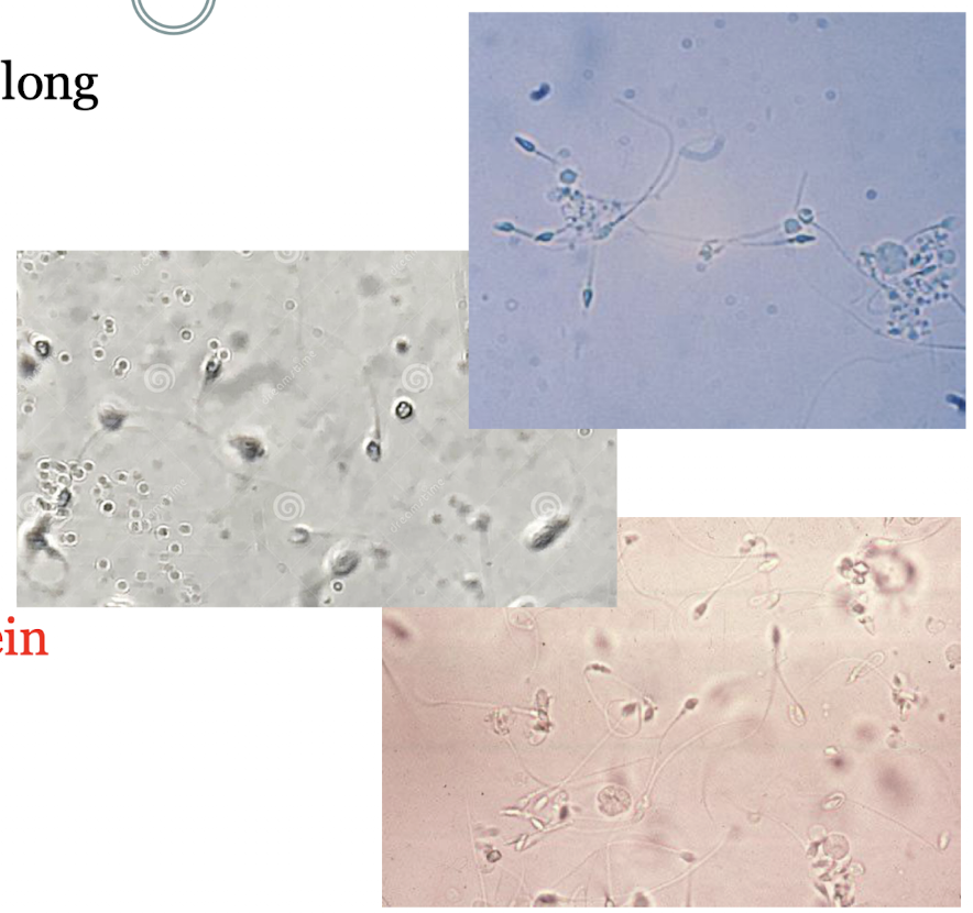
mucus
protein produced from RTE, glands, and epithelial cells
composed of Tamm-Horsfall protein (uromodulin)
Threadlike w/ low refractive index
confused w/ casts
irregular shape
UTIs, STDs, inflammation
mostly insignificant
rare, few, moderate, or many per hpf

casts
from the kidney
formed in distal convoluted tubules and collecting ducts
parallel sides, rounded ends, inclusions
reflect conditions of the nephron
detect under low power and identify under high power
report avg number per 10 lpf
scan edges of glass coverslip
composition, formation, and flow of casts
Tamm Horsfall (TH) protein secreted by renal tubular epithelial cells of the ascending loop of Henle, the distal convoluted tubule and upper portion of the collecting ducts
normally consistent excretion
increases w/ stress and exercise
TH protein gels
urine stasis, acid pH, Na and Ca
TH protein not detected by reagent strips
aggregated TH structure attached to RTEs
interweaving loose network which traps elements
more interweaving to form solid matrix
attachment of elements to matrix
detachment of structure from RTEs
excretion of cast
Hyaline casts
low refractive index
colorless when unstained
use low light or phase
normal parallel sides or convoluted, wrinkled, cylindroid, occasional adhering cell or granule

clinical significance of hyaline casts
most frequently seen cast
0-2 per lpf is normal
nonpathologic
stress, excercise, fever, heat exposure, dehydration
pathologic
glomerulonephritis, pyelonephritis, chronic renal disease, congestive heart failure

RBC casts
orange-red color
look for cast matrix to avoid mistaking a RBC clump for a cast
confirm seeing free RBCs and positive reagent strip for blood
report number seen under lpf

clinical significance of RBC casts
bleeding w/i the nephron
glomerular damage or nephron capillary damage
acute glomerulonephritis, nephritic syndrome
may be seen following strenuous exercise
cells begin to disintegrate w/ stasis of urine flow
hemoglobin degraded to methemoglobin
muddy brown casts
acute tubular necrosis
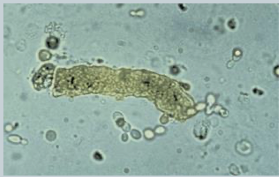
WBC casts
mostly neutrophils with multilobed nucleus and granules are seen
staining helps differentiate from RTE cells
may be tightly packed
look for cast matrix to distinguish from WBC clump
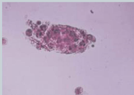
clinical significance of WBC casts
seen w/ infection and inflammation of the tubules
pyelonephritis
WBC casts and bacteria
acute interstitial nephritis
WBC casts and no bacteria
may accompany RBC casts
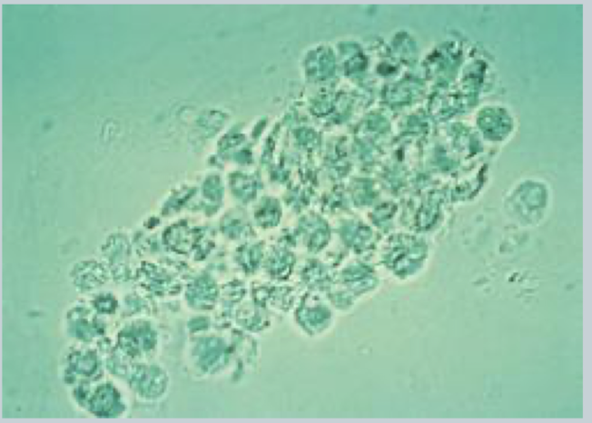
fatty casts
seen with oval fat bodies and free fat droplets
highly refractile
identify w/ polarized microscopy and lipid stains
nephrotic syndrome, diabetes mellitus, crush trauma, or tubular necrosis

granular casts
coarse and fine granular
detect with low power and identify with high power
pathologic or nonpathologic origin
granules disintegrate to form waxy casts
differentiate granular casts from clumps of debris and crystals
look for cast matrix

waxy cast
brittle, highly refractile
often fragmented with jagged ends and notches
well visualized w/ stain
degenerated hyaline and granular casts
represent extreme urine stasis
chronic renal failure
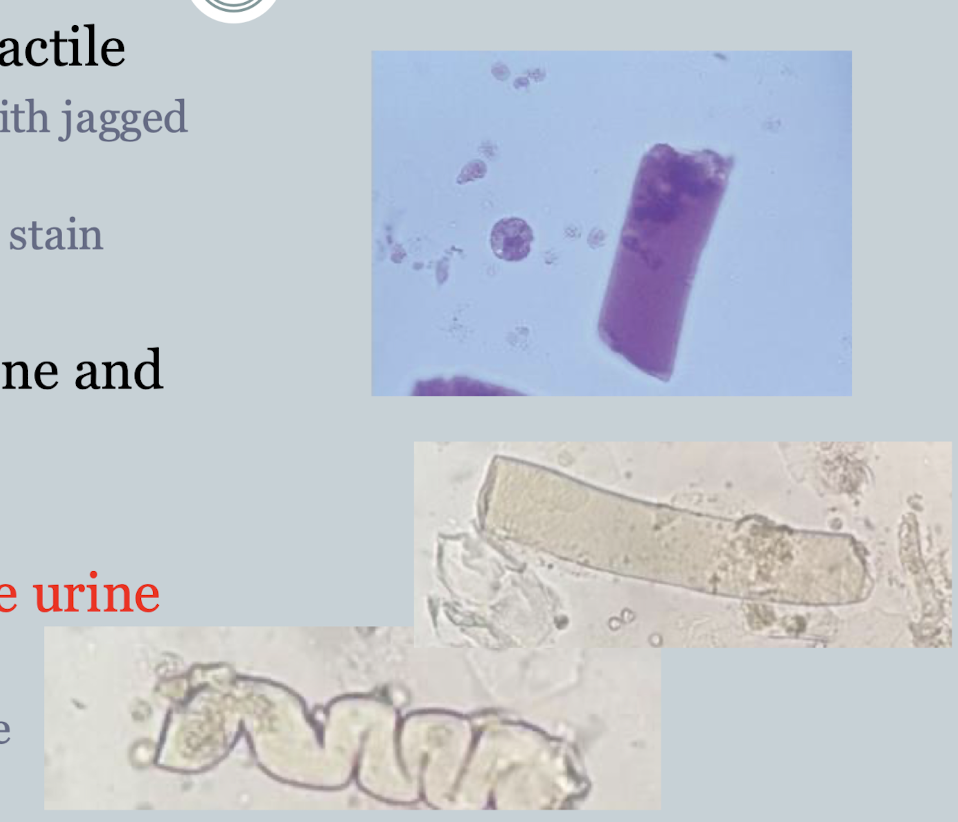
broad casts
chronic renal failure casts
destruction and widening of the tubule walls
formation in the upper collecting duct
all types of casts may be broad


material fibers, meat and vegetable fibers, hair, powder, pollen, oil droplets, fecal contamination
casts do not polarize except fatty casts
many fibers polarize
what artifacts resemble casts and how might they be distinguished
Lower UTI
what disease state do these results indicate:
RBC: ++
WBC: +++
Trans Epi: ++
Bacteria: +
upper UTI
what disease state do these results indicate:
RBC: +
WBC: +++
Renal epi: +
WBC cast: ++
RTE cast: +
Gran. cast: ++
acute glomerulonephritis
what disease state do these results indicate:
RBC: +++
WBC: +
Renal epi: +
RBC cast: +++
WBC cast: +
RTE cast: +
Gran. cast: ++
presence of dysmorphic RBCs (acanthocytes)
chronic glomerulonephritis
what disease state do these results indicate:
RBC: +
WBC: +
Renal epi: +
RTE cast: ++
Gran. cast: ++
waxy cast: ++
presence of broad casts
nephrotic syndrome
what disease state do these results indicate:
RBC: +
renal epi: +
gran cast: +
waxy cast: +
fatty cast: +++
presence of cholesterol crystals
acute tubular necrosis
what disease state do these results indicate:
RBC: ++
WBC: ++
renal epi: +++
RTE cast: ++
gran cast: ++
waxy cast: ++
presence of proximal RTE and collecting duct RTE
renal transplant
what disease state do these results indicate:
RBC: ++
WBC: ++
renal epi: +++
RTE cast: ++
gran cast: ++
waxy cast: ++
presence of lymphocytes
urinary crystals
significant in freshly voided urine
formed in vivo and can cause tubular damage
identified by microscopic appearance and pH
some indicate a pathologic process
report as rare, few, moderate, or many per hpf
crystal formation
precipitation of urine solutes
salts, organic compounds, and medications
formation based on:
temperature
solute conc.
urine pH
old urine
crystals in fresh specimens are associated w. high specific gravity
general identification of crystals
most have characteristic shapes and colors
most valuable identification is urine pH
polarized microscopy characteristics are valuable in identification
solubility characteristics of crystals
temperature and pH contribute to formation and solubility
amorphous urates form in refrigerated acid urine
dissolved w. heat
amorphous phosphates form in refrigerated alkaline urine
dissolve in acetic acid as will RBCs
amorphous urates
yellow-brown granules microscopically
urine sediment has pink color
often in clumps
pH is usually greater than 5.5

uric acid crystals
shapes
rhombic, whetstones, wedges, and rosettes
yellow-brown or colorless
may resemble cystine crystals but always polarize
seen in specimens from patients
with gout
undergoing chemotherapy for leukemia

calcium oxalate crystals
acid and neutral pH
colorless
envelope or two-pyramid-shaped
most common
monohydrate is oval or dumbbell-shaped
antifreeze poisoning
is a major component of renal calculi

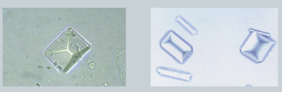
Triple phosphate
colorless
prism or coffin-lid shaped
polarize
no clinical significance
highly alkaline urine and urinary tract infections (UTIs)
amorphous phosphates
may appear similar to amorphous urates
differentiate
alkaline pH
heavy white precipitate after refrigeration

ammonium biurate crystals
yellow-brown, spicule-covered spheres
“thorny apples”
only urate in alkaline urine
found in old specimens and with urea-splitting bacteria producing ammonia

abnormal crystals - cystine
hexagonal, thin and thick plates
similar to uric acid
uric acid polarizes, but only thick cystine crystals polarize
seen in cystinuria
inherited ability to reabsorbed cystine
form renal calculi

abnormal crystals - cholesterol crystals
seen in refrigerated specimens
rectangular plates w. characteristic notched corners
identify by polarized light
seen in disorders w/ lipiduria
nephrotic syndrome
fatty casts and oval fat bodies

liver disease crystals
bilirubin crystals
tyrosine crystals
leucine crystals
bilirubin crystals
clumped needles or granules
characteristic yellow color
viral hepatitis w. tubular damage
positive reagent strip for bilirubin
liver disease crystal

tyrosine crystals
fine yellow needles in clumps or rosettes
seen w. leucine crystals
inherited amino acid disorders
liver disease crystals
will test positive on the bilirubin reagent strip

leucine crystals
yellow-brown spheres w/ concentric circles and radial striations
will test positive on the bilirubin reagent strip

ampicillin crystals
colorless needles that form bundles after refrigeration
massive dosage with inadequate hydration

sulfa crystals
most common to precipitate
variety of shapes
patient treated for UTI

radiographic dye
can form crystals in urine
