chap 9
 Call Kai
 Learn
 Practice Test
 Spaced Repetition
 Match
 Flashcards
 Knowt Play
1/105

There's no tags or description
Looks like no tags are added yet.
106 Terms

Joints (articulations)
places of union between bones, bones & cartilage, or bones & teeth
bones articulate at a joint
arthrology: study of joints
form by 6th week of development
categorized structurally on the basis of whether a space (called a joint cavity) occurs between the articulating bones & the type of CT that binds the articulating surfaces of the bones
classified by structure & function
structural (what fills between bones)
fibrous
cartilaginous
synovial
functional (extent of movement)
synarthroses
amphiarthroses
diarthroses
range of motion
no movement → extensive movement
structure of each determines its mobility & stability
inverse relationship (tradeoff) between mobility & stability
immobile = stable
mobile = unstable
ex. skull sutures are immobile but very stable
most stable → least stable / immobile → most mobile
suture → interosseous membrane → intervertebral joints → knee joint → glenohumeral joint (shoulder)

arthr- / arthro-
relating to joints

Fibrous joint
structural class
bones held together by dense regular CT (collagen)
primary function: hold 2 bones together
immobile or slightly mobile (synarthroses or amphiarthroses)
no joint cavity
ex. gomphosis, suture, syndesmosis
suture & syndesmosis has interosseous membrane

Cartilaginous joint
structural class
bones joined by cartilage
either hyaline cartilage or fibrocartilage
primary function: resist compression / tension stress, act as resilient shock absorbers
immobile or slightly mobile (synarthroses or amphiarthroses)
no joint cavity
ex. synchondrosis (hyaline), symphysis (intervertebral joints, pubic symphysis)

Synovial joint
structural class
bones joined by ligaments w fluid filled joint cavity separating bone surfaces
lined by synovial membrane
primary function: movement
freely mobile (diarthroses)
has joint cavity (articular cavity)
filled w synovial fluid
common features
articular capsule & joint cavity
synovial fluid / membrane
articular cartilage (end of each bone)
ligaments, nerves, blood vessels
also has bursae, tendon sheaths, & fat pads
classes by movement
uniaxial
bone moves in just 1 plane or axis
forward & back or up & down
biaxial
bone moves in 2 planes or axes
multiaxial / triaxial
bone moves in multiple planes or axes
classes by shape (least mobile → most mobile)
plane joints (uniaxial)
hinge joints (uniaxial)
pivot joints (uniaxial)
condylar joints (biaxial)
saddle joints (biaxial)
ball & socket joints (multiaxial / triaxial)
movements that occur at __ __ are
gliding
angular
rotational
special
ex. glenohumeral joint (shoulder), knee joint, elbow joint, temporomandibular joint
may be compared to levers

Articular cartilage
hyaline cartilage on bone surface at joint
lacks perichondrium
avascular
functions
reduces friction during movement
acts as a cushion to absorb joint compression
prevents damage to articulating ends of bones
articulating surface are enclosed within a CT capsule
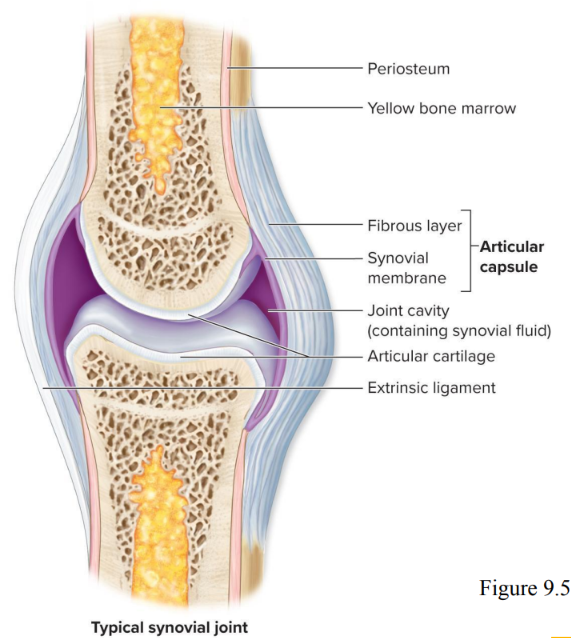
Joint capsule / Articular capsule
encloses a synovial joint
encapsulating joint cavity is double layered __
outer fibrous layer
dense CT
strengthens joints to prevent bones being pulled apart
inner synovial layer / membrane
composed primarily of areolar CT
covers internal joint surfaces not covered by cartilage
helps produce synovial fluid

Joint cavity / Articular cavity
space between articulating bones
separates articulating surfaces of the bones
lined by synovial membrane secreting synovial fluid
viscous, oily substance
functions
lubricate articular cartilage on articulating surfaces
nourishes the chondrocytes of articular cartilage
acts as a shock absorber (distributes stresses)
permits movement of the articulating bones
Ligaments
dense regular CT
connect one bone to another
stabilize, strengthen, & reinforce synovial joints
extrinsic ligaments
outside of & physically separate from articular capsule
intrinsic ligaments
thickening of the articular capsule itself (fibrous layer)
include ligaments outside (extracapsular ligaments) & within the articular capsule (intracapsular ligaments)
Sensory receptors & blood vessels
numerous in synovial joints
proprioceptors: report on movement, compressive forces, positioning, stretch,
nociceptors: detect painful stimul
Tendons
composed of dense regular CT
not part of the synovial joint itself
attach muscle to bone
help stabilize joints
sometimes limit the range of movement

Bursae
fibrous, saclike structures containing synovial fluid
lined internally by synovial membrane
either associated w joint cavity or completely separate from it
found in synovial joints where bones, ligaments, muscles, skin, or tendons rub together
large # in shoulder
alleviate friction

Tendon sheaths
elongated bursae
wrap around tendons where friction is excessive
common in wrist & ankle
Fat pads
act as protective packing material in joint periphery
can fill spaces when joint shape changes
Synarthroses / Synarthrosis
functional class
immobile joints
no joint cavity
stationary
can be fibrous (2) or cartilaginous (1) joints
Amphiarthroses / Amphiarthrosis
functional class
slightly mobile joints
no joint cavity
can be fibrous (1) or cartilaginous (1) joints
Diarthroses / Diarthrosis
functional class
freely mobile joints
has joint cavity
all synovial joints

Gomphosis
type of fibrous joint
“peg in a socket”
articulations of teeth w sockets of mandible & maxillae
tooth held in place by fibrous periodontal ligaments (periodontal membranes)
the only __ in the body
located within alveolar sockets
function as synarthroses / synarthrotic joints (immobile)
periodontal ligaments
peri = around / dontal = tooth
the only gomphoses in the body
specialized, dense fibrous connective tissue that suspends each tooth root inside its socket in the alveolar bone
rather than being fused directly to the bone like a dental implant, teeth are cushioned and held in place by thousands of tiny, shock-absorbing collagen fibers

Sutures
type of fibrous joint
found between some skull bones
very short collagen fibers
interlocking, irregular edges
increase stability & decrease risk of fracture
function as synarthrosis / synarthrotic joints (immobile)
allow growth in childhood
permit the skull to grow by new bone being deposited
become ossified synostoses in older adults
synostoses
the fusion or union of two or more separate bones into a single bone
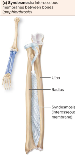
Syndesmosis / Syndesmoses
type of fibrous joint
bones bound by interosseous membrane (interosseous ligament) a broad ligamentous sheet
dense regular CT
function as amphiarthroses / amphiarthrotic joints (limited mobility)
provide pivot for 2 long bones
ex. found between radius & ulna / tibia & fibula

Synchondrosis / Synchondroses
type of cartilaginous joint
bones joined by hyaline cartilage
immobile (synarthroses)
ex. epiphyseal plate, costochondral joints (attach ribs to sternum), first sternocostal joint

Symphyses / Symphysis
type of cartilaginous joint
disc / pads of fibrocartilage between articulating bones
resist compression & act as shock absorbers
allow slight mobility (amphiarthroses)
ex. pubic symphysis / intervertebral discs / joints
Clinical: Costochondritis
inflammation of the costochondral joints
localized chest pain
cause usually unknown (trauma or infection)
may be mistaken for pain from a myocardial infarction (heart attack)
treated w nonsteroidal anti - inflammatory drugs

Plane joint
articular surfaces flat (or slightly curved)
simplest, least mobile synovial articulation
uniaxial: limited side-to-side gliding movement in a single plane
angle between bones do not change, only limited movement possible in any direction
ex.
intercarpal joints
intertarsal joints
acromioclavicular (AC) joint
temporomandibular (TMJ)
intervertebral (between superior & inferior articular processes of adjacent vertebrae)
vertebrocostal
lumbosacral (between articular processes)
sternocostal
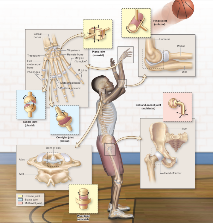

Hinge joint
convex surface within concave depression
uniaxial: like the hinge of a door
movement in one place, flexion/extension
ex.
elbow joint
humeroulnar joint
knee joint
IP (Interphalangeal joints)
temporomandibular (TMJ) (elevation & depression)

Pivot joint
bone w rounded surface fits into ligament ring
uniaxial: rotation of longitudinal axis
ex.
atlantoaxial joint (between first two cervical vertebrae)
radioulnar joint

Condylar / Condyloid joint
oval, convex surface articulating w oval, concave surface
biaxial (back & forth and side to side / flex & extend and abduct & adduct
ex.
MP (metacarpophalangeal or metatarsophalangeal) joints (also known as knuckles)
radiocarpal joint (wrists)
atlanto occipital joint

Saddle joint
both articulating bones have convex & concave surface resembling saddle shape
biaxial
allow movement across two perpendicular plane
ex.
articulation between a carpal bone (trapezium) & first metacarpal (carpometacarpal joint) also known as trapeziometacarpal
permits thumb opposition (the ability of the thumb to move toward the other fingers) so that we can grasp objects
sternoclavicular joint

Ball and socket joint
spherical head of one bone fitting into cuplike socket
multiaxial, permitting movement in 3 planes
the most freely mobile type of joint
ex.
glenohumeral (shoulder) joint
hip (coxal) joint
Costochondral joint
joint between each bony rib & its respective costal cartilage
is a synchondrosis
First sternocostal joint
attachment of the 1st rib to the sternum (manubrium) by costal cartilage
is a synchondrosis
Types of motion that occur at synovial joints
gliding
angular
rotational
special movements
motions that occur only at specific joints
Gliding motion
2 opposing articular surfaces slide past each other in almost any direction, the amount of movement is slight (limited)
back & forth or side to side
typically occurs along plane joints
ex. between carpals or tarsals
imagine sliding door
Angular motion
involve decreasing or increasing the angle of a joint
ex.
flexion
extension (+hyperextension)
lateral flexion
abduction
adduction
circumduction

Flexion
the angle between articulating bones decreases in an anterior posterior (AP) plane
brings bones closer together
ex. bending finger
opposing movement: extension
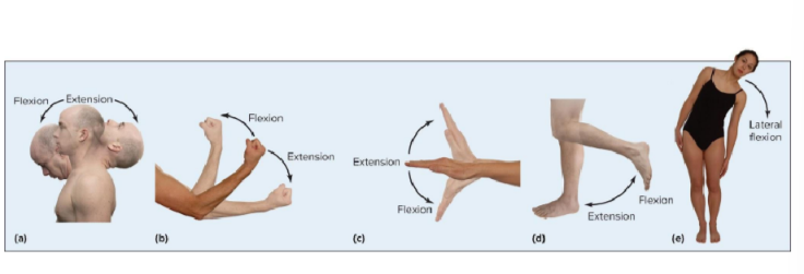

Extension
the angle between articulating bones increases in an anterior posterior (AP) plane
ex. straightening your fingers after making a fist
opposing movement: flexion

Hyperextension
extension beyond normal range of motion
possible w extensively mobile joints or an injury
Lateral flexion
the vertebral column / trunk moves (bends) in a lateral direction along a coronal plane
occurs between vertebrae in the cervical & lumbar region
angular movement
opposing movement: none


Abduction
lateral movement of a body part away from the midline
angular movement
ex. arm or thigh moved laterally from body midline
opposing movement: adduction


Adduction
medial movement of a body part toward the midline
angular movement
ex. arm or thigh brought back toward midline
opposing movement: abduction


Circumduction
a sequence of movements in which the proximal end of an appendage remains relatively stationary while the distal end makes a circular motion
movement makes an imaginary cone shape
complex movement that occurs as a result of a continuous sequence of flexion, abduction, extension & adduction
angular movement
ex. drawing a circle on the blackboard
opposing movement: none

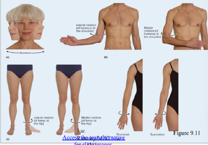
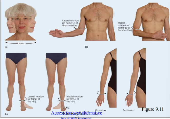
Rotational motion
a bone pivots around its own longitudinal axis
ex
pronation
supination
lateral rotation: turns anterior surface of bone laterally
medial rotation: turns anterior surface of bone medially

Pronation
rotation of the forearm where the palm is turned posteriorly or inferiorly
medial rotation of forearm so palm of hand posterior
opposing movement: supination
radius & ulna pivoting along interosseous membrane
palm of hand facing posteriorly
head of radius still on lateral side of elbow
distal end of radius has crossed over
movement does not happen in elbow, happens in forearm
“pour that out”

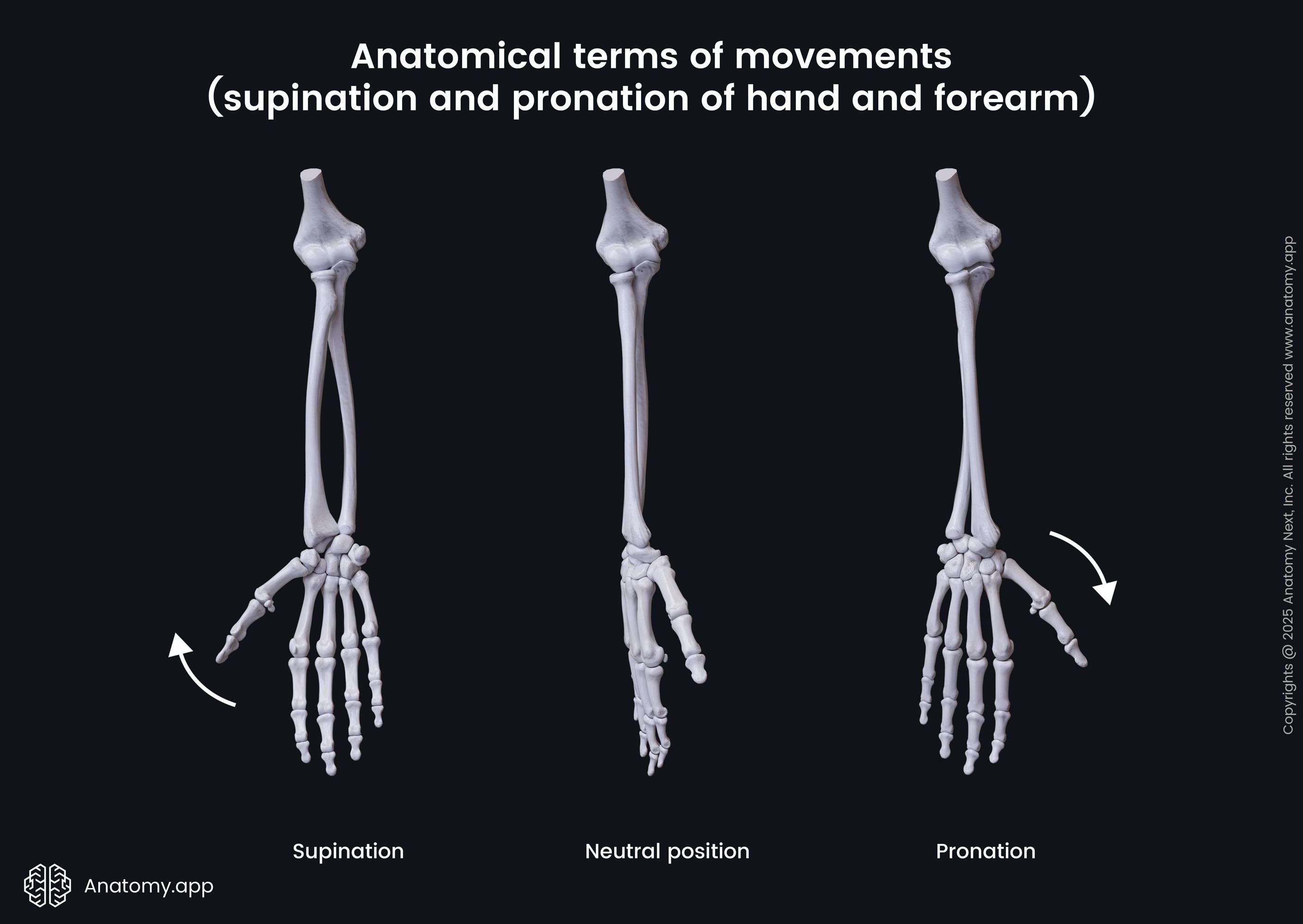
Supination
rotation of the forearm in which the palm is turned anteriorly or superiorly
lateral rotation of forearm so palm of hand anterior
opposing movement: pronation
in anatomic position, palm of hand facing anteriorly
radius & ulna parallel
radius on lateral side of forearm
ulna on medial side
movement does not happen in elbow, happens in forearm
“side by side” “how you carry soup”
Special movements
types of movement that do not fit into previous categories
ex.
depression
elevation
dorsiflexion
plantar flexion
eversion
inversion
protraction
retraction
opposition & reposition
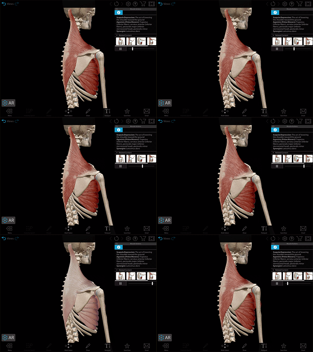
Depression
movement of a body part inferiorly
ex. movement of mandible while opening mouth
opposing movement: elevation


Elevation
movement of a body part superiorly
ex. movement of mandible when closing mouth
opposing movement: depression


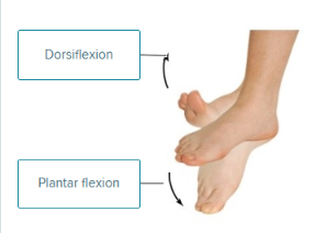
Dorsiflexion
ankle joint movement where the dorsum (superior surface) of the foot is brought toward the anterior surface of the leg
talocrural (ankle) joint bent so the dorsum of foot moves toward the leg
ex. digging in your heels
opposing movement: plantar flexion
“dig in your heels”
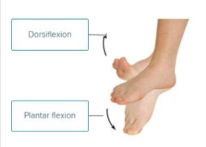

Plantar flexion
ankle joint movement where the sole of the foot is brought toward the posterior surface of the leg
talocrural (ankle) joint bent so dorsum pointed inferiorly
ex. ballet dancer on tiptoes in full plantar flexion
opposing movement: dorsiflexion
“push your toes into the plant”

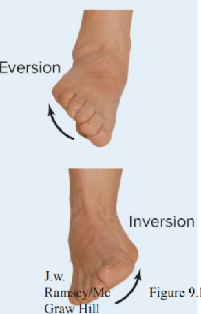
Eversion / foot pronation
twisting motion of the foot that turns the sole laterally or outward
occurs only at intertarsal joints (between tarsals) of foot
opposing movement: inversion
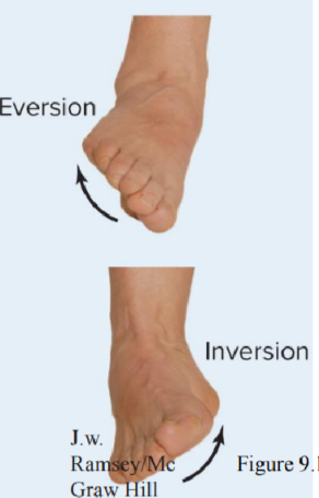

Inversion / foot supination
twisting motion of the foot that turns the sole medially or inward
occurs only at intertarsal joints (between tarsals) of foot
opposing movement: eversion

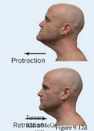
Protraction
anterior movement of a body part from anatomic position
ex. jutting jaw anteriorly at temporomandibular joint
opposing movement: retraction


Retraction
posterior movement of a body part from anatomic position
ex. pulling in jaw posteriorly at temporomandibular joint
opposing movement: protraction

Opposition
special movement of the thumb across the palm toward the tips of fingers to permit grasping & holding of an object
at carpometacarpal joint
opposing movement: reposition
back in anatomical position
biomechanics
practice of applying mechanical principles to biology

Levers
elongated, rigid object
rotates around fixed point, fulcrum
ability to alter the speed & distance of movement produced by a force, direction of an applied force, & force strength
effort applied to one point
resistance located at some other point
movement occurs if effort exceeds resistance
effort arm
part of lever from fulcrum to point of effort
resistance arm
part of lever from fulcrum to point of resistance
synovial joint movement can be compared to the mechanics of a lever system
FRE123 = the part near the middle of each type lever
Fulcrum in 1st class levers
Resistance in 2nd class lever
Effort in 3rd class lever
Class 1 = scissors, atlanto occipital joint
Class 2 = wheelbarrow, plantarflexion of ankle
Class 3 = tweezers, most common in our body
longer lever arms generate force
shorter level arms generate speed


(1st) First Class Levers
fulcrum is between effort & resistance
ex. pair of scissors / playground teeter totter
effort applied to handle of scissors
fulcrum along the middle of scissors
ex. atlanto-occipital joint of neck

(2nd) Second Class Levers
resistance is between fulcrum & effort
rare in body
small force able to balance larger weight
bc the effort is always farther from the fulcrum than the resistance
ex. wheelbarrow
fulcrum far from effort
ex. standing ontiptoe (plantarflexion of ankle)
ball of the foot is fulcrum, gravity trying to pull us down is resistance, muscle puling is effort

(3rd) Class Levers
effort applied between resistance & fulcrum
ex. tweezers / forceps
most common in body
ex. forceps
ex. elbow joint
joint is fulcrum, bicep tendon applies effort, weight in hand provides resistance (or by the weight of the forearm itself)
ex. mandible when you bite food
temporomandibular joint is fulcrum, temporalis muscle exerts effort, food eaten is resistance

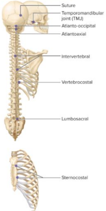
Axial skeleton joints
suture
temporomandibular
atlanto-occipital
atlantoaxial
intervertebral
vertebrocostal
lumbosacral
sternocostal
Suture joint
adjacent skull bones
fibrous joint
synarthrosis
no movement

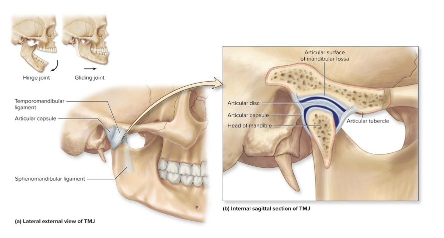
Temporomandibular joint (TMJ)
head of mandible & mandibular fossa of temporal bone
head of mandible & articular tubercle of temporal bone
synovial (hinge & plane) joints
diarthrosis
depression (opening mouth), elevation (biting down), lateral displacement (waggling jaw side to side), protraction (sticking jaw out), retraction (returning jaw back), gliding, & slight rotation of mandible
only mobile joint between bones in the skull
has loose articular capsule
surround joint
promotes extensive range of motion
has articular disc
thick pad of avascular dense regular CT
divides synovial cavity into 2 chambers
so tmj is 2 synovial joints, one between temporal bone & articular disc and one between articular disc and mandible
ligaments
sphenomandibular ligament (extracapsular)
extends anteriorly & inferiorly from sphenoid to mandible
temporomandibular ligament (lateral)
2 short bands that extends inferiorly & posteriorly from articular tubercle of temporal bone to mandible
movements
exhibits hinge, gliding, & some pivot joint movements
functions like a hinge during mandible depression & elevation (chewing)
glides during protraction of the mandible for biting, side to side grinding
articular disc
a thin, oval plate of fibrocartilage found within certain synovial joints
diff from intervertebral discs bc they have nucleus pulposus & annulus fibrosus and arent inside synovial joints
found within the TMJ, wrist, and the sternoclavicular joint
divides the joint (horizontally), directing flow of synovial fluid
absorbs shock
Clinical: TMJ Disorders
most commonly occurs due to alternation in the ligaments securing the joint
articular disc forced out of normal position
clicking or popping heard as person opens or closes mouth
possible paint in joint, paranasal sinuses, tympanic membrane, oral cavity, eyes, & teeth
Atlanto occipital joint
superior articular facets of atlas & occipital condyles of occipital bone
synovial (condylar) joint
diarthrosis
flexion & extension of the head
slight lateral flexion of the head

Atlantoaxial joint
anterior arch of atlas & dens of axis
synovial (pivot) joint
diarthrosis
rotation of the head
Intervertebral joint
vertebral bodies of adjacent vertebrae
cartilaginous joint (symphysis) between vertebral bodies
amphiarthrosis
superior & inferior articular processes of adjacent vertebrae
synovial (plane) joint between articular processes
diarthrosis
movements: flexion, extension, lateral flexion of vertebral column

Vertebrocostal joint
facets of heads of ribs & bodies of adjacent thoracic vertebrae & intervetebral discs between adjacent vertebrae
articular part of tubercles of ribs & facets of transverse processes of thoracic vertebra
synovial (plane) joint
diaarthrosis
some slight gliding of the ribs

Lumbosacral joint
body of the 5th lumbar vertebra & the base of the sacrum
cartilaginous joint (symphysis) between lumbar body & base of sacrum
amphiarthrosis
inferior articular facets of 5th lumbar vertebra & superior articular facets of 1st sacral vertebra
synovial (plane) joint between articular processes
diarthrosis
movements: flexion, extension, lateral flexion of vertebral column

Sternocostal joint
sternum (breastbone) and the costal cartilages of the first 7 ribs
cartilaginous joint (synchondrosis) between sternum & first ribs
synarthrosis
no movement
synovial (plane) joint between sternum & ribs 2-7
diarthrosis
some gliding movement


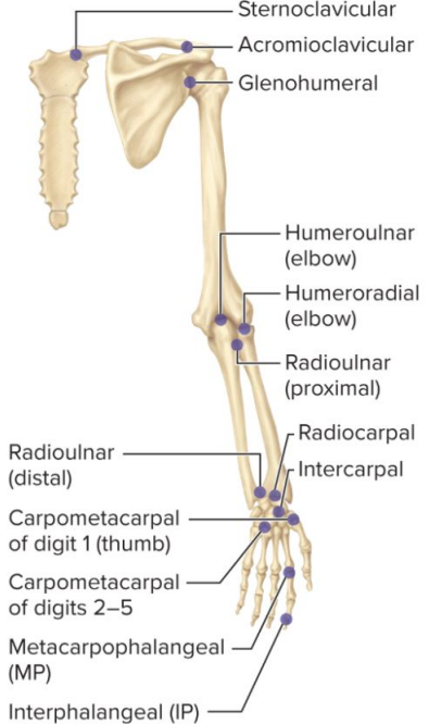
Pectoral girdle & upper limb joints
joints associated w movement at shoulder
sternoclavicular
acromioclavicular
glenohumeral
elbow (humeroulnar & humeroradial)
radioulnar (proximal & distal)
radiocarpal
intercarpal
carpometacarpal
metacarpophalangeal (MP, knuckles)
interphalangeal (IP)

Sternoclavicular joint
manubrium of sternum & sternal end of clavicle
synovial (saddle)
diarthrosis
depression, elevation, circumduction of clavicle
small-scale, precise circumduction during large arm movements
fibrocartilaginous articular disc
partitions joint into 2 parts forming 2 synovial cavities
stability provided by fibers & ligaments
makes this specific joint very stable & difficult to dislocate
sternoclavicular ligament & costoclavicular ligament


Acromioclavicular (AC) joint
acromion of scapula & acromial end of clavicle (lateral)
synovial (plane)
diarthrosis
gliding of scapula on clavicle
fibrocartilaginous articular disc within joint cavity
joint capsule strengthened superiorly by acromioclavicular ligament
connects clavicle to the acromion
clavicle bound to coracoid process by coracoclavicular ligament
if torn, acromion & clavicle no longer align (shoulder separation)

Glenohumeral (shoulder) joint
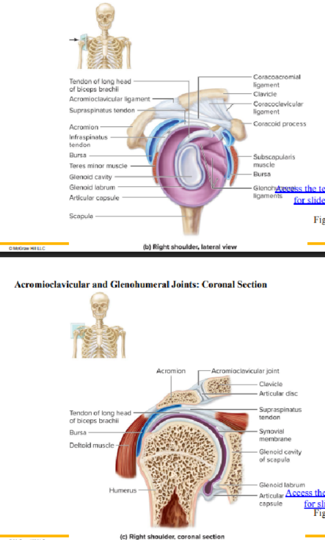
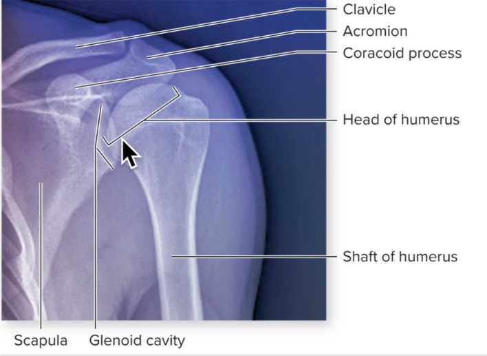
glenoid cavity of scapula & head of humerus
synovial (ball and socket)
diarthrosis
abduction, adduction, circumduction, flexion, extension, lateral rotation & medial rotation of arm
permits greatest range of motion of any joint in the body
most unstable & most frequently dislocated
shallow cup + minimal support from ligaments
fibrocartilaginous glenoid labrum encircles & covers the surface of the glenoid cavity
abundant bursae
decrease friction where tendons & muscles extend across capsule
supporting ligaments & tendons
coracoacromial ligament
extends across the space between the coracoid process & the acromion
coracohumeral ligament
thickening of the superior part of the articular capsule, extends from coracoid process to the humeral head
glenohumeral ligament
3 thickening of the anterior portion of the articular capsule, are often indistinct or absent & provide only minimal support
tendon of long head of biceps brachii
within articular capsule
helps stabilize the humeral head in the joint
most joint stability due to rotator cuff muscles
subscapularis, supraspinatus, infraspinatus, teres minor
work as a group to hold head of humerus in glenoid cavity
tendons encircle joint & fuse w articular capsule
loose articular capsule attaches to the surgical neck of the humerus

glenoid labrum
a ring of soft fibrocartilage attached to the rim of the shoulder blade's socket (the glenoid)
Clinical: Shoulder Joint Dislocations
common due to joint instability
shoulder separation refers to acromioclavicular joint dislocation
pain when arm abducted more than 90 degrees
acromion appearing prominent
glenohumeral joint dislocations usually occur when fully abducted humerus struck hard
should appears flattened and “squared off”
humeral head anterior & inferior to glenohumeral joint capsule

Elbow joint
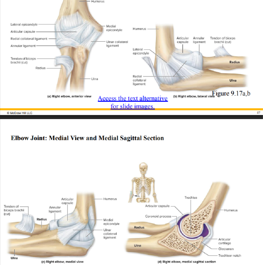
hinge joint composed of 2 articulations
flexion & extension
both enclosed within a single articular capsule
involves humerus, radius, & ulna
Humeroulnar joint
trochlea of humerus & trochlear notch of ulna
hinge joint
Humeroradial joint
capitulum of humerus & head of radius
diarthrosis
flexion & extension of forearm
is very stable (not as mobile) bc
articular capsule is thick (encloses both joints)
bony surfaces of humerus & ulna interlock closely
multiple, strong ligaments reinforce articular capsule
radial / lateral collateral ligament (RCL)
extends from lateral epicondyle (humerus) to head of radius
stabilizes joint at lateral surface
ulnar / medial collateral ligament (UCL)
extends from medial epicondyle (humerus) to coronoid & olecranon processes (ulna)
stabilizes medial side of the joint
annular ligament
surrounds neck of radius
binds head of the radius to the ulna

Clinical: Subluxation of the head of the radius
subluxation: refers to incomplete dislocation
head pulled out of annular ligament
occurs almost exclusively in children (usually < 5 yrs)
bc of the loose positioning of the still developing head of the radius, within a thin annular ligament
childs annular ligament thin & radial head not fully formed
pediatrician may maneuver radial head back into annular ligament
Radioulnar joint
proximal joint
proximal end / head of radius & radial notch of ulna
distal joint
distal end of ulna & ulnar notch of radius
synovial (pivot)
diarthrosis
pronation & supination of forearm

Radiocarpal joint
distal end of radius (scaphoid, lunate, & triquetrum)
synovial (condylar)
diarthrosis
abduction, adduction, circumduction, flexion & extension of wrist

Intercarpal joint
adjacent carpal bones
synovial (plane)
diarthrosis
gliding of the carpals

Carpometacarpal joint
thumb: trapezium & first metacarpal
synovial (saddle)
other digits: carpal & metacarpals II-V
synovial (plane)
diarthrosis
flexion, extension, abduction, adduction, circumduction, & opposition at thumb, gliding at other digits

Metacarpophalangeal (MP, knuckles) joint
head of metacarpals & bases of proximal phalanges
synovial (condylar)
diarthrosis
flexion, extension, abduction, adduction, circumduction of phalanges

Interphalangeal (IP) joint
heads of proximal & middle phalanges w bases of middle & distal phalanges, respectively
synovial (hinge)
diarthrosis
flexion & extension of phalanges

Pelvic girdle & lower limb joints
sacroiliac
hip (coxal)
pubic symphysis
knee (tibiofemoral & patellofemoral)
tibiofibular (superior & inferior)
talocrural (ankle)
intertarsal
tarsometatarsal
metatarsophalangeal (MP)
interphalangeal (IP)
Sacroiliac joint
auricular surfaces of sacrum & ilium
synovial (plane)
diarthrosis
slight gliding of the hip bones, more movement during pregnancy & childbirth

Hip joint (coxal)
head of femur & acetabulum of os coxae (hip)
synovial (ball and socket)
diarthrosis
flexion, extension, abduction, adduction, circumduction, medial, & lateral rotation of thigh
acetabular labrum
fibrocartilaginous ring that deepens socket
more stable, less mobile than glenohumeral joint
secured by articular capsule, ligaments & muscles
articular capsule
extends from acetabulum to trochanters
encloses femoral head & neck
retinacular fibers
ligamentous fibers of articular capsule
reflect around femur neck
retinacular arteries here
supply most blood to femur head & neck
intracapsular ligaments
iliofemoral ligament
Y shaped, provide support for anterior articular capsule
ischiofemoral ligament
spiral shaped, posteriorly located
pubofemoral ligament
triangular thickening of capsules inferior region
become taut when hip extended
so hip joint is most stable in its extended position
ligament of head of femur (ligamentum teres)
small ligament from acetabulum to fovea of head of femur
does not provide stability
typically contains small artery supplying head of femur
taut
stretched or pulled tight, rigid, tense, or concise

Clinical: Fracture of the femoral neck
fracture of femur often incorrectly referred to as fractured hip
intertrochanteric fractures (between the trochanters)
extracapsular fractures
usually in younger & middle aged individuals
subcapital fractures
occur within hip articular capsule (intracapsular)
usually in older individuals w osteoporosis
may result in tearing of retinacular arteries
may have avascular necrosis in the region
Pubic symphysis joint
symphyseal surface of both pubic bones
cartilaginous (symphysis)
amphiarthrosis
very slight gliding of the pubic bones, more movement during childbirth

Knee joint
largest & most complex diarthrosis
flexion, extension, lateral rotation of leg in flexed position, slight medial rotation
/ capable of slight rotation & lateral gliding when flexed
primarily hinge joint
tibiofemoral joint
medial condyle (femur), medial meniscus, & medial condyle (tibia)
synovial (hinge)
patellofemoral joint
patella & patellar surface of femur
synovial (hinge & plane)
articular capsule
encloses only medial, lateral, & posterior knee regions (NOT anterior)
no single unified capsule / joint cavity in the knee
quadriceps femoris muscle tendon
passes over knees anterior surface, surrounds patella
medial meniscus & lateral meniscus
deep to articular capsule within knee joint
C shaped fibrocartilage pads on top of tibial condyles
cushioning between articular surfaces
change shape to conform to articulating surfaces
partially stabilize joint medially & laterally
extracapsular ligaments
patellar ligament
extends from patella to tibial tuberosity
oblique popliteal ligament
protects against hyper extension
fibular collateral ligament (FCL / LCL)
extends from femur to fibula
reinforces lateral surface of joint
prevents hyperadduction
tibial collateral ligament (TCL /
MCL)
reinforces medial surface of joint
extends from femur to tibia
prevents hyperabduction
also attaches to medial meniscus
arcuate popliteal ligament
intracapsular ligaments
cruciate ligaments: cross each other X
deep to articular capsule
anterior cruciate ligament (ACL)
extends from posterior femur to anterior tibia
prevents hyperextension & anterior displacement of tibia
becomes taut on extension
posterior cruciate ligament (PCL)
extends from anteroinferior femur to posterior tibia
prevents hyperflexion & posterior displacement of tibia
becomes taut on flexion
Clinical: Knee injuries & Knee replacement surgery
knee is highly vulnerable to injury
tibial collateral ligament injury when leg forcible abducted
fibular collateral ligament injury when medial side of knee is struck
ACL injury when leg hyperextended
PCL injury when leg hyperflexed
meniscus injury due to trauma and/or overuse
unhappy triad: injury of tibial collateral ligament, medial meniscus, & ACL
occurs due to a lateral blow to the knee that abducts & laterally rotates leg
ligamentous injuries increase risk of osteoarthritis of knee
if sever enough, individual may opt for knee replacement surgery
arthritic parts of bones removed & replaced w metal or plastic compounds
how do i memorize fcl prevents hyperabduction and tcl prevents hyperabduction
the ligaments are located on the opposite side of the movement they are trying to stop
think of them like ropes or anchors. A rope can only stop something from moving away from it by pulling tight
collateral
a secondary, accessory, or side-by-side structure that runs alongside or supports a main pathway
Tibiofibular joint
superior joint
head of fibula & lateral condyle of tibia
synovial (plane)
inferior joint
distal end of fibula & fibular notch of tibia
fibrous (syndesmosis)
amphiarthrosis
slight rotation of fibula during dorsiflexion of foot

Talocrural (ankle) joint
distal end of tibia & medial malleolus of tibia with talus
lateral malleolus of fibula & talus
synovial (hinge)
highly modified
diarthrosis
dorsiflexion & plantar flexion of the foot
includes 2 articulations within 1 articular capsule
between distal end of tibia & talus
between distal end of fibula & lateral aspect of talus
medial & lateral malleoli of tibia & fibula
prevent talus from sliding medially or laterally
articular capsule
covers distal tibia, medial malleolus, lateral malleolus, & talus
deltoid / medial ligament
binds tibia to foot on medial side
prevents over eversion
lateral ligament
binds fibula to foot on lateral side
prevents over inversion
prone to sprain & tears
anterior & posterior tibiofibular ligaments
Clinical: Ankle Sprains & Pott fractures
sprain
stretching or tearing of ligaments without fracture or dislocation
results from twisting foot, usually over inversion
fibers of lateral ligament stretched or torn
localized swelling & tenderness anteroinferior to lateral malleolus
pott fracture
occurs with over eversion
medial malleolus avulsed off of tibia (fracture)
talus moves laterally and fractures fibula
Intertarsal
between the tarsal bones
synovial (plane)
diarthrosis
eversion & inversion of foot