The Central Nervous System
 Call Kai
 Learn
 Practice Test
 Spaced Repetition
 Match
 Flashcards
 Knowt Play
1/34

Earn XP
Description and Tags
Lecture 21
35 Terms
Central Nervous System ( CNS)
CNS- composed of the brain and spinal cord
Cephalization
Elaborization of the anterior portion of the CNS
Increase in the number of neurons in the head
Highest level is reached in the human brain
Basic Pattern of the Central Nervous System
Spinal Cord
central cavity surrounded by a gray matter core
External to which is white matter composed of myelinated fibers tracts
Brain
Similar to spinal cord but with additional areas of gray matter
Cerebellum has gray matter in nuclei
Cerebrum has nuclei and additional gray matter in the cortex
Basic Pattern of the Central Nervous System

Spinal Cord

Spinal Cord
Conus medullaris- terminal portion of the spinal cord
Filum terminale- fibrous extension of the pia mater; anchors the spinal cord to the coccyx
Denticulate ligaments- delicate shelves of pia mater; attach the spinal cord to the vertebrae
Spinal Cord
Spinal nerves- 31 pairs attach to the cord by paired roots
Cervical and lumbar enlargements- sites where nerves serving the upper and lower limbs emerge
Cauda equina- collection of nerve roots at the inferior end of the vertebral canal
Gray Matter and Spinal Roots

Ventricles of the Brain
Arise from expansion of the lumen of the neural tube
The ventricles are:
The pair C-shaped laterals ventricles
The third ventricle found in the diencephalon
The fourth ventricle found in the hindbrain dorsal to the pons
Ventricles of the Brain

Ventricles of the Brain

Ventricles of the Brain

Adult Neural Canal Regions
Adult structures derived from the neural canal
Telencephalon- lateral ventricles
Diencephalon- third ventricle
Mesencephalon- cerebral aqueduct
Metencephalon and myelencephalon- fourth ventricle
Cerebral Hemispheres
Form the superior part of the brain and make up 83% of its mass
Contains ridges (gyri) and shallow grooves (sulci)
Contains deep grooves called fissures
Are separated by the longitudinal fissure
Have three basic regions: cortex, white matter, and basal nuclei
Major Lobes, Gyri, and Sulci of the Cerebral Hemisphere
Deep sulci divide the hemispheres into five lobes:
Frontal, parietal, temporal, occipital, and insula
Central sulcus- separates the frontal and parietal lobes
Major Lobes, Gyri, and Sulci of the Cerebral Hemisphere
Parieto-occipital sulcus- separates the parietal and temporal lobes
Lateral sulcus- separates the parietal and temporal lobes
The precentral and postcentral gyri border the central sulcus
Functional Areas of the Cerebral Cortex
The three types of functional areas are:
motor areas- control voluntary movement
sensory areas- consious awareness of sensation
association areas- integrate diverse information
Functional Areas of the Cerebral Cortex

Functional Areas of the Cerebral Cortex

Cerebral Cortex: Motor Areas
Primary (somatic) motor cortex
Premotor cortex
Broca’s area
Frontal eye field
Primary Motor Cortex

Premotor Cortex
Located anterior to the precentral gyrus
Controls learned, repetitious, or patterned motor skills
Coordinates simultaneous or sequential actions
Involved in the planning of movements
Broca’s Area
Broca’s area
Located anterior to the inferior region og the premotor area
Present in one hemisphere (usually the left)
A motor speech area that directs the muscles of the tongue
Is active as one prepares to speak
Sensory Areas
Primary somatosensory cortex
Somatosensory association cortex
Visual and auditory areas
Olfactory, gustatory, and vestibular cortices
Sensory Areas

Primary Somatosensory Cortex
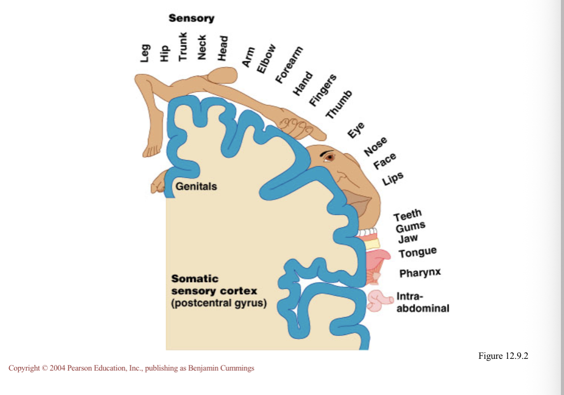
Located in the postcentral gyrus, this area:
Receives information from the skin and skeletal muscles
Exhibits spatial discrimination
Somatosensory homunculus- caricature of relative amounds of cortical tissue devoted to each sensory function
Primary Somatosensory Cortex
Somatosensory Association Cortex
Located posterior to the primary somatosensory cortex
Integrates sensory information
Forms comprehensive understanding of the stimulus
Determines size, texture, and relationship of parts
Visual Areas
Primary visual (striate) cortex
Seen on the extreme posterior tip of the occipital lobe
Most of it is buried in the calcarine sulcus
Receives visual information from the retinas
Visual association area
Surrounds the primary visual cortex
Interprets visual stimuli (e.g., color form, and movement)
Auditory Areas
Primary auditory cortex
Located at the superior margin of the temporal lobe
Receives information related to pitch, rhythm, and loudness
Auditory association area
Located posterior to the primary auditory cortex
Stores memories of sounds and permits perception of sounds
Wernicke’s area
Prefrontal Cortex
Located in the anterior portion of the frontal lobe
Involved with intellect, cognition, recall, and personality
Necessary for judgement, reasoning, persistence, and conscience
Closely linked to the limbic system ( emotional part of the brain)
Meninges

Dura Mater
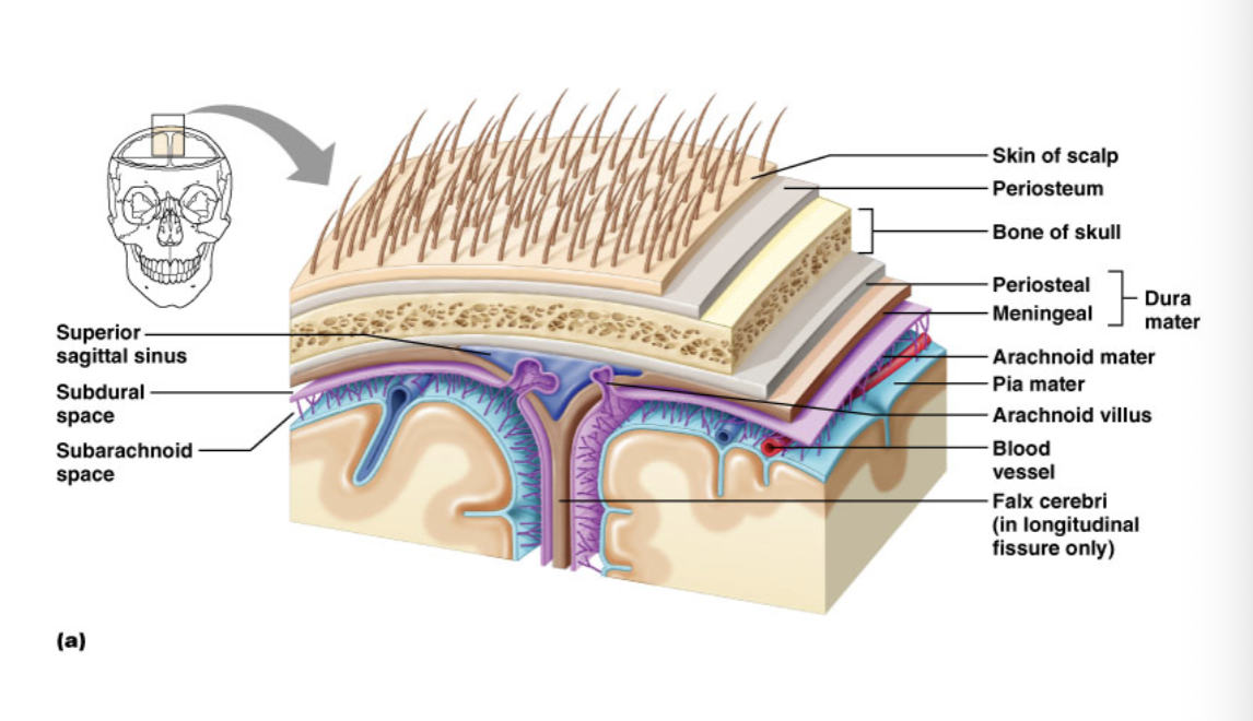
Leathery, strong meninx composed of two fibrous connective tissue layers
The two layers separate in certain areas and form dural sinuses
Arachnoid Mater
The middle meninx, which forms a loose brain covering
It is separated from the dura meter by the subdural space
Beneath the arachnoid is a wide subarachnoid space filled with CSF and large blood vessels
Arachnoid villi protrude superiorly and permit CSF to be absorbed into venous blood
Arachnoid Mater
Cerebrospinal Fluid (CSF)
Watery solution similar in composition to blood plasma
Contains less protein and different ion concentrations than plasma
Forms a liquid cushion that gives buoyancy to the CNS organs
Prevents the brain from crushing under its own weight
Protects the CNS from blows and other trauma
Nourishes the brain and carries chemical signals throughout it