Tibia, Fibula, Stifle Joint and Proximal Hindlimb Muscles
 Call Kai
 Learn
 Practice Test
 Spaced Repetition
 Match
 Flashcards
 Knowt Play
1/30

There's no tags or description
Looks like no tags are added yet.
31 Terms
What type of bones are the tibia and fibula
LONG bones
List the structures at the distal end of the tibia and fibula and list their function
Are these structures palpable?
Medial malleolus:
Attachment of medial collateral ligament of hock
Lateral malleolus:
Attachment of lateral collateral ligament of hock
Both palpable


List both cs, r, arrow and * in the image. List what bone this is and what anatomical location of the bone these structures are located at.
Tibial plateau / plate
Medial condyle (c)
Lateral condyle (c)
Smooth bone surface
Articulate with medial & lateral condyles of femur
Intercondylar ridge (r)
Non-articular rough bone surface
ligament attachment
Tibial crest
Tibial tuberosity
insertion of patellar ligament
Palpable
List the 4 centers of ossification of the Tibia
Proximal end (epiphysis)
Tibial tuberosity
Body (diaphysis)
Distal end (epiphysis)
List the 4 components that comprise the Stifle Joint
Femorotibial joint
Femoropatellar joint
Proximal tibiofibular (where fibula present)
Femur + paired sesamoids (carnivores)
List the articular surfaces of the femorotibial
Articular surfaces :
Lateral & medial femoral condyles
Lateral & medial tibial condyles
List the structures that provide the stifle joint with stability
Medial & lateral collateral ligaments
Cranial & Caudal Cruciate ligaments
What clinical test can you use to test for a cranial cruciate rupture?
Cranial draw test
tibia moves cranial relative to femur
List the components of the femoropatellar
Components
Trochlear groove (femur)
Patella
Stability
Medial and lateral trochlear ridges
Lateral & medial (collateral) femoro-patellar ligaments
Fascia / retinaculum

What happens when the patella is pulled proximally vs distally?
Patella pulled proximally
Produces stifle extension
Patella pulled distally
Allows stifle flexion
List the sesamoid bones of the stifle and how many of each there are
Patella (1)
Fabellae (2)
Popliteal sesamoid (1)
List the 3 compartments the stifle joint forms.
Describe the communication of these compartments in the canine vs. equine
Compartments
Femoro-patellar
Lateral femoro-tibial
Medial femoro-tibial
Communication
Dog – good
Horse – poor / none
Describe how the patellar locking mechanism works
Patella pulled proximally as stifle extended
Patella rotated medially
Engages horizontal / resting surface
Tubercle of medial trochlear ridge now sits between middle & medial patellar ligaments
Locks joint in extension
Describe what is happening to the patella when unlocking
What is the clinical significance of the patellar locking mechanism
Patella pulled proximally then back to midline to unlock
Clinical significance:
Upward fixation / ‘locked’ patella
List the nerve supply to the hind limb
Spinal nerves
L5, L6, L7, S1, S2.
Dorsal branches - dorsal structures
Ventral branches - lumbosacral plexus
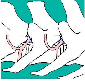
List the emerging peripheral nerves to the hindlimb
Gluteal
Obturator
Femoral
Sciatic – tibial
fibular / peroneal

Dorsal Thigh Muscles
Gluteal Muscles
Describe the gluteal muscles (how many parts, origin, insertion, function, and nerve supply)
3 parts
Origin
sacrum & pelvis
Insertion
greater trochanter of femur
acts as lever
Function
Hindlimb abductors - dog
Hindlimb retractor / hip extensors – horse
Nerve Supply
Gluteal nerve
Medial / Ventral Thigh Muscle
Gape Muscles
List the GAPE muscles and the insertion of G and P
Gracilis (+)
I = tibia
I = calcaneus (tarsus)
(via common calcanean tendon)
Adductor (*)
Pectineus (#)
Both I = femur
Function
adductors
Nerve Supply
obturator nerve
List the cranial thigh muscles
Quadriceps muscle
Sartorius muscle
Iliopsoas muscle
Describe the quadriceps muscle (4 heads, origin, insertion, function and nerve supply)
4 Heads and Origin
Rectus femoris: O-ilium
Vastus lateralis: O – lateral femur
Vastus medialis: O – medial femur
Vastus intermedius: O - cranial femur
Insertion
Tibial Tuberosity
Function
stifle extensor
(Hip flexor / limb protractor)
Nerve Supply
femoral nerve
Describe the Sartorius muscle (origin, insertion, function and nerve supply)
O - ilium
I - femur
I – tibial tuberosity
(via patellar ligament)
Function:
Stifle extensor
Hip flexor / limb protractor
Nerve Supply
femoral nerve
Describe the Iliopsoas muscle (origin, insertion, function and nerve supply)
O - lumbar vertebrae & ilium
I – medial proximal femur
Function:
Hip flexor / limb protractor
Nerve supply
femoral nerve
List the caudal thigh muscles (3) and their nerve supply
Biceps femoris
Semitendinosus
Semimembranosus
Nerve Supply
Sciatic Nerve
Describe the Biceps Femoris muscle (origin, insertion, function and nerve supply)
O – tuber ishium
I - fascia latae
I – calcaneus (tarsus)
(via common calcanean tendon)
Functions:
Hip extensor / hindlimb retractor
Stifle flexor
Hock extensor
Nerve supply
Sciatic nerve
Describe the Semitendinosus muscle (origin, insertion, function and nerve supply)
O – Tuber Ischium
I - Tibia
I – calcaneus (tarsus)
(via common calcanean tendon)
Function:
Hip extensor / hindlimb retractor
Stifle flexor
(Hock extensor)
Nerve Supply
sciatic nerve
Describe the Semimembranosus muscle (origin, insertion, function and nerve supply)
O – Tuber ischium
I - femur & tibia
Function:
Hip extensor / hindlimb retractor
Stifle flexor
Nerve Supply
sciatic nerve
Following transection of the patellar ligament, which major function of the stifle joint is most likely affected?
A. Abduction of the stifle joint
B. Adduction of the stifle joint
C. Extension of the stifle joint
D. Flexion of the stifle joint
Extension of the stifle joint
In dogs, the patella is embedded into the tendon of which muscle?
A. Biceps femoris
B. Gastrocnemius
C. Popliteal
D. Quadriceps femoris
Quadriceps femoris
Which head of the quadriceps femoris is a flexor of the hip joint and extensor of the stifle joint
A. Rectus femoris
B. Vastus intermedius
C.
D.
Rectus femoris