chapter 31: disorders of ventilation and gas exchange
 Call Kai
 Learn
 Practice Test
 Spaced Repetition
 Match
 Flashcards
 Knowt Play
1/90

There's no tags or description
Looks like no tags are added yet.
91 Terms
structurally respiratory system is divided into 2 parts:
upper respiratory system: nose, pharynx, and associated structures
lower respiratory system: larynx, trachea, bronchi, and lungs
functionally respiratory system is divided into 2 zones:
conducting zone
respiratory zone
conducting zone
carries air from the outside to the lungs (nasal cavity, pharynx, larynx, trachea, bronchi, bronchioles, terminal bronchioles)
respiratory zone
where O₂ and CO₂ are exchanged between the air and blood (respiratory bronchioles, alveolar ducts, alveolar sacs, alveoli)
alveoli
air sacs are found in lungs where oxygen and carbon dioxide exchanged btwn air and blood
surrounded by networks of pulmonary capillaries
two types of cells in alveoli
type I: allow gas exchange
type II: secrete surfactant
ventilation
mechanical movement of gas or air into and out of lungs
alveolar ventilation
volume of air that enters alveoli each minute
portion of total ventilation, that participates in exchange of oxygen and carbon dioxide
respiratory membrane
formed by shared alveolar and capillary walls, is composed of:
a layer of type I and type II alveolar cells
an epithelial basement membrane underlying alveolar wall
capillary basement membrane
capillary endothelium
lung compliance
ability of lungs to expand in response in pressure
describes how easily lungs inflate
lung compliance is affected by:
elastic recoil of lung tissue
surface tension
elastic recoil of lung tissue:
tendency of lungs to return to their original size
lungs naturally tend to recoil inward chest wall tends to spring outward
these opposing forces help maintain normal lung expansion
surface tension within alveoli
surfactant reduces surface tension
surfactant prevents alveolar collapse during expiration
surfactant increases lung compliance and reduces work of breathing
respiratory volumes represent:
amount of air present in lungs during different phases of breathing
they are measured using pulmonary function tests (PFTs)
tidal volume (TV)
vital capacity (VC)
residual volume (RV)
total lung capacity (TLC)
tidal volume (TV)
amount of air moved into and out of lung w/ each breath (~500ml)
vital capacity (VC)
max volume of air a person can forcefully expel from their lungs after maximum inhalation
residual volume (RV)
amount of air that always remains in lungs + needed to keep alveoli open
total lung capacity (TLC)
maximum volume of air lungs can hold
TLC = VC + RV (~6000ml)
picture of respiratory volumes

central chemoreceptors
located in medulla oblongata
reflect CO2
stimulated by hydrogen (H+) in cerebrospinal fluid (pH)
increases respiratory depth and rate
peripheral chemoreceptors
located in aorta and carotid bodies
stimulated by hypoxemia
responsible for increase in ventilation that occurs in response to arterial hypoxemia
hypercapnia
carbon dioxide levels in blood increased
carbon dioxide easily diffuses into CSF
lowers pH and stimulates central chemoreceptors
increased rate and depth of respirations - ventilation
hypoxemia
marked decrease in blood oxygen level
peripheral chemoreceptors stimulated and respond
important ctrl mechanism in individuals w/ chronic lung disease - move to hypoxic drive
picture of control of ventilation

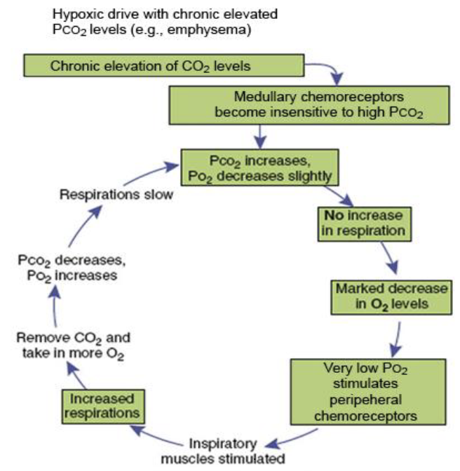
hypoxic drive
mechanism where body relies on low arterial oxygen lvls (PaO2) to stimulate breathing rather than primary stimulus - high arterial carbon dioxide levels (PaCO2)
w/ hypoxic drive, in chronic respiratory conditions (COPD):
cystic fibrosis hypoxic drive becomes primary ctrl of respiratory due to:
hypercapnic desensitization
shift to peripheral chemoreceptors
hypercapnic desensitization (COPD)
chronic hypercapnia makes central chemoreceptors become less sensitive to high carbon dioxide lvls
shift to peripheral chemoreceptors (COPD)
breathing ctrl shifts entirely to peripheral chemoreceptors located in carotid bodies and aortic arch
these receptors respond to decreased arterial oxygen (PaO2 < 60 mmHg)
picture of hypoxic drive
mild hypoxemia (manifestation of hypoxemia)
increase in heart rate
peripheral vasoconstriction
diaphoresis: sweating
increase in blood pressure
chronic hypoxemia
often presents w/ insidious manifestations
increased ventilation
increased production of RBC: hypoxia stimulates erythropoietin secretion
cyanosis: decreased oxygenation of RBC
eupnea
norm breathing pattern 14-20 bpm
types of abnormal breathing patterns:
tachypnea: abnorm fast breathing
bradypnea: abnorm slow breathing
apnea: cessation of breathing
kussmaul respirations (hyperpnea)
cheyne-stokes respiration
kussmaul respirations (hyperpnea)
slightly increased ventilatory rate, very large tidal volume, and no expiratory pause
commonly seen in diabetic ketoacidosis (DKA)
cheyne-stokes respiration
alternates btwn deeper and faster breathing and shallower breathing, culminating in period of apnea before cycle repeats
associated w/ heart failure + neurological conditions
dyspnea
subjective sensation of uncomfortable breathing described as SOB
severe dyspnea: flaring of nostrils, use of accessory muscles of respiration
dyspnea on exertion: shortness of breath with activity
orthopnea
dyspnea when lying down
wheezing
high-pitched whistling sound during breathing caused by narrowed airways
hemoptysis
coughing up blood or blood strained mucus from lower respiratory tract
cough
protective reflex that helps clear the airways by an explosive expiration
productive cough (wet): brings up sputum
non-productive cough (dry): does not bring mucus or sputum, characterized as “hacking”
abnormal sputum
changes in amount, consistency, color, and odor provide info about progression of disease and effectiveness of therapy
pneumothorax
air accumulation in pleural cavity causing lung to partially or completely collapse
manifestations: chest pain, dyspnea and tachypnea, reduced breath sounds
closed pneumothorax
air can enter pleural cavity from internal airways - no open chest wall
secondary pneumothorax
associated w/ underlying respiratory disease
rupture of an emphysematous bleb or erosion by tumor
open pneumothorax
traumatic pneumothorax
atmospheric air enters pleural cavity through an opening in chest wall - sucking wound on chest
tension pneumothorax
occurs when opening in chest wall or lung tissue allows air to enter pleural cavity during inhalation but prevents it from escaping during exhalation
trapped air accumulates, causing increasing pressure within pleural space
most serious form
pleural effusion
presence of excessive fluid in pleural cavity
cause increased pressure in pleural cavity
two main types: exudate effusions, transudative effusions
exudative effusions
response to inflammation
purulent drainage: empyema
blood: hemothorax
transudative effusions
result of increased hydrostatic pressure or decreased osmotic pressure in blood vessels
watery effusions: hydrothorax
manifestations of pleural effusions:
chest pain: unilateral, localized to lwr and lateral part of chest, usually becomes worst w/ chest movements
dullness to percussion and absence of breath sounds over affected area
dry cough: persistent cough without producing mucus
management: thoracocentesis - chest drainage performed to remove fluid and relieve pressure
atelectasis
incomplete expansion of lung or portion of lung
collapse of lung or part of lung leads to decreased gas exchange and hypoxia
alveoli becomes airless: inflammation or atrophy occur
atelectasis interferes with:
blood flow through the lung
when alveoli receive little or no oxygen, nearby pulmonary blood vessels constrict
both ventilation and perfusion are altered: diminished or absent gas exchange
atelactasis classified into 3 main types based on cause:
obstructive: caused by total obstruction of airway
compression: external mass or tumor exerts pressure on part of lung
postoperative: can occur after surgery
COPD
grp of chronic respiratory disorders that cause irreversible and progressive dmg to lungs
characterized by long-term breathing difficulties and airflow limitation
may lead to dev of respiratory failure and cor pulmonale (right sided heart failure)
causes and risk factors of COPD
tobacco smoke as well as second-hand smoke exposure
Environmental exposures: prolonged exposure to air pollution, chemical fumes, dust
genetic factors: 𝛼_1-antitrypsin deficiency a genetic disorder that leads to early-onset emphysema even in non-smokers
bronchial smooth muscle
found in walls of bronchi and bronchioles
ctrls diameter of airways, regulating how much air enters and leaves lungs
presence or absence in airway secretions can also influence airway patency
bronchial smooth muscle is innervated by the:
autonomic system:
parasympathetic vagal ctrl: bronchoconstrictor
sympathetic B2-adrenergic receptor ctrl: bronchodilator
asthma
bronchial obstruction occurs in persons w/ hypersensitive or hyperresponsive airways
most common chronic disease in children: 18.9 million in US, estimated 7.1 million are children
two categories: extrinsic asthma, intrinsic asthma
extrinsic asthma
acute episodes triggered by type I hypersensitivity reactions - common in children
intrinsic asthma
triggered by cold respiratory infections, stress - onset during adulthood
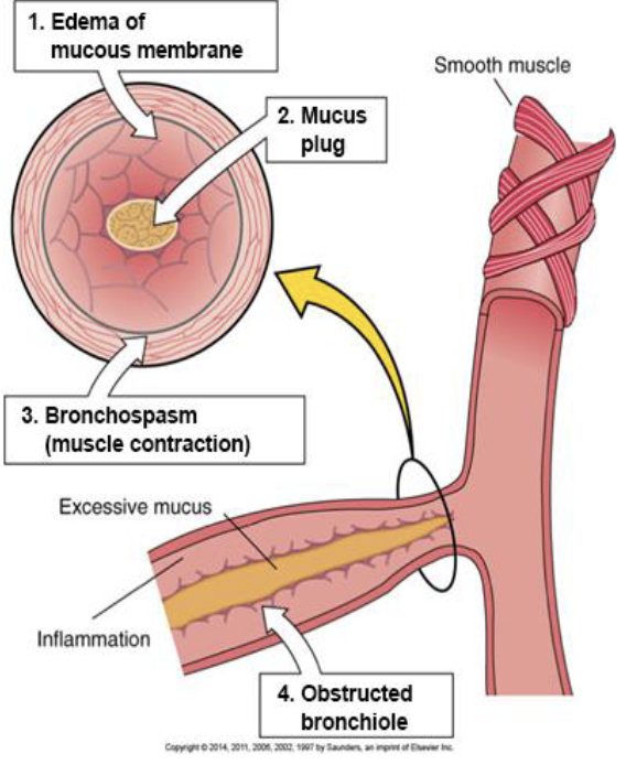
in asthma, pathological changes in bronchioles:
inflammation of the mucosa with edema
bronchoconstriction caused by contraction of smooth muscle
Increased secretion of thick mucus
pathogenesis of asthma

early phase pathogenesis of asthma
5-60 min: exposure to allergen, IgEs bind to mast cells, sensitized mast cells release histamine
bronchoconstriction: bronchial smooth muscle contraction
increased mucus secretion
attraction other immune cells
late phase pathogenesis of asthma
4-24 hrs: recruitment of eosinophils and sustained inflammation
repeated inflammatory episodes and injury
manifestations of asthma:
on inhalation narrowed bronchial lumen expands slightly
exhalation becomes prolonged due to obstruction: accessory muscles of expiration are used
forced expiratory volume decreased, residual volume increase:
dyspnea: difficult to exhale air from lungs, trapping it in smaller airway
wheezing: air forced through narrowed airways, creating high-pitched whistling sound on exhalation
asthma pH changes:
initially mild respiratory alkalosis caused by hyperventilation
prolonged severe asthma attack causes respiratory acidosis due to airway obstruction + air trapping
status asthmaticus
persistent severe attack on asthma
doesn’t respond to usual therapy fatal because of severe hypoxia and acidosis
management of asthma
pulmonary function studies to assess and monitor asthma
peak expiratory flow rate: measure speed of attack?
treatment: short and long acting B2 agonist (albuterol), corticosteroids
emphysema
abnormal permanent enlargement of air spaces distal to terminal bronchioles
destruction of alveolar walls and septae leads to large, permanently inflated alveolar air spaces
hypercapnia becomes marked
hypoxia becomes driving force of respiration (hypoxic drive)
contributing factors of emphysema
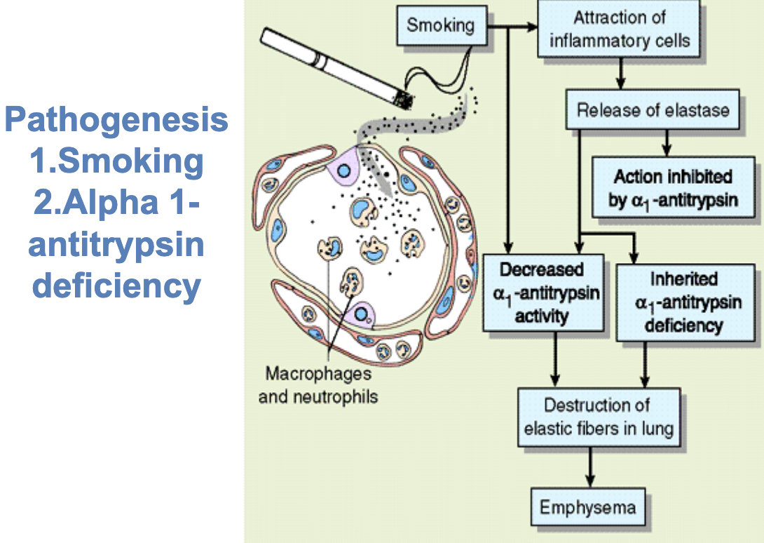
genetic deficiency
smoking
pathogenic bacteria
picture of pathogenesis w/ smoking
alpha-1 antitrypsin (AAT)
protein produced mainly by liver
main function to protect lungs from dmg caused by enzyme called neutrophil elastate
process occurs in following steps involving AAT:
genetic deficiency: abnorm genes produce insufficient AAT
inflammation attract neutrophils
neutrophils release elastate to breakdown harmful bacteria + potentially dmging elastic fibers in lungs
normally AAT inhibits elastase + coats lungs protecting them from neutrophil elastase
AAT deficiency: uninhibited elastase cause destruction of elastic fibers in lungs
breakdown of alveolar walls results in: (pathologic changes in emphysema)
loss of surface area for gas exchange
loss of pulmonary capillaries
altered ventilation-perfusion ratio: mismatch btwn air reaching alveoli and blood flow in pulmonary capillaries
fibrosis (pathologic changes in emphysema)
cause weakening of walls and narrowing in airways
interference w/ passive expiratory airflow - difficult to remove air from lungs
manifestations of emphysema
progressive difficulty w/ expiration cause air trapping + increased residual volume:
overinflation of lungs
barrel chest: fixation of ribs in respiratory position, increased anterior-posterior diameter of thorax
flattened diaphragm (on radiographs)
“pink puffer”: SOB and pink color of skin: no cyanosis until late stage of disease
pursed-lip breathing: prolonged expiration accessory muscles
management of emphysema
injections of donor AAT
anti-inflam drugs
bronchodilators: medications relax bronchial smooth muscles, widen airways, and improve airflow
oxygen therapy as condition advances
in late stages: lung reduction surgery and transplant
chronic bronchitis
inflammation obstruction, and repeated infection in bronchi with excessive mucus production, resulting in persistent coughing and difficulty breathing
clinical definition: productive cough lasting st least 3 months per 2 consecutive yrs, after excluding other possible causes
cause and risk factors of chronic bronchitis
increased risk with history of cigarette smoking or living in urban or industrial area
chronic irritation from smoke cause inflammation and swelling in mucosa in large and small bronchi and overproduction of mucus
chronic inflammation cause obstructive narrowing of airways:
in large bronchi: increase in goblet cells and squamous metaplasia of ciliary epithelial cells
impaired ciliary function: build-up of mucus plugs in airways
in small bronchi: smooth muscle hyperplasia and fibrosis: thickening walls and narrowing of bronchial lumen: increased airway resistance
manifestations of chronic bronchitis
manifestations develop gradually, chronic productive cough in early stages
in advanced stages dyspnea and wheezing
pulmonary hypertension due to fibrosis in pulmonary capillaries cause cor pulmonale - right sided heart fialure
peripheral edema: back up effects of right-sided heart failure
“blue bloater”: cyanosis and fluid retention due to right-sided heart failure
picture of symptoms for chronic bronchitis and emphysema

bronchiectasis
abnorm irrev dilation of medium-sized bronchi
result of weakening of muscle and elastic fibers in bronchial walls, causing perm dmg and abnorm dilation
bronchiectasis develops when repeated:
inflammation or infection dmgs bronchial walls
impaired bronchial drainage cause recurrent infections by streptococci, staphylococci, pneumococci, H. influenzae
manifestations of bronchiectasis
chronic productive cough w/ large amnt of sputum
rales and rhonchi in lungs: light and harsh sounds
foul breath: due to persistent bacterial infection
dyspnea
hemoptysis: due to dmgd bronchial blood vessels
cystic fibrosis (CF)
inherited genetic disorder that affects lungs, pancreas, digestive system, and other organs
causes body to produce thick, sticky mucus
tenacious thick mucus from exocrine glands obstruct airways, pancreatic, bile ducts
cause of cystic fibrosis
autosomal recessive disorder, abnorm CFTR gene located on chromosome 7
most common inherited disease in caucasians
1 in 25 caucasian americans is carrier of gene
in cystic fibrosis, the CFTR gene provides:
instructions to protein channel on surface of cells to ctrl flow of chloride ions + water
mutations in CFTR cause to abnorm transport of chloride ions and water w/ excessive sodium secretion and formation of thick mucus
for cystic fibrosis, within airways and lungs, thick mucus obstructs:
airflow and impairs drainage leading to chronic, persistent lung infections
results in perm dmg to bronchial walls development of cor pulmonale
in digestive tract:
bowel obstruction in newborns
pancreatitis: caused by blockage of pancreatic ducts
cirrhosis of liver: due to obstruction of bile ducts
sweat glands: high sodium chloride content
manifestations of cystic fibrosis:
meconium ileus may occur at birth: due to intestinal obstruction
salty skin: due to increased salt concentration in sweat
signs of malabsorption, steatorrhea: fatty stool abdominal distention
chronic cough and freq respiratory infections
dehydration: excessive salt loss through sweating
failure to meet norm growth milestone
diagnosis of cystic fibrosis with screening in newborn:
specific pancreatic protein: blood spot test
sweat test: 2 positive test w/ elevated NaCl
require confirmation
genetic testing