chapter 17: blood
 Call Kai
 Learn
Learn Practice Test
 Spaced Repetition
 Match
Match Flashcards
 Knowt Play
1/24

There's no tags or description
Looks like no tags are added yet.
Name | Mastery | Learn | Test | Matching | Spaced | Call with Kai | Chat |
|---|
No analytics yet
Send a link to your students to track their progress
25 Terms
plasma
Plasma is the fluid matrix of the blood, within which cellular elements are suspended (FIG. 17.1). Water is the main component of plasma, accounting for about 92%
of its weight.

plasma protein(s)
Plasma is identical in composition to interstitial fluid except for the presence of plasma proteins
The liver makes most plasma proteins and secretes them into the blood. Some globulins, known as immunoglobulins or antibodies, are synthesized and secreted by specialized blood cells rather than by the liver.
albumin(s)
Albumins are the most prevalent type of protein in the plasma, making up about 90% of the total. Albumins and nine other proteins—including globulins, the clotting protein fibrinogen, and the iron-transporting protein transferrin—make up more than of all plasma proteins
erythrocyte
Three main cellular elements are found in blood (Fig. 17.1): red blood cells (RBCs), also called erythrocytes {erythros, red};
Red blood cells have lost their nuclei by the time they enter the bloodstream, and platelets, which also lack a nucleus, are cell fragments that have split off a relatively large parent cell known as a megakaryocyte
Red blood cells play a key role in transporting oxygen from lungs to tissues, and carbon dioxide from tissues to lungs.
leukocyte
Three main cellular elements are found in blood (Fig. 17.1): white blood cells (WBCs), also called leukocytes {leukos, white
White blood cells are the only fully functional cells in the circulation.
White blood cells play a key role in the body’s immune responses, defending the body against foreign invaders, such as parasites, bacteria, and viruses. Most white blood cells circulate through the body in the blood, but their work is usually carried out in the tissues rather than in the circulatory system.
ex. Blood contains five types of mature white blood cells: (1) lymphocytes, (2) monocytes, (3) neutrophils, (4) eosinophils, and (5) basophils. Monocytes that leave the circulation and enter the tissues develop into macrophages. Tissue basophils are called mast cells
thrombocyte (platelet)
Three main cellular elements are found in blood (Fig. 17.1): and platelets or thrombocytes {, lump, clot}.
Platelets are instrumental in coagulation, the process by which blood clots prevent blood loss in damaged vessels.
hematopoiesis
Hematopoiesis {haima, , formation}, the synthesis of blood cells, begins early in embryonic development and continues throughout a person’s life.
The common embryological origin of the endothelium and blood cells perhaps explains why many cytokines that control hematopoiesis are released by the vascular endothelium.
Hematopoiesis continues in the marrow of all the bones of the skeleton until age five.
Hematopoiesis Is Controlled by Cytokines

erythropoietin
Erythropoietin is usually called a hormone, but technically it fits the definition of a cytokine because it is made on demand rather than stored in vesicles like peptide hormones are.
Another hematopoietic cytokine is erythropoietin, which controls red blood cell synthesis.
reticulocyte
The final immature cell form, called a reticulocyte, leaves the marrow and enters the circulation, where it matures into an erythrocyte in about 24hrs
hematocrit
The ratio of red blood cells to plasma is indicated clinically by the hematocrit and is expressed as a percentage of the total blood volume (Fig. 17.3).
Hematocrit is determined by drawing a blood sample into a narrow capillary tube and spinning it in a centrifuge so that the heavier red blood cells go to the bottom of the sealed tube, leaving the thin “buffy layer” of lighter leukocytes and platelets in the middle, and plasma on top.
the column of packed red cells is measured, and the hematocrit value is reported as a percentage of the total sample volume
chat: Hematocrit is the percentage of red blood cells in your blood, reflecting your blood’s capacity to carry oxygen.
hemoglobin
Hemoglobin, the main component of red blood cells, is best known for its role in oxygen transport.
Hemoglobin (Hb) is a large, complex protein with four globular protein chains, each of which is wrapped around an iron-containing heme group (FIG. 17.6a). There are several isoforms of globin proteins in hemoglobin.

anemia
Because hemoglobin plays a critical role in oxygen transport, the red blood cell count and hemoglobin content of the body are important. If hemoglobin content is too low—a condition known as anemia—the blood cannot transport enough oxygen to the tissues. People with anemia are usually tired and weak, especially during exercise.
polycythemia
Although the anemias are common, it is also possible to have too many red blood cells, or polycythemia.
Polycythemia vera {vera, true} is a stem cell dysfunction that produces too many blood cells, white as well as red.
In relative polycythemia, the person’s red blood cell number is normal, but the hematocrit is elevated because of low plasma volume.
hemostasis
Hemostasis {haima, , stoppage} is the process of keeping blood within a damaged blood vessel (FIG. 17.9). (The opposite of hemostasis is hemorrhage {, abnormal flow}.)
Hemostasis has three major steps: ❶ vasoconstriction, ❷ temporary blockage of a break by a platelet plug, and ❸ coagulation, the formation of a clot that seals the hole until tissues are repaired.
The first step in hemostasis is immediate constriction of damaged vessels to decrease blood flow and pressure within the vessel temporarily. When you put pressure on a bleeding wound, you also decrease flow within the damaged vessel. Vasoconstriction normally is caused by paracrine molecules released from the endothelium.

heparin
In addition, endothelial cells release chemicals known as anticoagulants, which prevent coagulation from taking place. Most act by blocking one or more of the reactions in the coagulation cascade. The body produces two anticoagulants, heparin and antithrombin III, which work together to block active factors IX, X, XI, and XII. Protein C, another anticoagulant in the body, inhibits clotting factors V and VIII.
Identify what percentage of extracellular fluid is plasma and the approximate
blood volume in an adult human
Plasma makes up one-fourth of the extracellular fluid, the internal environment that bathes cells and acts as a buffer between cells and the external environment.
Total blood volume in a 70kg man is equal to about 7% of his total body weight, or .07 × 70kg = 4.9kg.
ex. Thus, if we assume that 1kg of blood occupies a volume of 1 liter, a 70kg
man has about 5 liters of blood.
Identify the major components of plasma (water, proteins, organic molecules
and ions) and their approximate percentage contributions by weight
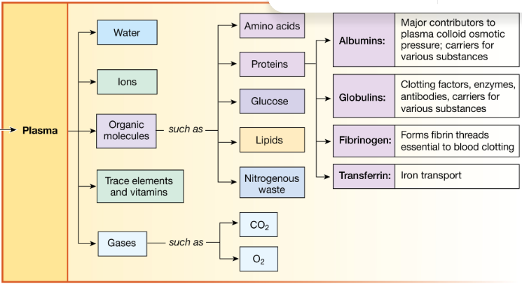
Water is the main component of plasma, accounting for about 92% of its weight. Proteins account for another 7%. The remaining 1% is dissolved organic molecules (amino acids, glucose, lipids, and nitrogenous wastes), ions, and trace elements and vitamins, and dissolved oxygen and carbon dioxide .
Describe the major functions of plasma proteins and give several examples of
them with their specific functions
Plasma proteins participate in many functions, including blood clotting and defense against foreign invaders. In addition, they act as carriers for steroid hormones, cholesterol, drugs, and certain ions such as iron. Finally, some plasma proteins act as hormones or as extracellular enzymes
ex in pictures
Identify the major cellular elements of blood, counting platelets as cells, and
provide their major functions (for now, you only need to identify the function of
leukocytes as a whole, not individually)
Three main cellular elements are found in blood (Fig. 17.1): red blood cells (RBCs), also called erythrocytes {erythros, red};white blood cells (WBCs), also called leukocytes {leukos, white}; and platelets or thrombocytes {, lump, clot}.

Describe where and from what precursor all blood cells originate
Where do these different blood cells come from? They are all descendants of a single precursor cell type known as the pluripotent hematopoietic stem cell (FIG. 17.2).
This cell type is found primarily in bone marrow, a soft tissue that fills the hollow center of bones.
Pluripotent stem cells have the remarkable ability to develop into many different cell types.
Identify the major cytokines (just the ones named in Table 17.1) involved in
hematopoiesis and their origins and roles (basically, learn Table 17.1!); identify
hypoxia as the major stimulus for EPO production
Red blood cell production (erythropoiesis) is controlled by the glycoprotein erythropoietin (EPO), assisted by several cytokines. Erythropoietin is made primarily in the kidneys of adults.
Thrombopoietin (TPO) is a glycoprotein that regulates the growth and maturation of megakaryocytes, the parent cells of platelets. (Recall that thrombocyte is an alternative name for platelet.) TPO is produced primarily in the liver.
Some of the best-known cytokines in hematopoiesis are the colony-stimulating factors, molecules made by endothelial cells and white blood cells. Others are the interleukins {white}, such as IL-3. The name interleukin was first given to cytokines released by one white blood cell to act on another white blood cell.
Hypoxia stimulates production of a transcription factor called hypoxia-inducible factor, which turns on the EPO gene to increase EPO synthesis. This pathway, like other endocrine pathways, helps the body maintain homeostasis. By stimulating the synthesis of red blood cells, EPO puts more hemoglobin into the circulation to carry oxygen.

Recognize that mature RBCs lack a nucleus and most organelles, and are
essentially short-lived “sacks of hemoglobin” and a few enzymes rather than true
cells
ok
Identify or describe the basic structure of human hemoglobin
Hemoglobin (Hb) is a large, complex protein with four globular protein chains, each of which is wrapped around an iron-containing heme group (FIG. 17.6a). There are several isoforms of globin proteins in hemoglobin
The most common isoforms are designated alpha α, beta β, gamma, and delta δ depending on the structure of the chain. Most adult hemoglobin (designated HbA) has two alpha chains and two beta chains, as shown. However, a small portion of adult hemoglobin (about 2.5%) has two alpha chains and two delta chains (HbA2)
The four heme groups in a hemoglobin molecule are identical. Each heme group consists of a carbon-hydrogen-nitrogen porphyrin ring with an iron atom (Fe) in the center
Recognize that, although millions of RBCs die every day in each person, the iron
and amino acids from these dead cells are recycled into new compounds
(including new hemoglobin).
ok
Describe the process of hemostasis, including the basic process of, and
importance of, each of the three step
Hemostasis has three major steps: ❶ vasoconstriction, ❷ temporary blockage of a break by a platelet plug, and ❸ coagulation, the formation of a clot that seals the hole until tissues are repaired.
The first step in hemostasis is immediate constriction of damaged vessels to decrease blood flow and pressure within the vessel temporarily. When you put pressure on a bleeding wound, you also decrease flow within the damaged vessel. Vasoconstriction normally is caused by paracrine molecules released from the endothelium.
Vasoconstriction is rapidly followed by the second step, mechanical blockage of the hole by a loose platelet plug. Plug formation begins with platelet adhesion, when platelets adhere or stick to exposed collagen in the damaged area. The adhered platelets become activated, releasing cytokines into the area around the injury. These platelet factors reinforce local vasoconstriction and activate more platelets, which aggregate or stick to one another to form a loose platelet plug. Platelets activating more platelets are an example of a positive feedback loop
Simultaneously, exposed collagen and tissue factor (a protein-phospholipid mixture) initiate the third step, the formation of a fibrin protein mesh that stabilizes the platelet plug to form a clot. Fibrin is the end product of a series of enzymatic reactions known as the coagulation cascade. Some chemical factors involved in the coagulation cascade also promote platelet adhesion and aggregation in the damaged region. Eventually, as the damaged vessel repairs itself, the clot retracts when fibrin is slowly dissolved by the enzyme plasmin.