BISC205 Topic 7
 Call Kai
 Learn
 Practice Test
 Spaced Repetition
 Match
 Flashcards
 Knowt Play
1/45

Earn XP
Description and Tags
Muscular System - Fall 2026
46 Terms
myofibrils vs muscle fibers
muscle fibers: a single long multinucleate muscle cell, which contains hundred to thousands of smaller cylindrical units called myofibrils
myofibrils: filamentous structures that make up the muscle fibers. they are composed of:
repeating sarcomeres
two types of myofilaments: thin filaments of actin and thick filaments of myosin

what are sarcomeres
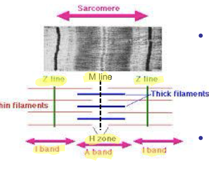
the functional unit of a muscle. they are the basic contractile unit of muscle fibre that is defined by alternating light and dark bands that create striations
parts of a sarcomere
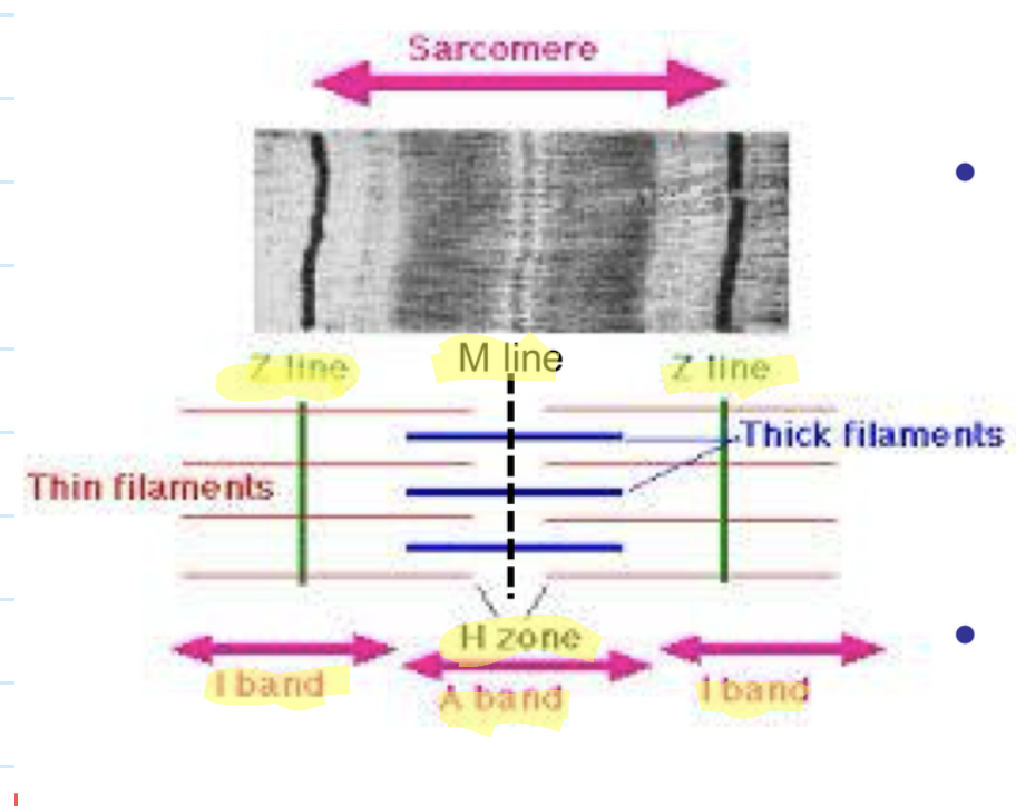
Z-line = boundary
M line = midline of sarcomere
H zone = contains myosin only
I band = contains actin only
A band = contains myosin and overlapping portions of actin

what parts of the sarcomere is not present when the sarcomere is fully contracted
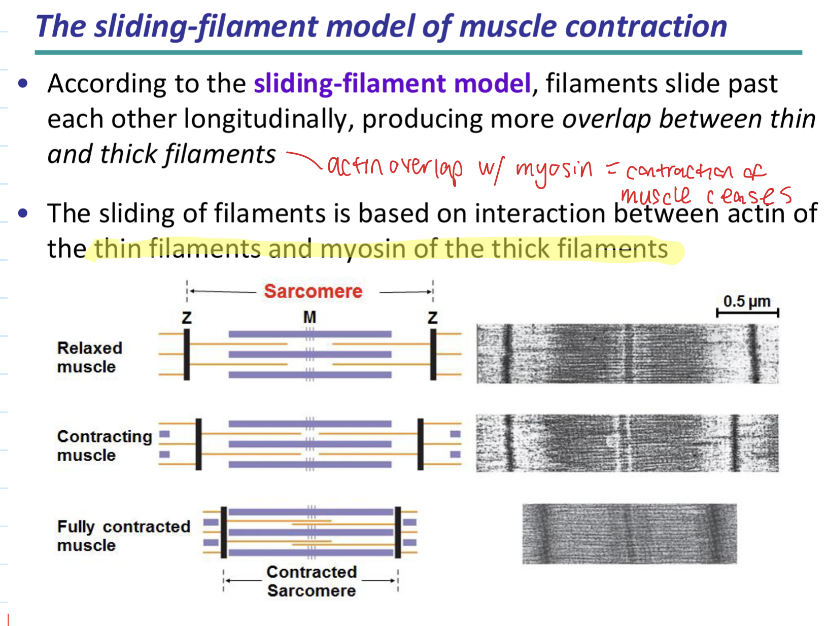
the H zone and I bands
which parts of the sarcomere do not change in length in a fully contracted sarcomere
the A band and M line

what happens to the Z lines in a fully contracted sarcomere
the Z lines move closer together
what is the sliding filament model
when filaments slide past each other longitudinally, producing more overlap between thin and thick filaments. they are based on interaction between actin of the thin filaments and myosin of the thick filaments
step one of the sliding filament model
ATP binds to the myosin head, causing the conformation change that releases the head from the actin

step two of the sliding filament model
when ATP is hydrolyzed to ADP and inorganic phosphate (Pi), the myosin head pivots.
The ADP and Pi are still bound to the myosin head, but the myosin head then binds to a new actin subunit farther down the actin filament (a cross-bridge is formed).
this causes the myosin head to be in a high energy position, and ready for the power stroke

what is the power stroke
key part of muscle contraction where a myosin head pulls on an actin filament, causing it to slide (and the sarcomere to shorten)

step three of the sliding filament model
when inorganic phosphate is released, the myosin head pivots back to its original conformation.
the power stroke is generated as the myosin head forcefully pull the actin filament toward the M line of sarcomere (thereby shortening the sarcomere)

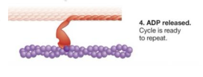
step four
ADP is released, and the myosin filament is ready for binding to another ATP molecule.
As ATP binding and hydrolysis continue, myosin continues to bind actin, move the thin filament, release actin, and bind again - if ATP is available, the two ends of the sarcomere continue to be pulled closer together
now recite all the four steps of the sliding filament model
ATP binds to the myosin head, causing the conformation change that releases the head from the actin
when ATP is hydrolyzed to ADP and inorganic phosphate (Pi), the myosin head pivots.
The ADP and Pi are still bound to the myosin head, but the myosin head then binds to a new actin subunit farther down the actin filament (a cross-bridge is formed).
this causes the myosin head to be in a high energy position, and ready for the power stroke
when inorganic phosphate is released, the myosin head pivots back to its original conformation.
the power stroke is generated as the myosin head forcefully pull the actin filament toward the M line of sarcomere (thereby shortening the sarcomere)
ADP is released, and the myosin filament is ready for binding to another ATP molecule.
As ATP binding and hydrolysis continue, myosin continues to bind actin, move the thin filament, release actin, and bind again - if ATP is available, the two ends of the sarcomere continue to be pulled closer together
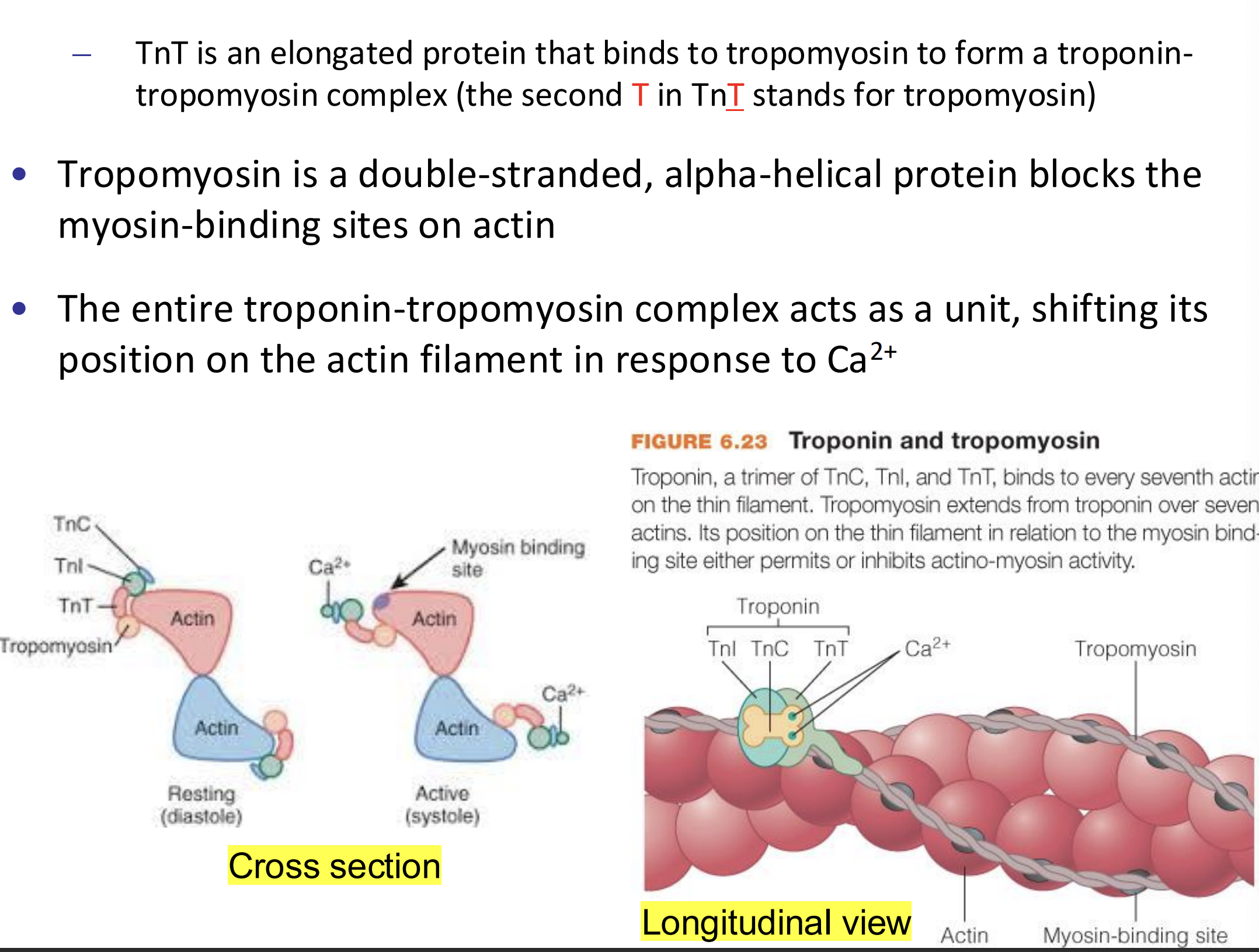
how do tropomyosin and troponin work together to block muscle contraction
when a muscle is at rest, myosin binding sites on the thin filament are blocked by the regulatory protein tropomyosin
Ca2+ is released into the muscle cell
Ca2+ ions bins to the troponin complex, causing the troponin-tropomyosin to move. this exposes the myosin head binding sites
muscle fibers contract because because the myosin binding sites are uncovered

how does the concentration of Ca2+ affect muscle fibre contraction
concentration of Ca2+ is high = muscle fibers contract
concentration of Ca2+ is low = muscle fibers do not contract

structure and function of troponin
three subunits: TnC, TnI, and TnT
function: works with tropomyosin to block muscle contraction by binding to Ca2+ and causing the troponin-tropomyosin complex to move. it binds to every seventh actin on the thin filament

structure of troponin and function of its subunits
composed of three subunits:
TnC: dumbbell shaped protein that acts as a Ca2+ sensor. it can bind four calcium ions
TnI: inhibits interaction of myosin and actin, keeping muscle in a relaxed state when Ca2+ levels are low.
binding of Ca2+ by TnC induces a conformational change in the troponin complex, which causes the TnI to release actin and expose myosin binding site.
this allows actin to form cross-bridge with myosin, resulting in muscle contraction as ATPase activity is activated.
TnC drives away TnI allowing interaction between myosin head and actin
TnT: elongated protein that bind to tropomyosin to form a troponin-tropomyosin complex. tropom

what do the letters in each subunit stand for
TnC: the C in TnC stands for calcium
TnI: the I in TnI stands for inhibitory
TnT: the second T in TnT stands for tropomyos

structure and function of tropomyosin
structure: it is a double stranded, alpha helical protein
function: blocks the myosin binding sites on actin. this causes the muscle fibers to not contract. when there is high Ca2+, it shifts in a troponin-tropomyosun complex to uncover myosin binding sites
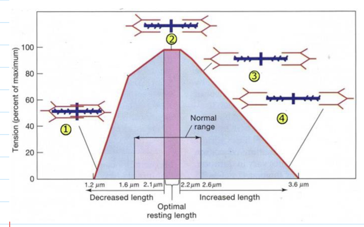
relationship between sarcomere length an force (figure)
some important notes: magnitude of tension depends on length, shortening velocity, and stimulation rate. however, muscle shortening velocity and stimulation rate are constant, so they are not shown in the graph

why do certain sarcomere lengths generate high force
at shorter sarcomere lengths, cross-bridges form. This also means that H zone and I bands are shorter in length.
At maximum tension/force, sarcomeres are 2.1-2.2 micro meters long

why do certain sarcomere lengths generate low force/tension
sarcomere lengths generate low tension when
sarcomeres are at their longest length. this means filaments are not interacting, meaning cross bridges fail to form. As a result, H zone and I bands are at their longest length
sarcomeres are at their shortest length. this means the actin filaments slide towards and over each other, reducing the number of cross bridges that forms. as a result, H zone and I bands disappears
what is the neuromuscular junction
a specialized chemical synapse that serves to transmit electrical impulses from the motion neuron nerve terminal to the skeletal muscle. its primary neurotransmitter is acetylcholine

7 steps associated with activation of skeletal muscle from action potentials of a motor neuron
the action potential reaches presynaptic axon terminal of a motor neuron
at this time voltage-gated calcium channels open to facilitate the influx of Ca2+ ions into the presynaptic nerve terminal
the increase in intracellular Ca2+ ion concentration triggers fusion of synaptic vesicles containing ACh with the nerve terminal membrane
vesicles release ACh into synaptic cleft
ACh binds to its nicotinic receptors on motor end plate. Binding of ACh causes nicotinic receptors to open as ligand gated channels. This causes more Na+ to rush into the muscle cells than K+ leave
the opening of these ligand gated channels will lead to the opening of voltage gated Na+ channels to initiate the action potential in muscle fibre
acetylcholine located on the motor end plate degrades ACh into acetic acid and choline. choline is reabsorbed back into the axon terminal of the neuron and is combined with acetyl coenzyme A so that more ACh can be synthesized while acetic acid diffuses away into the surrounding medium

steps associated with regulation of skeletal muscle contraction
at the neuromuscular junction, acetylcholine (ACh) is released at synaptic terminal diffuses across synaptic cleft and binds to receptor proteins on muscle fibers sarcolemma. this triggers action potential (AP) in muscle fiber
the AP is propagated along the sarcolemma and down invaginations called transverse tubules (T-tubules)
the AP triggers CA2+ ions to be released from sarcoplasmic reticulum (SR) which increases intracellular Ca2+ concentration
CA2+ ions bind to troponin associated with the thin filament (actin) the tropomyosin slides to expose the myosin head binding sites on actin
the sliding filament cycling begins with myosin cross-bridge formation and breakdown (coupled with ATP hydrolysis). as a result, the actin slides toward the centre of the sarcomere
cystolic Ca2+ ions are removed by the calcium pump (via active transport also known as Ca2+ ATPase) into the SR after the action potential ends. this decreases intracellular Ca2+ concentration
tropomyosin blockage of myosin head binding sites is restored, and the contraption ends. at this state, the muscle fibre relaxes

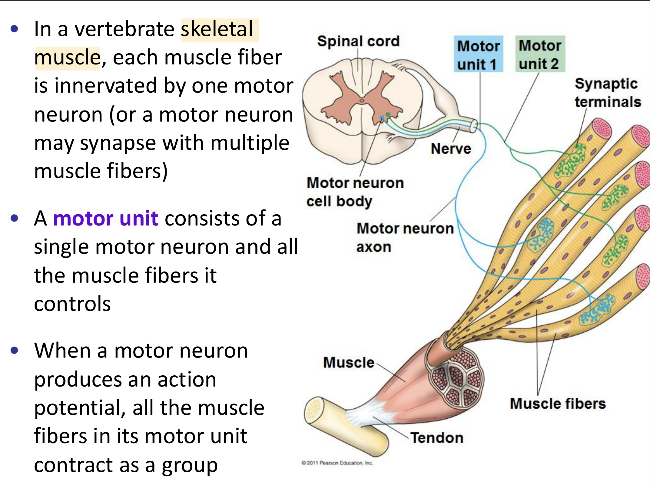
what is a motor unit?
a single motor neuron and all the muscle fibers it controls
what is muscle tension?
a force generated by a muscle as it contracts. more motor units and higher firing rates lead to greater tension, allowing for a wide range of forces from fine movements to heavy lifting

three outcomes of muscle tension
twitch: results from a single action potential in a motor neuron. this results in brief contraction and relaxation cycle of muscle fiber
wave summation: results from addition of multiple twitches before muscle can fully relax. (ex/ a second action potential is applied before the muscle has fully relaxed from the first twitch)
tetanus: a state of smooth and sustained contraction produced when motor neurons deliver a volley of action potentials. these action potentials cause increasingly strong contractions before the muscle fibre does not have time to fully relax.

incomplete vs complete tetanus
incomplete tetanus: a sustained muscle contraction with visible relaxation periods between stimuli
complete tetanus: a sustained maximum contraction with no relaxation between stimuli because the stimulation is so frequent

what is tetanus?
a medical condition characterized by a prolonged contraction of skeletal muscle fibers
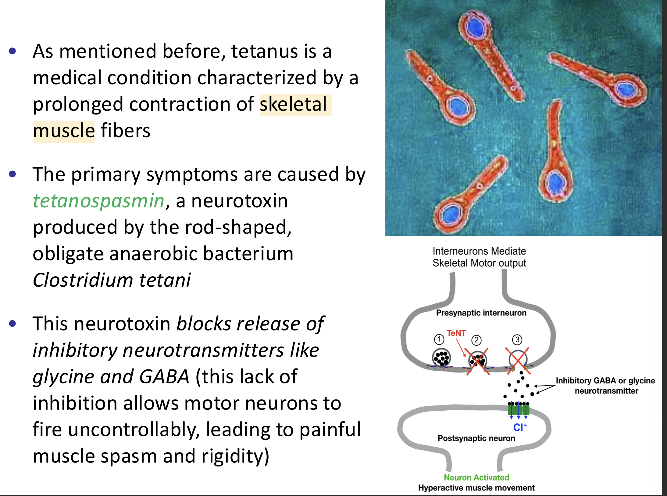
what is tetanospasmin? how does it cause symptoms of tetanus?
tetanospasmin: a neurotoxin produced by the rod shaped, obligate anaerobic bacterium clostridium tetani.
function: the neurotoxin blocks release of inhibitory neurotransmitters like glycine and GABA. this lack of inhibition allows motor neurons to fire uncontrollably, leading to painful muscle spasm and rigidity.

how does infection with tetanus occur?
infection occurs through wound contamination and involves a cut or deep puncture wound. as the infection progresses, muscle spasms develop in the jaw and elsewhere in the body

two types of skeletal muscle contractions
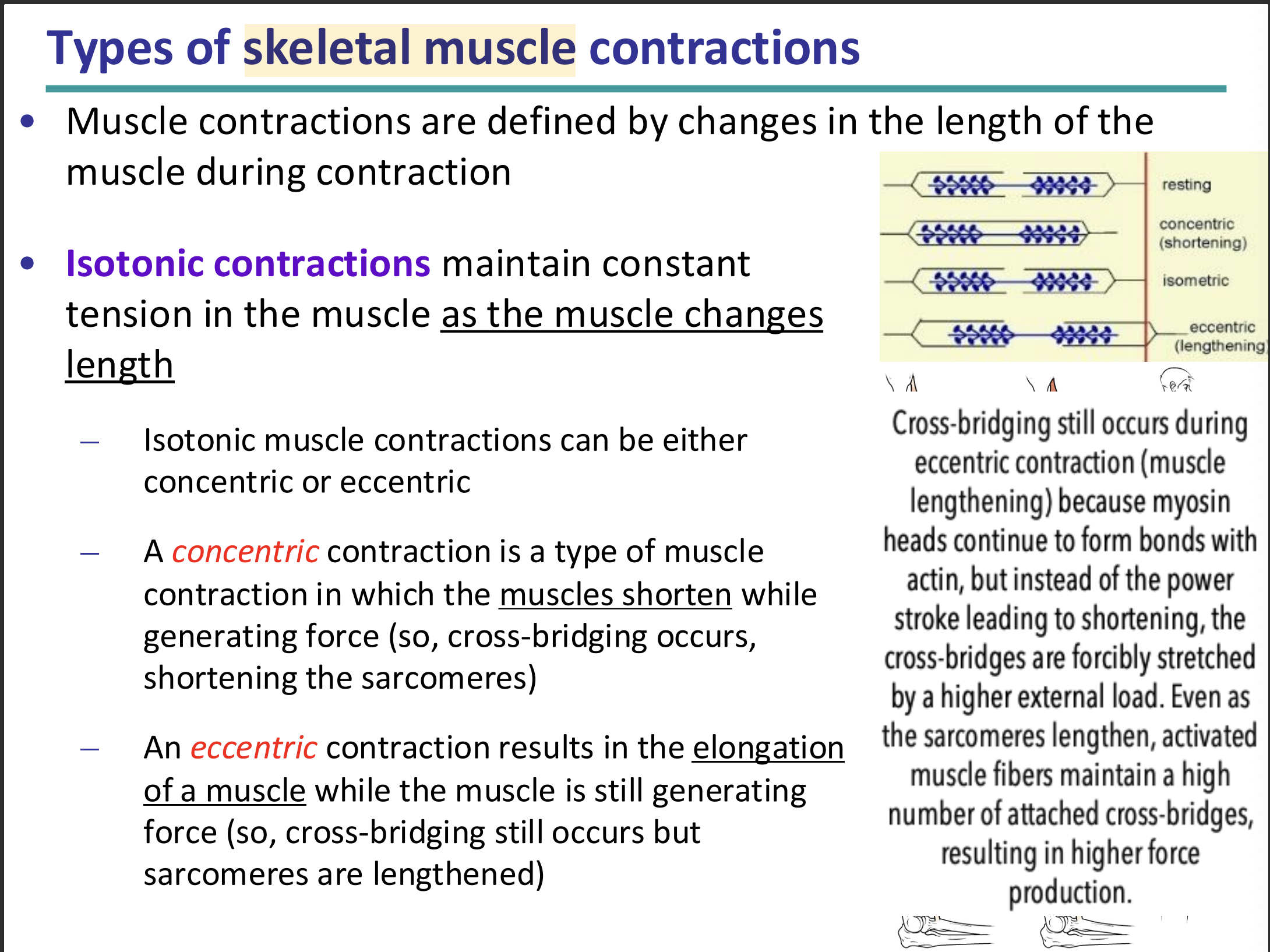
isotonic contractions: maintain constant tension in the muscle as the muscle changes length. isotonic contractions are either concentric or eccentric
isometric contractions: generate force/tension without changing length of the muscle. this does not require sliding of myofibrils. they are either yielding or overcoming

isotonic contractions: concentric vs essentric
concentric contraction: a type of muscle contraction in which the muscles shorten while generating force. cross bridging occurs, shortening the sarcomeres
eccentric contraction: results in elongation of a muscle while the muscle is still generating force. cross bridging still occurs, but sarcomeres are lengthened.
isometric contractions: yielding and overcoming
yielding contraction: occurs when a muscle contraction is opposed by resistance (ex/ holding a heavy weight steadily in mid-position where the weight is neither raised or lowered)
overcoming contraction: occurs when a muscle contraction is opposed by an immovable object (ex/ contraction generated while pushing against an immovable object is an example of this contraction)
in both instances, cross-bridge cycling is maintaining tension in the muscle, but sarcomere is not changing length

types of skeletal muscle fibers
oxidative fibers: rely mostly on aerobic respiration to generate ATP. these fibers have high density of mitochondria, a rich blood supply, and a higher content of myoglobin. myoglobin binds to oxygen more tightly than hemoglobin. oxidative fibers are more resistant to fatigue
glycolytic fibers: use anaerobic glycolysis as their primary source of ATP. they have lower density of mitochondria, poor blood supply, lower myoglobin content, and fatigues easily compared to oxidative fibers.

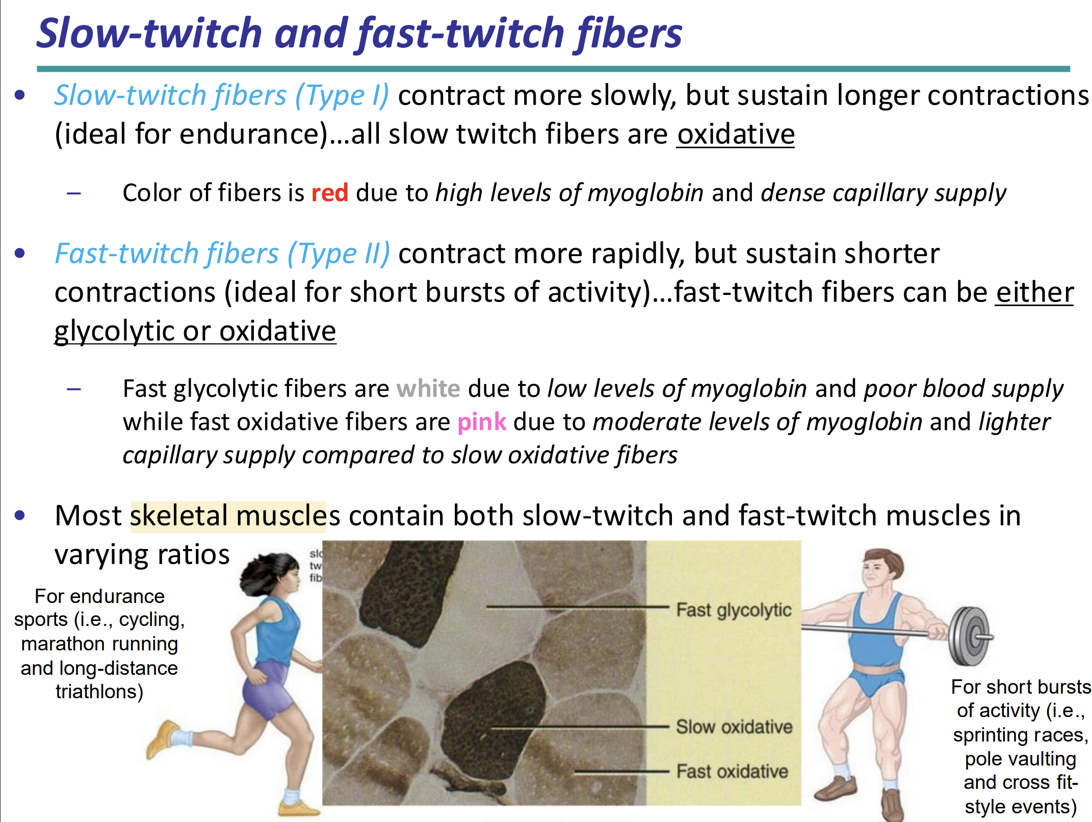
slow twitch fibers (Type I) vs fast twitch fibers (Type 2)
slow twitch fibers: contract more slowly, but sustain longer contractions (ideal for endurance). all slow twitch fibers are oxidative. the colour of thee divers are red due to high levels of myoglobin and dense capillary supply
fast twitch fibers: contract more rapidly, but sustain shorter contractions (ideal for short bursts of activity). fast twitch fibers can be either glycolytic or oxidative.
fast glycolytic fibers: white due to low levels of myoglobin and poor blood supply
fast oxidative fibers: pink due to moderate levels of myoglobin and lighter capillary supply compared to slow oxidative fibers
neurogenic control vs myogenic control
myogenic control
contraction in response to mechanical stretch or pressure changes as noted in some organs/tissues (urinary bladder, stomach, blood vessels)
presence of specialized pacemaker cells can generate their own action potentials to initiate peristalsis in gastrointestinal tract
neurogenic control
contraction can be under influence of autonomic nervous system
nervous system can modulate contraction in terms of strength and rate (regulation)

why are vertebrate skeletal muscles neurogenic while cardiac muscles are myogenic
skeletal muscles
neurogenic because they are activated by neuronal stimulation
its primary neurotransmitter is ACh
cardiac muscles
myogenic control because they can generate action potential without neural input.
instead, pacemaker cells transmit their electrical signal throughout the heart and cause other cardiomyocytes to depolarize and contract

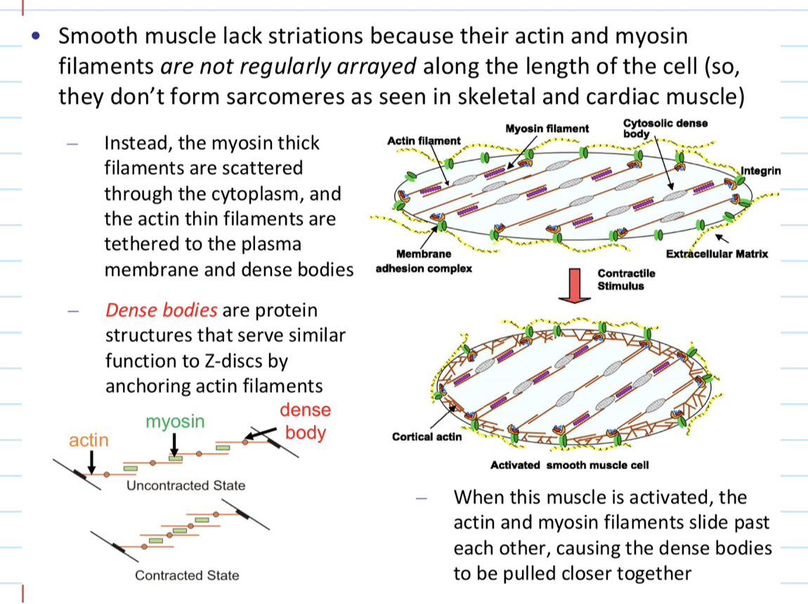
4 characteristics of the arrangement of actin and myosin in smooth muscle
smooth muscle’s actin and myosin filaments are not regularly arrayed along the length of the cell. this means they do not form sarcomeres or striations.
myosin thick filaments are scattered throughout the cytoplasm and the actin filaments are tethered to the plasma membrane and dense bodies (protein structures that serve similar function to Z discs by anchoring actin filaments)
when smooth muscle is activated, actin and myosin slide past each other, causing the dense bodies to be pulled closer together
why does smooth muscle have calmodulin?
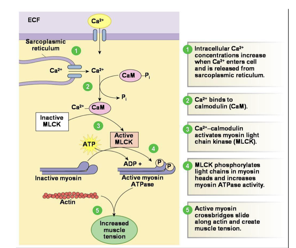
Smooth muscle does not have a troponin complex, T-tubules, or a developed sarcoplasmic reticulum. They use calmodulin so that the smooth muscle can contract/create cross bridges.
in other words, they use calmodulin as a calcium transducer for smooth muscle

steps in cross bridge formation/muscle contraction in smooth muscle
intracellular Ca2+ concentrations increase when Ca2+ enters cell and is released from sarcoplasmic reticulum
Ca2+ binds to calmodulin (CaM)
CA2+-calmodulin complex activates myosin light chain kinase (MLCK)
LMCK phosphorylates light chains in myosin heads and increases myosin ATPase activity
Active myosin cross bridges slide alone actin and create muscle tension
how is muscle contraction inhibited? how can calmodulin reverse this?
inhibition:
calponin and caldesmon bind to actin and block the interaction with myosin heads
reverse
high levels of calcium-calmodulin reverse the inhibition caused by calponis and caldesmon
this repositions the tropomyosin and expose the myosin head binding sites on actin
cross bridging can now occur

what is the neuroeffector junction
the chemical synapse involving smooth muscle or cardiac muscle. It is less structured than the neuromuscular junction


7 structural/functional differences between neuromuscular junction and neuroeffector junction. fill in the blanks

4 steps in which rigor mortis arise after death
stiffening of the body begins 3-4 hours after death. during this time, the sarcoplasmic reticulum deteriorates and releases Ca2+ ions
Ca2+ ions activate myosin-actin cross-bridging and muscle contracts, but muscle cannot relax afterwards
muscle relaxation requires ATP, but ATP production is no longer produced after death
fibers remain contracted until myofilaments decay
