Extremity Venous Anatomy (copy)
 Call Kai
 Learn
 Practice Test
 Spaced Repetition
 Match
 Flashcards
 Knowt Play
1/108

There's no tags or description
Looks like no tags are added yet.
109 Terms
How can the gastrocnemius veins and the posterior tibial veins be differentiated on an ultrasound exam?
A. Document the direction of flow relative to the knee
B. Document the presence of a corresponding artery
C. Follow them distally to locate their point of origination
D. Document the direction of flow relative to the foot
C. Follow them distally to locate their point of origination
Gastrocnemius veins originate deep within the calf muscle and extend to the popliteal fossa to join the popliteal vein. The posterior tibial veins originate at the ankle and extend to join the tibioperoneal trunk near the popliteal fossa. If you are progressing down the leg for a venous evaluation, when you reach the popliteal fossa and begin noting the calf vessels, pick a set to follow. If they dive deep into the calf = gastrocnemius veins, if they extend to the ankle = PTVs.
Both sets of veins have a corresponding artery and flow moving toward the heart. These characteristics would not differentiate them from each other.

The image displayed is from the right groin in a patient with suspected venous insufficiency. Use your mouse to position the cursor over the superficial epigastric vein and click to select it.
The superficial epigastric vein is a tributary of the great saphenous vein. It drains the skin of the lower abdomen and can cause superficial varicosities in the lower abdomen if the valves are incompetent.

When a patient performs the Valsalva strain, the intra-abdominal pressure _____ and the intrathoracic pressure _____
A. increases, increases
B. decreases. increases
C. increases, decreases
D. does not change, increases
A. increases, increases
When evaluating the GSV for use as a bypass graft, what is the preferred diameter for the vein?
A. >7mm
B. >5mm
C. >10mm
D. >3mm
D. >3mm
When evaluating a vein for use as a bypass graft, a 3mm diameter or greater is preferred
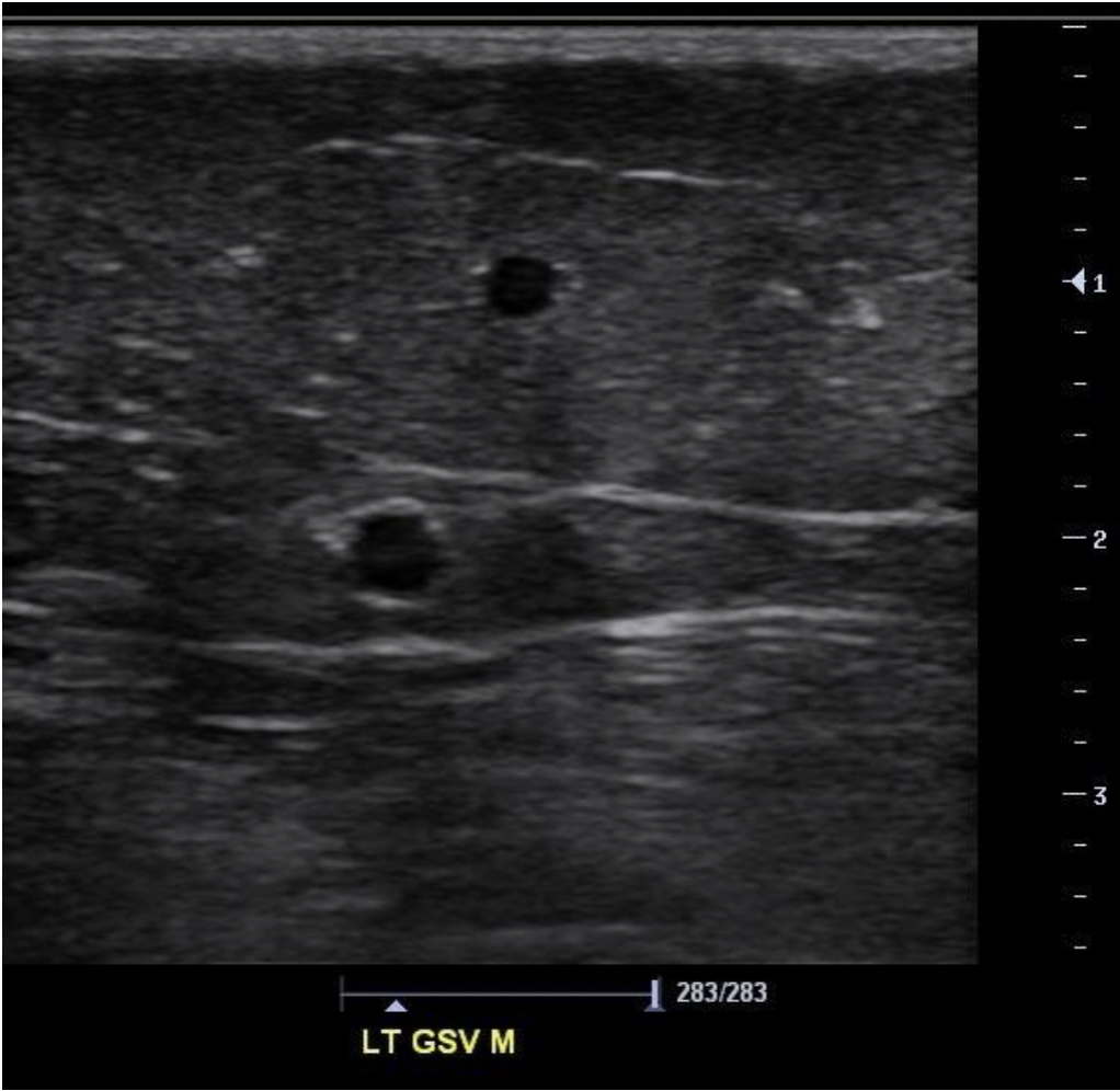
Find the great saphenous vein
The saphenous veins course within the saphenous compartment that is formed by the two layers of fascia. The fascia appears as a linear echogenic reflection separating the layers of tissue. Tributary veins are found superficial to the superficial fascia.


If the image displayed is a transverse view of the posterior right lower leg, Letter D indicates the:
A. lateral gastrocnemius veins
B. posterior tibial veins
C. medial gastrocnemius veins
D. popliteal vein
D. popliteal vein
When imaging the posterior leg, the notch on the transducer should be placed toward the right side of the patient, just like imaging the anterior leg. The right side of the image is lateral and the left side of the image is medial in both imaging views.
A. small saphenous vein
B. lateral gastrocnemius veins
C. medial gastrocnemius veins
D. popliteal vein
There are normally _____ perforator veins in each leg.
A. about 25-30
B. about 100
C. about 300
D. over 500
B. about 100
The great saphenous vein originates ______
A. at the great toe and courses along the ventral aspect of the foot to reach the lateral ankle and extend up the leg.
B. at the dorsum of the foot and courses anterior to the lateral malleolus.
C. at the dorsum of the foot and courses anterior to the medial malleolus.
D. at the great toe and courses along the ventral aspect of the foot to reach the medial ankle and extend up the leg.
C. at the dorsum of the foot and courses anterior to the medial malleolus.
Normal perforator valves allow blood flow in the perforator to move:
A. toward the heart
B. toward the ankle
C. from the femoral vein to the GSV
D. from the GSV to femoral vein
D. from the GSV to femoral vein
Normal perforator valves allow blood flow in the perforator to move from the superficial system to the deep system. Dilated perforators will allow bidirectional blood blow.
Which of the following normal vessels will collapse when you ask the patient to sniff quickly with moderate exertion?
A. common femoral vein
B. basilic vein
C. subclavian vein
D. cephalic vein
C. subclavian vein
The quick change in intrathoracic pressure with a sniff should cause the normal subclavian vein to collapse quickly due to the increased outflow into the innominate vein. The cephalic vein is a superficial vein that courses outside the musculature of the arm to enter the subclavian vein at the shoulder. A quick sniff would not affect the flow in a superficial vein. The common femoral vein is outside the chest/abdomen cavity. The change in pressure in these two areas with a quick sniff would not affect a vessel in the extremity.
The left and right innominate veins merge to form the ____
A. Brachiocephalic vein
B. inferior vena cava (IVC)
C. superior vena cava (SVC)
D. Right atrium
C. superior vena cava (SVC)
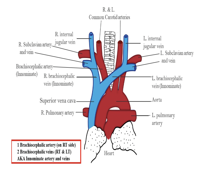
How can you differentiate the anterior accessory saphenous vein (AASV) from the great saphenous vein (GSV)?
A. look for the alignment sign that is only associated with the AASV
B. the AASV courses through the lateral thigh and the GSV courses through the medial thigh
C. the GSV travels in the saphenous compartment but the AASV does not
D. the AASV travels in the saphenous compartment but the GSV does not
A. look for the alignment sign that is only associated with the AASV
Alignment Sign - the AASV is aligned with the femoral artery and it courses anterior to the femoral artery within the saphenous compartment. This characteristic differentiates it from the GSV because the GSV is medial to the femoral artery/vein in the saphenous compartment.

Which of the following statements regarding venous perforators is correct?
A. Flow in the perforators normally moves from the deep to the superficial system.
B. Thrombosis rarely occurs within the perforators and it cannot to be easily demonstrated on ultrasound evaluation.
C. They each contain a single valve.
D. Perforator flow is normally unidirectional, but becomes bidirectional with exercise.
C. They each contain a single valve.
Venous valves are designed to allow unidirectional flow. If the valves are damaged or the vein is dilated to prevent proper valve movement, flow reversal may occur. Flow in the perforators normally moves from the superficial to the deep system. They each contain a single valve. Thrombosis can occur within the perforators, which can lead to the formation of varicosities.
While performing a lower extremity venous exam, you identify a small pair of veins with an accompanying artery deep in the calf muscle, that connect to the popliteal vein. What veins are these?
A. anterior tibial veins
B. sural veins
C. posterior arch veins
D. veins of Giacomini
B. sural veins
Sural veins are also called the gastrocnemius veins. They are located deep in the calf muscle and course alongside their accompanying artery. The sural veins empty their blood into the popliteal vein.
Which vein(s) originates at the ankle and courses along the medial border of the fibula to join the posterior tibial veins just distal to the popliteal fossa?
A. deep femoral veins
B. peroneal veins
C. great saphenous vein
D. small saphenous vein
B. peroneal veins
The peroneal vein originates at the ankle and courses along the medial border of the fibula to join the PTV just distal to the popliteal fossa.

Which of the following is a limitation of a CW Doppler evaluation of the lower extremity veins?
A. Augmentation cannot be demonstrated on a CW Doppler tracing
B. Can only be performed on patients in the supine position
C. Cannot be certain which vein you are evaluating
D. Flow velocities in veins are too low for detection and display on CW Doppler evaluation
C. Cannot be certain which vein you are evaluating
It is difficult to differentiate deep veins from superficial veins and superficial veins from perforators due to lack of range resolution with CW Doppler. The Sonographer cannot be certain which vein they are evaluating because the CW Doppler samples everything in its path. This would make the evaluation of systems with duplicated veins even more difficult.

If the image displayed is a transverse view of the posterior right lower leg, Letter A indicates the:
A. small saphenous vein
B. medial gastrocnemius vein
C. great saphenous vein
D. posterior tibial vein
A. small saphenous vein
When imaging the posterior leg, the notch on the transducer should be placed toward the right side of the patient, just like imaging the anterior leg. The right side of the image is lateral and the left side of the image is medial in both imaging views.
A. small saphenous vein
B. lateral gastrocnemius veins
C. medial gastrocnemius veins
D. popliteal vein
During a color evaluation of the common femoral vein, the patient is asked to perform the Valsalva maneuver. The cephalic flow indicated by the blue color in the CFV immediately disappears until the patient releases their muscles. The blue color returns and completely fills the vessel. What do these color Doppler findings indicate?
A. The response described is a normal result for the CFV.
B. Superficial venous insufficiency is present.
C. There is most likely an occlusive DVT in the superficial femoral vein causing the venous turbulence proximally.
D. A significant level of insufficiency is present and should be further evaluated with PW or CW Doppler techniques.
A. The response described is a normal result for the CFV.
When the Valsalva maneuver is applied, flow within the lower extremities should decrease significantly and/or stop completely. The loss of color flow during the Valsalva maneuver is a normal response of cessation of flow.

The image is a transverse view of the right antecubital fossa. What vessel is indicated by letter B?
A. brachial vein
B. median cubital vein
C. cephalic vein
D. basilic vein
D. basilic vein
If the image is of the right arm, the left side of the image corresponds with the lateral right arm. The veins of the arm listed from lateral to medial: cephalic, brachial, basilic
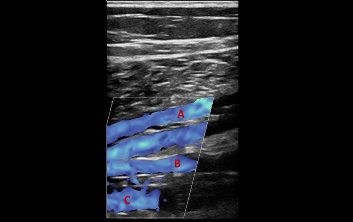
The transducer is placed in the longitudinal position slightly anterior to midline of the medial upper left calf. The beam is angled posterior until the image displayed appears. What vessel is indicated by letter B?
A. posterior tibial vein
B. peroneal vein
C. anterior tibial vein
D. gastrocnemius vein
B. peroneal vein
The image displays the confluence of the posterior tibial and peroneal veins. The peroneal veins are deeper on the image.
Flow through venous perforators is:
A. normally unidirectional from the superficial to the deep venous system
B. normally unidirectional from the deep venous to the superficial system
C. normally bidirectional to help equalize pressures in the deep and superficial systems
D. normally absent unless the deep system becomes overloaded and forces the excess blood into the superficial system
A. normally unidirectional from the superficial to the deep venous system
Once the pressure in the deep venous system consistently exceeds the pressure in the superficial system,
A. perforators will dilate leading to bi-directional flow
B. perforators will exhibit stasis of flow and thrombus formation
C. perforators will dilate and allow flow to only move into the deep system
D. perforators will constrict forcing flow reversal into the superficial system
A. perforators will dilate leading to bi-directional flow
Once the pressure in the deep venous system consistently exceeds the pressure in the superficial system, perforators will dilate leading to bi-directional flow.
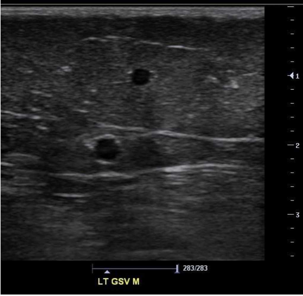
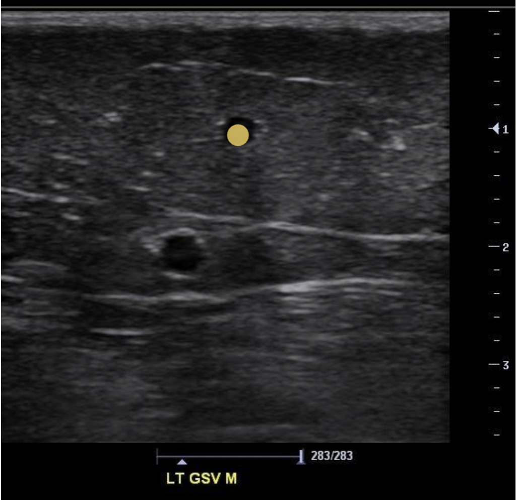
Find the saphenous tributary vein
The saphenous veins course within the saphenous compartment that is formed by the two layers of fascia. Tributary veins are found superficial to the superficial fascia.

The transducer is placed in the longitudinal position slightly anterior to midline of the medial upper left calf. The beam is angled posterior until the image displayed appears. What vessel is indicated by letter A?
A. posterior tibial vein
B. peroneal vein
C. anterior tibial vein
D. gastrocnemius vein
A. posterior tibial vein
The image displays the confluence of the posterior tibial and peroneal veins. The peroneal veins are deeper on the image.
Which vein below is usually the longest vein in the body?
A. small saphenous vein
B. inferior vena cava
C. great saphenous vein
D. femoral vein
C. great saphenous vein
The great saphenous vein originates at the ankle and courses cephalad to join the common femoral vein in the groin.
Which of the following veins is located deep to the muscular fascia in the proximal arm and superficial to the fascia in the forearm?
A. cephalic
B. median cubital
C. basilic
D. brachial
C. basilic
The basilic vein originates at the wrist and courses above to the muscular fascia. It penetrates the muscular fascia just above the elbow and courses cephalad, beneath the fascia, to join the axillary vein.

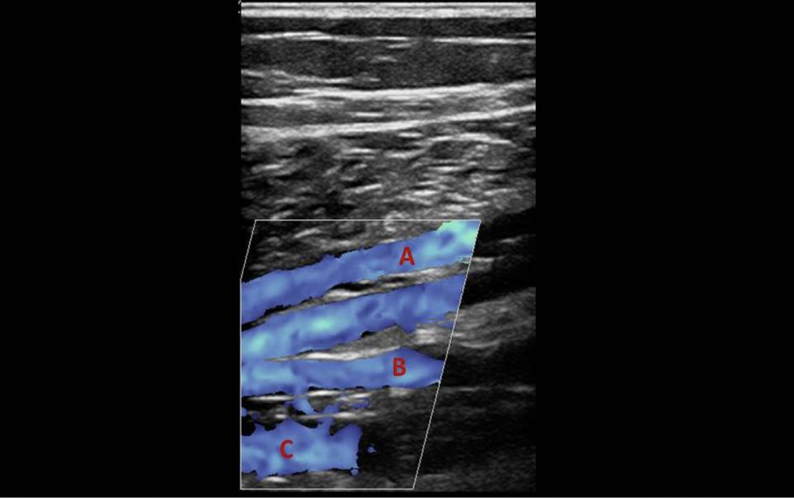
The transducer is placed in the longitudinal position slightly anterior to midline of the medial upper left calf. The beam is angled posterior until the image displayed appears. What vessel is indicated by letter A?
A. gastrocnemius vein
B. posterior tibial vein
C. peroneal vein
D. anterior tibial vein
B. posterior tibial vein
Venous valve leaflets:
A. are composed of muscle cells.
B. are extensions of the vessel's intimal layer.
C. are small flaps of connective tissue within the veins.
D. are muscular flaps that control backflow within the venous lumen
B. are extensions of the vessel's intimal layer.

The image is a transverse view of the right antecubital fossa. What vessel is indicated by letter A?
A. median cubital vein
B. cephalic vein
C. basilic vein
D. brachial vein
B. cephalic vein
If the image is of the right arm, the left side of the image corresponds with the lateral right arm. The veins of the arm are listed from lateral to medial: cephalic, brachial, basilic.
Which of the following veins are part of the superficial venous system in the lower extremity?
A. small saphenous vein and peroneal veins
B. posterior tibial vein
C. great saphenous vein and femoral vein
D. great and small saphenous veins
D. great and small saphenous veins
The great and small saphenous veins make up the superficial venous drainage system of the lower extremity.
Which of the following veins normally do not demonstrate spontaneous flow on Doppler evaluation?
A. posterior tibial and popliteal veins
B. peroneal and femoral veins
C. posterior tibial, peroneal and great saphenous vein
D. All lower extremity veins should normally demonstrate spontaneous flow on Doppler evaluation. Loss of spontaneity indicates flow stasis.
C. posterior tibial, peroneal and great saphenous vein
Calf veins and superficial veins do not normally demonstrate flow without augmentation. (Lack spontaneity) Normally the flow is very low velocity and not easily detected without augmentation.
Which of the following veins courses anterior to the tibial bone in the calf?
A. great saphenous vein
B. great saphenous vein and anterior tibial vein
C. anterior tibial vein
D. peroneal vein
C. anterior tibial veins
Which of the following occurs during calf muscle relaxation?
A. blood flows through the perforators from the superficial to the deep veins
B. the superficial veins fill with blood
C. blood flows through the perforators from the deep to the superficial veins
D. The gastrocnemius veins fill with blood from the popliteal vein
A. blood flows through the perforators from the superficial to the deep veins
When the muscle contracts, flow moves from the deep calf veins into the popliteal and femoral vein. As the muscle relaxes, blood is essentially sucked through the perforators from the superficial system into the deep system to play a part in refilling the deep vessels.
2D transverse views of the lower extremity veins are preferred for:
A. PW Doppler evaluation
B. evaluating compressibility
C. color Doppler evaluation
D. demonstrating the extent of thrombus
B. evaluating compressibility
Sagittal views are preferred for evaluating the extent of thrombus formation, demonstrating color and PW Doppler exams. Transverse views are used to obtain 2D images or clips of vein compression.
During inspiration the flow in the veins of the lower extremities ______
A. increases
B. decreases
C. remains unchanged
D. reverses direction
B. decreases
During inspiration the pressure within the chest cavity decreases allowing venous flow from the upper extremities to increase. During expiration, the pressure within the chest cavity increases causing the venous flow from the upper extremities to decrease. THE OPPOSITE IS TRUE WITH THE FLOW FROM THE LOWER EXTREMITIES.
During inspiration the pressure within the abdominal cavity increases causing venous flow from the lower extremities to decrease. During expiration, the pressure within the abdominal cavity decreases allowing the venous flow from the lower extremities to increase.
The femoral vein ______ the femoral artery.
A. is harder to compress than
B. courses posterior to
C. courses anterior to
D. is usually much longer than
B. courses posterior to

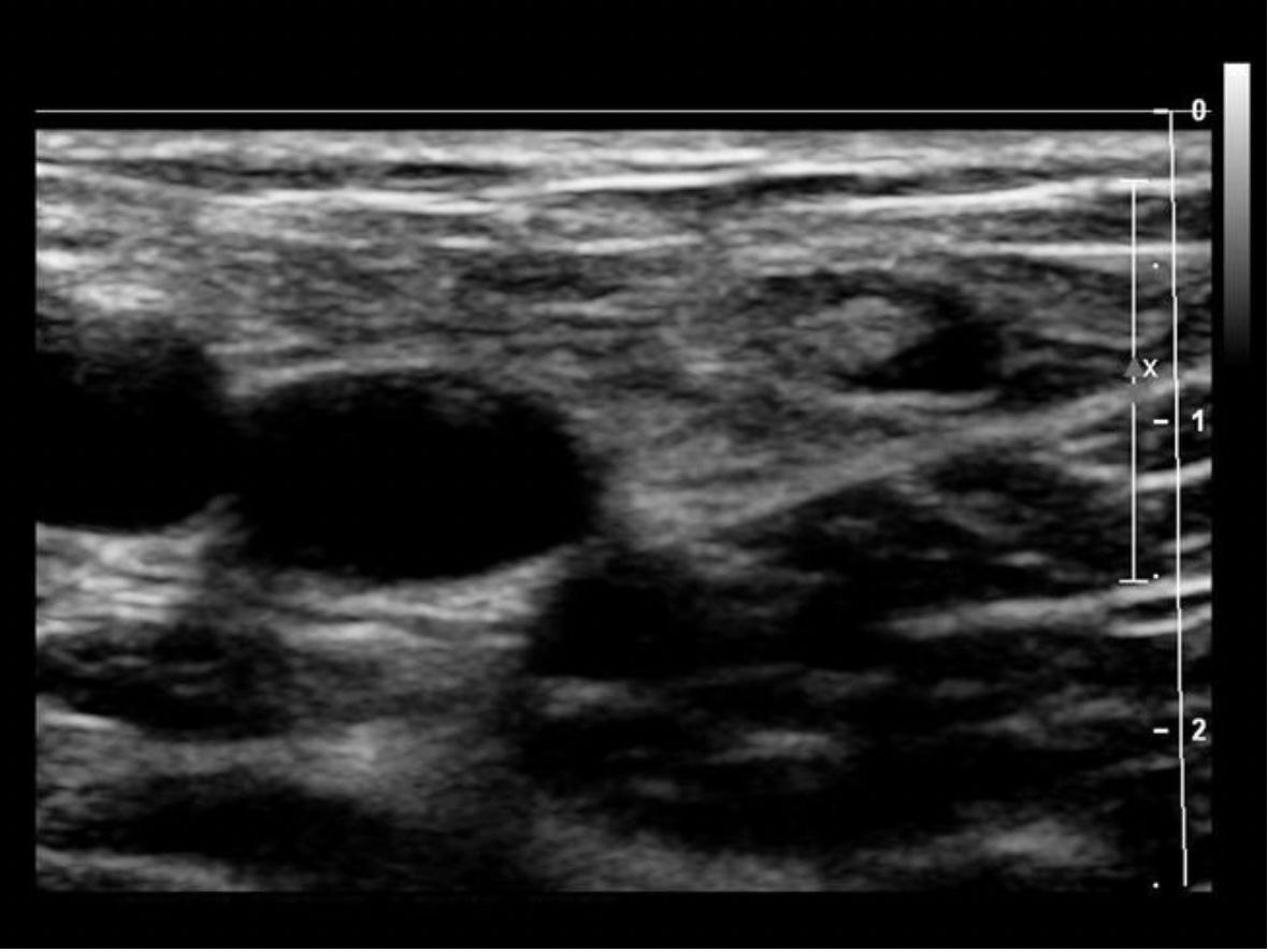
Find the normal lymph node
A normal lymph node is oval in shape and demonstrates a thin hypochoic rim of tissue around an echogenic hilum. Abnormal lymph nodes demonstrate a more rounded shape, thickened cortex that can appear anechoic. and there is loss of differentiation of the hilum.

Which of the following locations has a direct connection between the deep and superficial veins of the leg?
A. adductor canal
B. groin
C. hunter canal
D. ankle
B. groin
The greater saphenous vein connects directly to the common femoral vein in the groin area, adjacent to the inguinal ligament. The superficial system connects to the deep system through perforator veins located throughout the leg.

Which of the following can be found immediately posterior to the inguinal
A. saphenopopliteal junction
B. profunda femoral vein
C. popliteal vein and artery
D. common femoral vein and artery
D. common femoral vein and artery
The external iliac artery becomes the common femoral artery at the inguinal ligament. The CFA and CFV course posterior to the inguinal ligament. The GSV joins the CFV near the ligament. The FV and PFV join to form the CFV just distal to the ligament. The saphenopopliteal junction is located in the upper calf or popliteal fossa in most patients
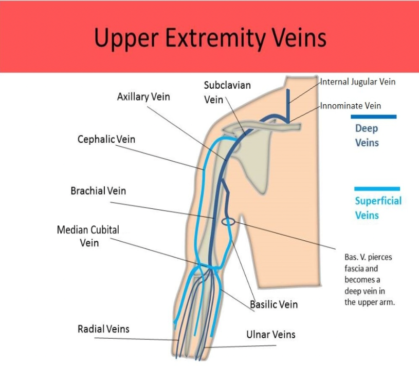
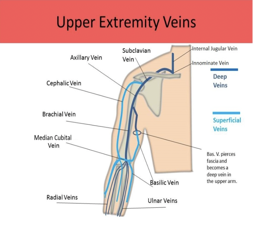
Which of the following veins originates at the medial wrist and courses up the medial arm to join the axillary vein?
A. Ulnar vein
B. Basilic vein
C. Cephalic vein
D. Radial vein
B. Basilic vein
CONSIDER THE ARM IS IN ANATOMIC POSITION FOR ALL ANATOMY QUESTIONS. The radial veins join the ulnar veins at the elbow to form the brachial vein(s). The cephalic vein originates near the base of the wrist and travels up the lateral side of the arm to empty into the subclavian vein. The basilic vein originates at the medial wrist and courses up the medial arm to join the axillary vein,
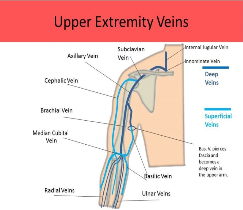
Which of the following statements regarding a Duplex exam of the upper extremity veins is correct?
A. The cephalic vein is easily compressed with light probe pressure, which can be an exam limitation.
B. The subclavian and innominate veins normally demonstrate continuous flow.
C. Inspiration will cause a decrease in flow within the larger upper extremity veins.
D. The internal jugular vein cannot be evaluated by ultrasound due to its course through bony structures of the neck.
A. The cephalic vein is easily compressed with light probe pressure, which can be an exam limitation.
The cephalic vein is easily compressed with light probe pressure, which can be an exam limitation. The basilic vein is also easily compressible due to its location.
Proper compression of the subclavian vein is normally inhibited by the clavicle. The subclavian and innominate veins usually demonstrate pulsatile, phasic flow due to their proximity to the heart/lungs. The IJV should be evaluated on all upper extremity venous duplex exams. It courses through the soft tissue/muscle of the neck, adjacent to the carotid artery. Inspiration will cause an increase in flow within the larger upper extremity veins.
Venous perforators connect the deep and superficial systems of the leg at many locations including:
A. groin only
B. groin and knee only
C. groin and ankle only
D. groin, knee, ankle
D. groin, knee, ankle
Venous perforators connect the deep and superficial systems of the leg at many locations including the groin, thigh, knee, calf, and ankle.

The attached image is taken from a lower extremity venous Doppler exam. This flow pattern would be abnormal for which of the following veins?
A. anterior tibial veins
B. popliteal vein
C. great saphenous vein
D. posterior tibial veins
B. popliteal vein
The deep calf veins and superficial veins do not normally exhibit spontaneous flow patterns. Augmentation is usually necessary to demonstrate patency of these veins. Lack of spontaneous flow in the popliteal vein is abnormal and a sign of flow stasis.
Where is the eustachian valve found?
A. near the junction of the IVC with the right atrium
B. popliteal fossa
C. antecubital fossa
D. thoracic outlet
A. near the junction of the IVC with the right atrium
A Eustachian valve is a single flap of tissue at the junction of the IVC with the right atrium that is necessary for fetal circulation. The Eustachian valve may persist in some adults, but it has no effect on circulation. It is not a TRUE venous valve in an adult and the IVC is described as having no valves.

If letter A is the basilic vein, what extremity is being evaluated?
A. left forearm
B. right distal upper arm
C. right forearm
D. left distal upper arm
D. left distal upper arm
If the image is of the left arm, the left side of the image corresponds with the medial arm. The basilic vein is the medial vein in the left arm. There is only one set of deep veins and an artery located centrally on the image. This would indicate the brachial vessels. The image is of the upper arm.

The image is a transverse view of the right antecubital fossa. What vessel is indicated by letter C?
A. cephalic vein
B. brachial artery
C. basilic vein
D. brachial vein
B. brachial artery
If the image is of the right arm, the left side of the image corresponds with the lateral right arm. The veins of the arm listed from lateral to medial: cephalic, brachial, basilic. The red color in the vessel labeled C indicates flow in the opposite direction of the other vessels on the screen. Letter C is the brachial artery.
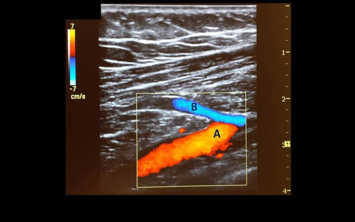
The image is a transverse view of right shoulder. What vessel is indicated by letter B?
A. axillary vein
B. subclavian vein
C. cephalic vein
D. basilic vein
C. cephalic vein
The cephalic vein joins the axillary vein to form the subclavian vein.
Which of the following is part of the superficial venous system of the lower extremity?
A. vein of Giacomini
B. peroneal veins
C. gastrocnemius veins
D. sural sinuses
A. vein of Giacomini
The vein of Giacomini is a superficial communicator vein connecting the GSV and SSV. The gastrocnemius veins and sural sinus are located deep in the calf muscle. The peroneal veins are deep veins of the calf.
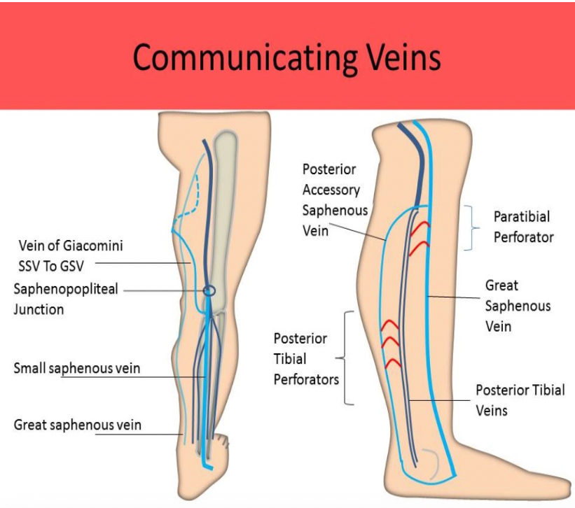
The Achilles tendon is a landmark for which deep calf vein?
A. anterior tibial vein
B. posterior tibial vein
C. peroneal vein
D. small saphenous vein
B. posterior tibial vein
The posterior tibial veins are identified at the area of the medial malleolus. The posterior tibial veins drain blood from the plantar arches and then course cephalad laying anterior to the tendon and posterior to the medial malleolus/tibial bone. The SSV is located posterior to the tendon, but it is a superficial vein.
______ receives the blood from the posterior tibial perforators and plays a major role in venous stasis of the lower extremity.
A. The anterior accessory saphenous vein
B. The vein of Giacomini
C. The lateral perforator
D. The posterior venous arch
D. The posterior venous arch
Posterior arch vein (AKA posterior accessory saphenous vein) extends superior from the ankle to the GSV in the mid-calf; plays a major role in venous stasis; connected to 3 ankle perforators called posterior tibial perforators (formerly Cockett's Perforators.)

Which vein is best evaluated for compressibility by asking the patient to perform a quick, deep sniff?
A. subclavian vein
B. distal IVC
C. great saphenous vein
D. axillary vein
A. subclavian vein
Manual compression of the subclavian vein is limited due to its position beneath the clavicle. If the patient performs a quick, deep sniff, the normal subclavian vein should demonstrate compression.
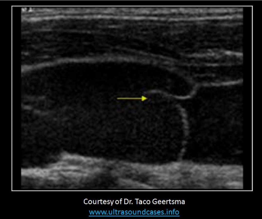
The image attached demonstrates which of the following?
A. Venous sclerosis
B. Chronic DVT
C. Venous dissection
D. Venous valve
D. Venous valve

If the image displayed is a transverse view of the posterior right lower leg, Letter C indicates the:
A. posterior tibial veins
B. lateral gastrocnemius veins
C. peroneal veins
D. medial gastrocnemius veins
D. medial gastrocnemius veins
When imaging the posterior leg, the notch on the transducer should be placed toward the right side of the patient, just like imaging the anterior leg. The right side of the image is lateral and the left side of the image is medial in both imaging views.
A. small saphenous vein
B. lateral gastrocnemius veins
C. medial gastrocnemius veins
D. popliteal vein
Which of the following are typically characterized as concomitant veins?
A. posterior tibial veins
B. soleal veins
C. popliteal vein
D. subclavian vein
A. posterior tibial veins
Concomitant veins - refers to a pair of veins that are coursing on both sides of a single artery. Radial, ulnar, PTV, ATV, peroneal and gastrochemius veins are examples of concomitant veins. The soleal veins/sinuses have no accompanying artery.
Which of the following terms would be used to describe abnormal venous flow in the deep veins of the thigh?
A. spontaneous
B. phasic
C. cardiac pulsatility
D. low velocity
C. cardiac pulsatility
The term pulsatility refers to repetitive flow changes demonstrated on the Doppler tracing. Veins closest to the heart will demonstrate cardiac pulsatility due to the changes in right atrial pressure. It can be equated to a "ripple effect", like a stone being thrown into a pool of water. The variations in flow are demonstrated in vessels further from the heart as they become engorged with blood. If venous flow is pulsatile in the lower extremities, cardiac complications like CHF or severe tricuspid regurgitation should be suspected.
Normal venous flow varies with respiration and has very low velocity. Spontaneous flow should also be present in most veins in the body (without any augmentation maneuvers applied).

When performing a lower extremity venous exam for insufficiency, you locate an incompetent vein that extends from the ankle to connect to the GSV near the popliteal fossa. Incompetent perforators are also identified in the mid-calf that allow flow between this vein and the GSV. Identify the incompetent vein.
A. Paratibial perforator
B. Posterior arch vein
C. Vein of Giacomini
D. Small saphenous vein
B. Posterior arch vein
The posterior arch vein (AKA posterior accessory saphenous vein) originates at the ankle and extends to connect to the GSV near the popliteal fossa. It is connected to the GSV in the mid to upper calf by the posterior tibial perforators (Cockett's perforators). This vein usually plays a major role in disease caused by venous stasis.

Which of the following is correct regarding the brachiocephalic veins?
A. Right and left brachiocephalic veins merge to form the superior vena cava
B. Formed by the junction of the external jugular and subclavian veins
C. Cardiac pulsatility is an abnormal Doppler finding in the brachiocephalic veins
D. Right and left brachiocephalic veins course anterior and medial to the right and left brachiocephalic arteries
A. Right and left brachiocephalic veins merge to form the superior vena cava
The brachiocephalic veins are formed by the junction of the internal jugular and subclavian veins. Right and left brachiocephalic veins merge to form the superior vena cava. There is no brachiocephalic artery on the left side. Cardiac pulsatility is a normal Doppler finding in the brachiocephalic veins due to their proximity to the heart.
Which of the following is true regarding the superficial veins of the lower extremity?
A. They are located 1-2cm below skin surface
B. They are more difficult to compress than the deep system
C. They do not contain valves
D. The superficial veins course cephalad to meet the profunda vein to form the common femoral vein
A. They are located 1-2cm below skin surface
The superficial system of the legs contains numerous valves and the veins are very easy to compress because they are located 1-2cm below the skin surface.
The weight of the column of blood within the veins when standing is called?
A. Systemic Pressure
B. Hydrostatic Pressure
C. Pulmonary Pressure
D. Transmural Pressure
B. Hydrostatic Pressure
The systemic blood pressure is related to the arterial pressures within the body. The pulmonary pressure is related to the pressure within the pulmonary arteries.
Proximal augmentation during an extremity Doppler evaluation is used to assess:
A. for the presence of CHF
B. venous insufficiency
C. arterial insufficiency
D. vein compressibility
B. venous insufficiency
Proximal augmentation and distal augmentation techniques are used to assess for venous insufficiency. Distal augmentation is also used in DVT evaluation.
Which vein courses from the lateral aspect of the wrist extending up the lateral arm to the shoulder to join the axillary vein?
A. cephalic
B. brachial
C. axillary
D. basilic
A. cephalic
The cephalic vein courses from the lateral aspect of the wrist extending up the lateral arm to the shoulder to join the axillary vein to form the subclavian vein.

Which of the following correctly describes the venous system of the upper extremity?
A. The superficial venous system of the arm is the primary drainage system of the extremity
B. The veins of the upper extremity have nearly twice the number of valves compared to the lower extremity
C. Effort thrombosis refers to clot in the veins of the hand caused by repetitive wrist motion
D. Patients with May Thurner syndrome have an increased risk of developing upper extremity thrombosis
A. The superficial venous system of the arm is the primary drainage system of the extremity
The superficial venous system of the arm is the primary drainage system of the extremity. The deep system is the primary drainage system in the legs. The veins of the upper extremity have far fewer valves than the lower extremity. They do not need as many because the arm is closer to the heart and experiences lower hydrostatic pressures. May Thurner syndrome affects the left common iliac vein. Effort thrombosis refers to thrombus formation at the shoulder caused by repetitive shoulder movement that compresses the vein in the thoracic outlet.
During inspiration the flow in the veins of the upper extremities _____
A. increases
B. decreases
C. remains unchanged
D. reverses direction
A. increases
During inspiration the pressure within the chest cavity decreases allowing venous flow from the upper extremities to increase. During expiration, the pressure within the chest cavity increases causing the venous flow from the upper extremities to decrease. THE OPPOSITE IS TRUE WITH THE FLOW FROM THE LOWER EXTREMITIES.
During inspiration the pressure within the abdominal cavity increases causing venous flow from the lower extremities to decrease. During expiration, the pressure within the abdominal cavity decreases allowing the venous flow from the lower extremities to increase.
The superficial venous system of the lower extremities contains vessels that are located
A. on the lateral aspect of each leg
B. within the connective tissue between the muscle tissue
C. superficial to the deep muscular fascia
D. adjacent to an artery with the same name
C. superficial to the deep muscular fascia
The saphenous veins and their tributaries course within the subcutaneous tissue, between the fascial layers.

The paratibial perforators connect:
A. the small saphenous vein and posterior tibial veins
B. the great saphenous vein and the anterior tibial veins
C. the small saphenous vein and popliteal vein
D. the great saphenous vein and posterior tibial veins
D. the great saphenous vein and posterior tibial veins

Hydrostatic venous pressure ______ when a patient goes from standing to the supine position.
A. shows a quick decrease before returning to 100mmHg
B. will show little change
C. increases
D. decreases
D. decreases
Hydrostatic pressure refers to the weight of the column of blood within the veins when a patient is standing. The pressure is 0mmHg at the level of the heart and increases more distally to a level of 100mmHg at the ankles. When a patient lies down, the pressure in the "column" is dispersed.
_____ drain(s) blood from the GSV in the proximal thigh into the proximal and mid femoral vein.
A. Posterior tibial perforators
B. Posterior venous arch
C. Paratibial perforators
D. Thigh perforators
D. Thigh perforators
The Hunterian perforators drain blood from the GSV in the proximal/mid-thigh into the femoral vein. They are also referred to as proximal thigh perforators.
Which of the following is part of the deep venous system of the forearm?
A. cephalic vein
B. basilic vein
C. radial vein
D. median cubital vein
C. radial vein
The basilic, cephalic and median cubital veins are superficial veins of the arm. The radial, ulnar, brachial and axillary veins are deep veins of the arm.

Which of the following explains the increased incidence of DVT in the left leg when compared to the right leg?
A. Left iliac vein is a common location for DVT formation with pregnancy
B. There are more valves present in the left iliac vein which can lead to increased venous stasis.
C. The left iliac vein is usually smaller than the right.
D. The anatomic course of the left iliac vein leads to increased risk of extrinsic compression.
D. The anatomic course of the left iliac vein leads to increased risk of extrinsic compression.
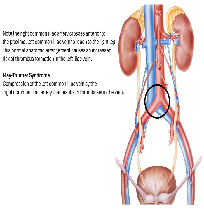
Left iliac vein courses beneath the right iliac artery at the aortic bifurcation. Extrinsic compression has been linked to increased incidence of left leg DVT. This condition is called May Thurner syndrome.
Pressure in the venules does not normally exceed _____
A. 20mmHg
B. 0mmHg
C. 80mmHg
D. 50mmHg
A. 20mmHg
Which of the following is unaffected by vasoconstriction and vasodilation in the arterioles?
A. hydrostatic pressure
B. peripheral resistance
C. the amount of diastolic flow in an artery
D. the number of phases of flow in the arterial waveform
A. hydrostatic pressure
Hydrostatic pressure is the weight of the column of blood in the vessels when standing. Vasoconstriction will increase peripheral resistance and increase resistance in the feeding arteries. Higher resistance flow will have less antegrade flow in diastole. Vasodilation will decrease peripheral resistance and decrease resistance in the feeding arteries. Lower resistance flow will have more antegrade flow in diastole. A biphasic waveform can become monophasic.
Respiration and _____ are responsible for venous blood flow.
A. vessel recoil
B. Valsalva maneuvers
C. the calf muscle pump
D. the tunica media layer of the wall
C. the calf muscle pump
Respiration and the calf muscle pump are responsible for moving
venous blood through the body.
Which of the following veins does not normally contain any functioning valves?
A. Innominate vein
B. Axillary vein
C. Peroneal veins
D. Small saphenous
A. Innominate vein
Lower extremity veins that do NOT contain valves = IVC, iliac veins and soleal sinuses
Upper extremity veins that do NOT contain valves = SVC and innominate veins.

The ____ course(s) between the tibial and fibular head to empty
blood into the popliteal vein.
A. peroneal veins
B. small saphenous vein
C. anterior tibial veins
D. posterior tibial veins
C. anterior tibial veins
ATVs course anterolaterally to the tibial bone across the interosseous membrane of the calf and through a space between the tibial and fibular head to empty into the popliteal vein.
The great saphenous vein is attached to the ____ aspect of the common femoral vein.
A. anterior
B. medial
C. lateral
D. posterior
B. medial


Find the great saphenous vein
The great saphenous vein travels medial to the femoral vein to join the common femoral vein near the groin. When visualized on an exam in a patient with symptoms of venous insufficiency, it should be evaluated with color and PW Doppler for signs of reflux.

Bank tellers, teachers, cashiers and assembly line workers have an increased risk of all of the following, except:
A. varicose veins
B. jugular vein thrombosis
C. thrombus formation in the soleal sinuses
D. venous hypertension
B. jugular vein thrombosis
Bank tellers, teachers, cashiers and assembly line workers are on their feet, standing still for several hours of the day. This will cause venous blood to pool in the legs and increase pressure in the veins (venous hypertension). Stasis of venous flow can lead to thrombus formation. Flow in the jugular veins would flow easily into the vena cava with no stasis or increased risk of thrombus formation.
Which of the following is a limitation of the 2D evaluation of the extremity veins?
A. location of the brachial vein
B. patient cannot lay flat for exam
C. increased amount of fibrous tissues and ligaments in the adductor canal
D. inability to visualize the vessels immediately posterior to the inguinal ligament
C. increased amount of fibrous tissues and ligaments in the adductor canal
Limitations of venous duplex evaluations:
Non-visualization of veins due to excessive transducer pressure
Location of the subclavian vein beneath the clavicle
increased amount of fibrous tissues and ligaments in the adductor canal
Patient that cannot be semi-erect or standing for exam
The inguinal ligament is not normally visible on ultrasound evaluation and it does not cause non-visualization of adjacent anatomy.
The adductor canal contains several ligaments and connective tissues that limit compression of the distal femoral vein. Increased transducer pressure can cause non-visualization of normal veins.
The subclavian vein courses posterior to the clavicle, which limits manual compression.
The superficial venous systems of the upper and lower extremities can be easily compressed causing non-visualization
Patients for DVT exams should be scanned in the semierect position to dilate the veins for easier visualization. A supine patient can cause the veins to be difficult to identify.
Insufficiency exams should be performed with the patient standing, to engorge the veins with blood and encourage the display of reflux. A supine patient could cause false negative results for reflux.
Which superficial vein listed forms a connection at the elbow between the two main upper extremity superficial veins?
A. cephalic vein
B. median cubital vein
C. radial vein
D. basilic vein
B. median cubital vein
The antecubital vein (AKA median cubital vein) forms a connection at the elbow between the basilic and cephalic veins.
What patient position should be used when mapping the great saphenous vein for harvest?
A. left lateral decubitus
B. supine
C. Trendelenburg
D. reverse Trendelenburg
D. reverse Trendelenburg
Place the extremity in a dependent position.
Leg: reverse Trendelenburg for the GSV and decubitus for the SSV
Arms: supine with arms extended at side at a level lower than the heart or seated position with hand on lap; tourniquets can be applied in cases where the veins are very small
An effective calf pump causes _____ in the pressure in the deep veins of the calf.
A. an increase
B. an increase in cardiac output and an increase
C. little change
D. a decrease
D. a decrease
Effective calf pump reduces blood stasis/pooling, decreases venous volume and pressure in the calf veins. Ineffective calf pump leads to increased stasis, venous volume and pressure in the calf veins; reflux is a contributing factor
The small saphenous vein:
A. originates posterior to the lateral malleolus and ascends along the midline aspect of posterior calf
B. originates immediately posterior to the Achilles' tendon attachment at the heel and ascends along the midline aspect of posterior calf
C. originates anterior to the lateral malleolus and ascends along the midline aspect of posterior calf
D. originates posterior to the medial malleolus and ascends along the midline aspect of posterior calf
A. originates posterior to the lateral malleolus and ascends along the midline aspect of posterior calf

Assuming proper transducer orientation, which of the following normal vessels could be displayed on the image?
A. right axillary vein and cephalic vein
B. left subclavian vein and cephalic vein
C. left common femoral vein and greater saphenous vein
D. superior vena cava and right innominate vein
A. right axillary vein and cephalic vein
The flow in the blue vessel is moving into the red vessel. The flow in the red vessel is moving toward the right side of the image. This is a transverse image of the right shoulder. The left side of the image is the lateral arm. The right side of the image is the medial arm. The vessels would be reversed on the image of this was a left shoulder image. If this were a longitudinal view of the groin it would demonstrate the greater saphenous vein extending upward on the left side of the image.
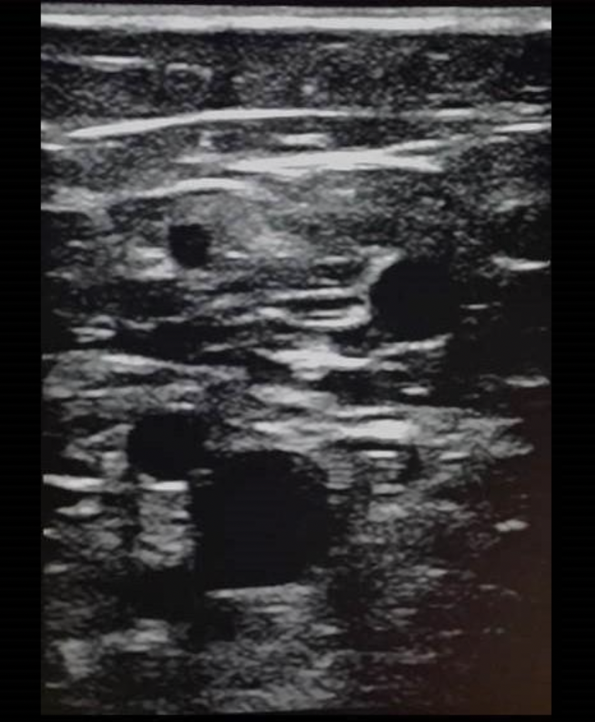
Find the anterior accessory saphenous vein
The anterior accessory saphenous vein travels anterior to the femoral artery to join the great saphenous vein near its junction with the common femoral vein at the groin. When visualized on an exam in a patient with symptoms of venous insufficiency, it should be evaluated with color and PW Doppler for signs of reflux.

Which calf vessel's anatomic course has been referred to as a "stocking seam"?
A. posterior tibial vein (PTV)
B. great saphenous vein (GSV)
C. small saphenous vein (SSV)
D. superficial femoral vein (SFV)
C. small saphenous vein (SSV)
The small saphenous vein originates very superficially in the distal posterior calf. It courses midline up the calf to reach the popliteal vein at or near the popliteal fossa. Its course has been said to resemble the seam of a type of pantyhose or stockings.
______ carry(s) the blood from the GSV in the distal thigh into the distal femoral vein.
A. Paratibial perforators
B. Posterior venous arch
C. Lateral perforators
D. Thigh perforators
D. Thigh perforators
The distal thigh perforators, formerly known as Dodd's perforators, drain blood from the GSV in the distal thigh into the distal femoral vein.

An ineffective calf muscle pump causes:
A. flow to move through the perforators into the deep system
B. the pressure in the deep calf veins to increase
C. the pressure in the deep calf veins to drop
D. the pressure in the superficial veins to drop
B. the pressure in the deep calf veins to increase
Effective calf pump reduces blood stasis/pooling, decreases venous pressure. Ineffective calf pump leads to increased stasis, venous volume and pressure; reflux is a contributing factor
The gastrocnemius plexus is usually connected to the:
A. superficial femoral vein
B. popliteal vein
C. profunda vein
D. great saphenous vein
B. popliteal vein
The gastrocnemius plexus is located in the calf muscle and drains into the popliteal veins. It acts as a reservoir for blood when sitting and standing etc.

The transducer is placed over the mid femoral vein while calf compression is performed. The Doppler waveform demonstrates spontaneous flow with an increase in forward flow with the calf compression. Which of the following describes the findings?
A. The flow response is normal for the femoral vein.
B. Heart disease is most likely present which causes the abnormal flow response.
C. The popliteal and distal femoral veins have incompetent valves.
D. The popliteal vein has incompetent valves.
A. The flow response is normal for the femoral vein.
A normal response to distal augmentation is an increase in forward flow with the calf compression and NO flow reversal after the increase in flow.
Paired veins that accompany an artery of the same name are called:
A. venae comitantes
B. posterior arch veins
C. Klippel Trenauney Weber syndrome
D. duplicated veins
A. venae comitantes
Concomitant veins or venae comitantes refers to a vein that is usually paired, with a vein lying on either side of a single artery of the same name. Radial, ulnar, PTV, ATV, peroneal and gastrocnemius veins are examples of concomitant veins.
The radial and ulnar veins merge to form the _____ at the _____ fossa
A. axillary vein, axillary
B. axillary vein, antecubital
C. brachial vein, popliteal
D. brachial vein, antecubital
D. brachial vein, antecubital
Increased pressure in the venous system results in:
A. decreased arterial pressure
B. edema
C. an increased risk of stroke
D. Baker cyst formation
B. edema
Increased pressure in the venous system results in edema as fluid is forced outward into surrounding tissues.
Where are the venous sinusoids in the lower extremity?
A. popliteal fossa
B. within the adductor canal
C. within the quadriceps muscle
D. between the soleal and gastrocnemius muscle
D. between the soleal and gastrocnemius muscle
The venous sinusoids serve as blood reservoirs in the calf and are an important part of the calf pump mechanism. They are located between the soleal and gastrocnemius muscle.

What is the arrow pointing to on the image of the right groin?
A. great saphenous vein
B. femoral vein
C. superficial femoral artery
D. common femoral vein
A. great saphenous vein
Note the thrombus in the most anterior vessel. This vessel courses anterior to the deep fascia. Note the linear echogenicity that courses posterior to the thrombosed vein. The image demonstrates the femoral vein and the profunda femoral vein merging to form the common femoral vein. The great saphenous vein joins the common femoral vein at the groin. The GSV demonstrates thrombosis.
When transmural pressure in a vein is high:
A. the associated artery will distend and flow velocity decreases
B. the vein becomes flattened and dumbbell shaped
C. the associated artery will flatten and flow velocity increases
D. the vein becomes enlarged and rounded
D. the vein becomes enlarged and rounded
Transmural pressure is the pressure within the veins pushing outward and is normally low. It increases with increasing venous volume. When the pressure inside the vein is high compared to the surrounding pressure outside the vein, the vessel distends. When the pressure inside the vein is low compared to the surrounding pressure outside the vein, the vessel collapses to a dumbbell shape.

The image demonstrates a venous perforator connected to the greater saphenous vein. Which of the following is true regarding the image and venous perforators?
A. Perforators are normally identified in most venous exams.
B. The flow within the perforator is moving in the normal direction.
C. Flow within the perforator system is normally bidirectional.
D. Compression techniques are non-diagnostic for thrombus formation in the perforator system because it is not possible thrombus to form within them due to their small size.
B. The flow within the perforator is moving in the normal direction.
The flow demonstrated is moving away from the probe, according to the color map. This indicates normal flow direction from the superficial to the deep system.
Which of the following veins has a direct connection to both superficial and deep veins?
A. popliteal vein and inferior vena cava
B. brachial vein and small saphenous vein
C. femoral vein and axillary vein
D. subclavian and popliteal vein
D. subclavian and popliteal vein
The subclavian vein receives blood from the cephalic vein and the axillary vein. The popliteal vein receives blood from the small saphenous and deep calf veins.
What vein contains blood with a very high oxygen content?
A. portal veins
B. pulmonary veins
C. IVC
D. hepatic veins
B. pulmonary veins
The pulmonary veins are the only veins in the body that carry highly oxygenated blood. These veins drain the blood from the lungs into the left atrium for distribution to the body through the left ventricle/aorta. All other veins carry blood that have depleted levels of oxygen because the organs that they are draining require the oxygen to function.

The image is a transverse view of right shoulder. What vessel is indicated by letter A?
A. subclavian vein
B. innominate vein
C. superior vena cava
D. axillary vein
D. axillary vein
Which of the following describes a normal lower extremity response to the Valsalva maneuver?
A. flow in the iliac vein will reverse for at least 1 second before normalizing
B. flow in the subclavian vein will be augmented toward the heart with slight reversal upon release
C. flow in the femoral vein will stop at onset of the maneuver
D. the femoral vein will demonstrate cardiac pulsatility
C. flow in the femoral vein will stop at onset of the maneuver
The Valsalva maneuver will significantly decrease or stop flow toward the heart in the deep veins of the legs. Upon release of the maneuver, flow will be briefly augmented toward the heart and then normalize.