Digestive System - Lecture Notes
 Call Kai
 Learn
Learn Practice Test
 Spaced Repetition
 Match
Match Flashcards
 Knowt Play
1/78

There's no tags or description
Looks like no tags are added yet.
Name | Mastery | Learn | Test | Matching | Spaced | Call with Kai | Chat |
|---|
No analytics yet
Send a link to your students to track their progress
79 Terms
Digestive System Components: Gastrointestinal Tract (GI)
Continuous tube for processing food
Lined with a mucous membrane
Includes: Oral cavity, pharynx, esophagus, stomach, small intestine, large intestine, and anus
Within the lumen, food is broken down into smaller components to be absorbed

Digestive System Components: Accessory Digestive Organs
Assist in digestion
Some produce secretions that empty into the GI tract
Include: teeth, tongue, salivary glands, liver, gallbladder, and pancreas

Functions of the Digestive System
Ingestion
Motility
Secretion
Digestion:
Mechanical and Chemical digestion
Absorption
Elimination
Function: Ingestion
Introduction of solid and liquid nutrients into the oral cavity
First Step in process of digesting and absorbing nutrients
Function: Motility
Voluntary and involuntary muscular contractions
Mixing and moving materials through the GI tract
Function: Secretion
Process of producing and releasing fluid products facilitating digestion
Example: digestive enzymes, acid, and bile
Function: Digestion
Breakdown of ingested food into smaller structures
Mechanical Digestion:
Material physically broken down by chewing and mixing
Chemical Digestion:
Involves specific enzymes to break chemical bonds
Change large complex molecules into smaller molecules
Function: Absorption
Transport of digested molecules, electrolytes, vitamins, water
Move from G tract into blood or lymph
Function: Elimination
Expulsion of indigestible components that are not absorbed
GI Tract Wall: Innermost to Outermost
Hollow tube of 4 concentric layers, tunics
Mucosa
Submucosa
Muscularis
Adventitia (or serosa)

GI Tract Wall: Mucosa
Epithelium in contact with food; secretes mucus & absorbs nutrients
Lamina propria (connective tissue) with blood and lymph capillaries
Muscularis mucosae facilitates secretion and absorption

GI Tract Wall: Submucosa
Connective tissue, blood vessels, lymphatic tissue (MALT), and nerves
Submuscosal nerve plexus are nerves ganglia innervting smooth muscle and glands
Peyer patches are larger aggregates of lymphatic nodules in distal small intestine
GI Tract Wall: Muscularis
Inner circular for peristalsis & segmentation
Contraction constricts tube lumen
Outer longitudinal layers for peristalsis & segmentation
Contraction shortens tube
Myenteric nerve plexus controls contractions
Sphincters regulate passage of food
GI Tract Wall: Muscularis - Motility
Functions to mix and propel contents within GI tract
Mixing backward-and-forward motion that lacks directional movement
Blends ingested materials with secretions
Propulsion: Occurs by Peristalsis
Sequential contraction of muscularis, GI tract wall moves like a wave

GI Tract Wall: Adventitia (Serosa)
Adventitia: Found outside peritoneal cavity
Serosa: Found within peritoneal cavity (covered by visceral peritoneum)
Enteric Nervous System (ENS)
Sensory and motor neurons within submucosal plexus and myenteric plexus
Innervates smooth muscle and glands of GI tract
Coordinates mixing and propul
Autonomic Nervous System (ANS)
Parasympathetic innervation promotes GI tract activity (Increases digestion)
Sympathetic innervation opposes GI tract activity (Decreases digestion)
Hormonal Regulation
Gastrin: Stimulates gastric activity
Secretion & Cholecystokinin (CCK): Inhibit gastric activity and stimulate accessory organs
Receptors
Initiates reflexes
Baroreceptors detect stretch in GI tract wall
Chemoreceptors monitor chemical contents in lumen
Serous Membranes: Peritoneum
Serous membrane associated with abdominopelvic cavity
Parietal peritoneum: Lines inside surface of abdominal wall
Visceral peritoneum: Serous membrane reflecting over and covering the internal organs surface
Peritoneal cavity: Lubricating serous fluid secreted between both peritoneum.
Allows abdominal organs to move freely
Serous Membranes: Mesentery
Supports, suspends, stabilizes intraperitoneal GI tract organs
Falciform ligament: Flat, thin crescent-shaped fold that attaches the liver to internal surface of anterior abdominal wall
Mesentery proper: Suspends most of small intestine from posterir abdominal wall
Mesocolon: Fold of peritoneum; attaches part of large intestine to posterior abdominal wall
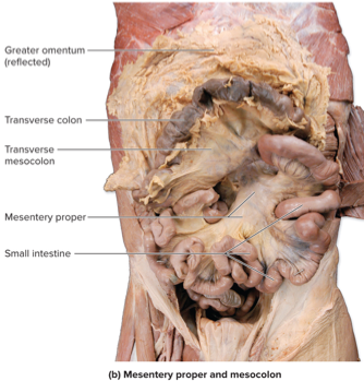
Serous Membranes: Omenta
Greater omentum
Extends inferiorly from inferolateral stomach
Covers most abdominal organs
Accumulates large amounts of adipose tissue (fatty apron)
Lesser omentum
Connects superomedial surface of stomach and proximal end of duodenum to the liver

Clinical View: Peritonitis
Inflammation of peritoneum
Abdominal pain
Most common cause: GI tract perforation
Allows contents to contact peritoneum
May result within (ulcer, ruptured appendix) or without (gunshot wound, surgery)
Upper GI tract organs and accessory
Oral cavity and salivary glands
Pharynx & Esophagus
Stomach
Gastric Motility
Oral Cavity and Salivary Glands
Functions: Mechanical digestion (chewing) and enzymatic breakdown
Saliva Components: Water mucus, salivary amylase (starch digestion), and lingual lipase
Teeth: Incisors (cutting), canines (tearing), premolars/molars (grinding)
Salivary Glands
Intrinsic Salivary Glands: Within oral cavity
Contain lingual lipase, enzyme that begins digestion
Extrinsic Salivary Glands: Outside of oral cavity
Produce most saliva
Parotid, submandibular, and sublingual glands

Parotid salivary glands
Largest salivary glands
25%-30% of saliva
Conducted through parotid duct to oral cavity
Extends from gland across masseter, opening near second upper molar
Infection of the parotid gland causes mumps
Submandibular and Sublingual Salivary Gland
Submandibular:
Produces 60% to 70% of saliva
Submandibular duct opens from each gland to floor of cavity
Sublingual
Extends tiny ducts opening into inferior surface of cavity
Contribute only 3% to 5% of saliva
Oral Cavity - Anterior View

Oral Cavity and Pharynx - Sagittal Section

Pharynx & Esophagus
Swallowing (Deglutition) Phases:
Voluntary Phase: Tongue pushes bolus to oropharynx
Pharyngeal Phase: Involuntary; soft palate & epiglottis close passageways
Esophageal Phase: Peristalsis moves food to stomach
Esophageal Sphincters: Prevent air entry & acid reflux

Phases of Swallowing

Stomach
Regions: Cardia, fundus, body pylorus (controls chyme entry to small intestine)
Gastric Secretions:
Mucous Cells: Produce mucus for protection
Parietal Cells: Secrete intrinsic factor (B12 absorption) & HCL
Chief Cells: Secrete pepsinogen (activated to pepsin for protein digestion)
G Cells: Release gastrin, stimulating stomach motility & secretion)
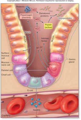
Gross Anatomy of the Stomach

Stomach Mucosa

Stomach: Gastric Motility
Mixing: Forms chyme
Emptying: Peristalsis moves chyme into duodenum
Clinical View: Reflux Esophagitis and Gastroesophageal Reflux Disease
Inflammation of esophagus due to acidic chyme refluxing into esophagus
Pain posterior to sternum, heartburn
Seen frequently
Overweight individuals, smokers, after large meals
Hiatal hernias: portion of stomach protruding through disaphragm
Treatment
Lifestyle changes, head elevation, limiting meal size
Clinical View: Reflux Esophagitis and Gastroesophageal Reflux Disease (continue)
Can result from chronic reflux esophagitis
Erodes esophageal tissue
Scar tissue buildup, leading to narrowed lumen
May change from stratified squamous to columnar secretory epithelium, Barrett esophagus
Increases risk of cancerous growth
May be treated with multiple medications
Includes proton pump inhibitors, histamine blockers
Motility in the Stomach: Gastric Mixing and Gastric Emptying

Stomach: Cephalic Phase
Cephalic reflex, initiated by the thought of food
Nerve signals from higher regions of the brain are sent to the hypothalamus, which relays nerve signals to medulla oblongata
Causes increase in motility and secretory activity
Stomach “growls”
Stomach: Gastric Phase
Process following bolus reaching stomach
Regulated via gastric reflex and through release of gastrin hormone
Gastric reflex is initiated as baroreceptors detect stomach stretch
Chemoreceptors detect protein and increased pH
Nerve signals relayed to medulla oblongata
Results in increased stomach motility and secretory activity
Presence of food (especially protein) in stomach causes release of hormone gastrin
Stomach: Intestinal phase
Process following chyme reaching small intestine
Involves intestinal reflex
Intestinal reflex opposing cephalic and gastric reflexes
Chyme in duodenum signals the medulla oblongata
Causes decrease in motility and secretory activity of stomach
Cholecystokinin (CCK) and secretin also decrease stomach motility and secretory activity
Slows emptying stomach
Regulation of Digestive Processes in the Stomach

Clinical View: Gastric Bypass
Surgical treatment for obesity
Small part of stomach sectioned off and attached to lower part of small intestine
Less nutrients absorbed
Reduced appetite
Altered response to hormones like insulin
Cann induce remission of type 2 diabetes within a few days of surgery
Clinical View: Peptic Ulcers
Peptic ulcer
Solitary erosion of portion of stomach or duodenum
Gastric ulcers in stomach, duodenal ulcers in duodenum
Symptoms of gnawing, burning pain in epigastric region
May erode and cause perforation, medical emergency
Helicobacter pylori causes erosion of gastric lining
Treatment similar to those of gastric reflux
Clinical View: Vomiting
Rapid expulsion of gastric contents through oral cavity
Controlled by vomiting center in the mdeulla oblongata
Responds to head injury, motion sickness, infection, toxicity, food irritation
Closure of nasal passages and the glottis
Skeletal muscle contraction increasing intragastric pressure
Gastric contents forced into and through the esophagus
Danger of aspiration
Lower GI Tract Organs and Associated Accessory Digestive Organs

Clinical View: Inflammatory Bowel Disease (IBD)
Autoimmune disorders, both with a region of inflamed intestine
Clinical View: Inflammatory Bowel Syndrome (IBS)
More common, affecting 1 in 5 Americans
Abnormal function of colon
More common in women than men
Cramps, bloating, constipation, diarrhea
Treatment with diet, medication, stress reduction
Clinical View: Crohn Disease and Ulcerative Colitis
Chron Disease:
Young adults
Intermittent and relapsing episodes of cramping and diarrhea
Ulcerative Colitis:
Similar to Crohn’s but only involves large intestine
Small Intestine: Regions
Duodenum: Receives chyme and secretions from liver & pancreas
Jejunum: Primary site of digestion and absorption
Ileum: Final absorption; connects to large intestine
Small Intestine: Surface Area Enhancements
Circular Folds: Slow movement for more absorption
Villi: Finger-like projections containing capillaries & lacteals
Microvilli (Brush Border): Contain enzymes for final digestion

Small Intestine: Accessory Digestive Organs
Liver: Produces bile (fat emulsification), detoxifies blood, stores nutrients
Gallbladder: Stores & concentrates bile, releases it via the bile duct
Pancreas: Produces digestive enzymes & bicarbonate to neutralize stomach acid

Gross Anatomy of the Liver: Posterior inferior View

Large Intestine Regions
Cecum, colon (ascending, transverse, descending, sigmoid), rectum, anal canal

Large Intestine: Functions
Water & electrolyte absorption
Compacts feces
Houses gut microbiota for vitamin production & fermentation
Defection Reflex: Triggered by rectal distension; controlled by internal (involuntary) and external (voluntary) and sphincter
Anal Canal
Lined by stratified squamous epithelium
Passes through opening in levator ani muscle and terminates at anus
Longitudinal ridges, anal columns
Depression between, anal sinuses
Release mucus when pressure exerted
Internal anal sphincter
Involuntary smooth muscle at base of anal canal
External anal sphincter
Voluntary skeletal muscle
Sphincters normally closed off but relax during defacation
Small Intestine Tunics
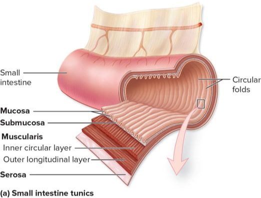
Section of Small Intestine

Bile
Secreted by liver, no digestive enzymes
Contains: Water, bicarbonate ions, bile salts and pigments, cholesterol, lecithin, mucin
Bile salts and lecithin help emulsify lipids
Hepatic Lobules: Structure and Function
Hepatic lobules are hexagonal structural and functional units.
They are composed of hepatocytes, which are liver cells that filter and process nutrient-rich blood.

Hepatic Lobules: Components
Each hepatic lobule contains a central vein.
In each corner of the lobule, there is a portal triad, which consists of:
Hepatic portal vein
Hepatic artery
Bile duct

Liver Lobule

Hepatic Portal System

Gallbladder
Stores, concentrates, and releases bile produced in liver
3 tunics: Inner mucosa, middle muscularis, external serosa
Mucosa with folds that allow distension of wall
Connected to the common bile duct by cystic duct
Sphincter valve
Controls flow of bile into and out of gallbladder
Clinical View: Gallstones
In Gallbladder or biliary apparatus
Risk factors: Female sex, obesity age, caucasian
Condensation of cholesterol or calcium and bile salts
Majority asymptomatic until lodged in neck of cystic duct: cholecystitis
Severe pain in right hypochondriac region
Symptoms worse after fatty meal
May require surgical removal, cholecystectomy
Pancreas
Endocrine function: Produces and secretes insulin and glucagon
Exocrine function: Produces pancreatic juice to assist with digestive activities

Pancreatic Juice
Formed from secretions of acinar cells and pancreatic duct cells
Alkaline fluid
Mostly water: HCO3-, digestive enzymes
Pancreatic amylase to digest starch
Pancreatic lipase to digest triglycerides
Inactive proteases that digest proteins when activated
Nucleases for digestion of nucleic acids
Cholecystokinin (CCK)
Hormone released from small intestine in response to fatty chyme
Stimulates gallbladder to strongly contract and release bile
Stimulates pancreas to release pancreatic juice
Relaxes smooth muscle within hepatopancreatic ampulla
Allows entry of bile and pancreatic juice into small intestine
Inhibits stomach motility and release of gastric secretions
Defacation
Gastrocolic Reflex
Initiated by stomach and causes a mass movement
Defecation Reflex
Filling of rectum initiates urge to defecate
Receptors signal to spinal cord
Increases parasympathetic output to sigmoid colon and rectum
Decreases output to internal (involuntary) anal sphincter
Voluntary defecation
Involves Valsalva Maneuver and relaxation of external (voluntary) anal sphincter

Clinical View: Constipation
Temporary impaired ability to defecate
Compacted feces difficult to eliminate
From low fiber, dehydration, lack of exercise, poor bowel habits, anesthesia
Clinical View: Diarrhea
Disrupted in normal mechanism to absorb intestinal water
Example: from osmotically active solutes that move large volumes of water into colon
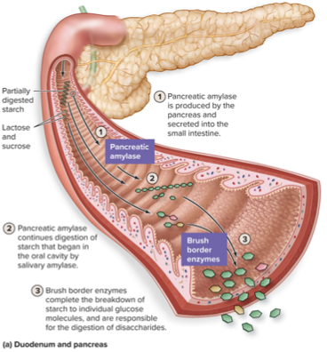
Carbohydrate Digestion
Starts in mouth: Salivary amylase
Continues in small intestine: Pancreatic amylase & brush border enzymes
Protein Digestion
Starts in stomach: Pepsin breaks proteins into peptides
Continues in small intestine: Trypsin, chymotrypsin, and peptidases
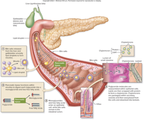
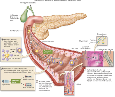
Lipid Digestion
Starts in stomach: lingual and gastric lipase
Major digestion in small intestine: Bile emulsifies fats pancreatic lipase digests triglycerides
Digestion of Startch
Brush border enzymes complete starch breakdown
Includes dextrinase and glucoamylase
Break bond between glucose subunits of oligosaccharides
Maltase
Breaks bonds between the two glucose molecules

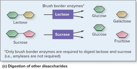
Digestion of Other Disaccharides
Digestion of other disaccharides requires one enzyme each
Named for substrate it digests
Example: lactase digests lactose into glucose and galactose
Reduced amount or lack of lactase = lactose intolerance
Clinical View: Celiac Disease
Gluten-Sensitive Enteropathy
Autoimmune disorders affect 1% of the U.S. population
Gluten = protein in wheat, rye, barley
Stimulates immune response, damages villi of small intestine, interferes with absorption
Symptoms:
Abdominal pain, diarrhea, nutrient deficiencies
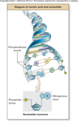
Nucleic Acid Digestion
Small intestine: Pancreatic nucleases break down DNA & RNA into nucleotides
Brush border enzymes:
Phosphatase: breaks bonds holding phosphate
Nucleosidase: breaks bond between sugar and nitrogenous base
Components absorbed across epithelium of small intestine
Phosphate, sugar, nitrogenous base