triage and stabilisation
 Call Kai
 Learn
 Practice Test
 Spaced Repetition
 Match
 Flashcards
 Knowt Play
1/37

There's no tags or description
Looks like no tags are added yet.
38 Terms
triage
Triage examinations can be used to sort cases into order of priority
OR
Recognise genuinely sick patients who need further interventions to prevent deterioration
common triage systems
ABCD
ATT - animal trauma triage system
ABCD system
easy and commonly used
A = airway
B = breathing
C = circulation
D = disability
ATT system
The Animal Trauma Triage Score (ATT) is a veterinary illness severity score used to classify degree of trauma. This scoring system has 6 categories: perfusion, cardiac, respiratory, eye/muscle/integument, skeletal, and neurological.
Each category is given a score of 0 to 3 and total score is calculated ranging from 0 to 18. A higher score indicates greater severity of injury
timeline

emergency - immediate assessment
distended abdomen with non-productive retching
breathing difficulties
seizure lasting > 5 mins
collapsed animal
major bleeding
male cat unable to urinate
dystocia with pup/kitten stuck
toxin ingestion within last 2hrs
not exhaustive - if unsure assess and have them wait if patient stable
potentially serious - triage when available
persistent vomiting and diarrhoea with lethargy
wounds without major bleeding
acute lameness
acute onset painful eye
not exhaustive
if stable can wait for vet appointment
assessment of respiratory system
RR
effort
pattern
open mouth breathing in cats
inspiratory vs expiratory dyspnoea
parasoxical patterns
noise
auscultations
areas of dullness
lung sounds - normal, increased, decreased
abnormal sounds in thorax e.g gut sounds in a diaphragmatic hernia case
pulmonary auscultation

cardiovascular system
HR
rhythm
pulse quality
consistent or variable
synchronous with HR
mucous membrane colour
CRT
auscultation
murmur present
normal or muffled heart sounds
CRT

mucous membrane colour
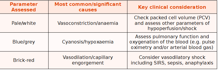
muffled heart sounds - causes and key clinical considerations
pleural or pericardial effusion
consider emergency thorax POCUS scan
pulse quality - weak - causes and key clinical considerations
hypoperfusion/ vasoconstriction/ thrombus
assess other parameters of perfusion
pulse quality - bounding - causes and key clinical considerations
anaemia, hyperdynamic shock
check PCV and look for supporting evidence of hyperdynamic shock
pulse quality - irregular - causes and key clinical considerations
arrhythmia
ECG recommended
what is hyperdynamic shock
early stage of shock characterized by fast HR, high cardiac output and vasodilation leading to brick-red mm and bounding pulses. Often seen in sepsis
neurological assessment
mentation
alert and responsive
quiet
dull
obtunded
comatose
seizuring
ambulatory
not a full neurology examination - checking CNS for basic signs
if signs of significant dysfunction then consider more detailed assessment
modified Glasgow coma scale
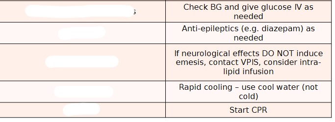
neurological stabilisation for these problems

respiratory stabilisation for dyspnoea
reduce stress - hands off, consider sedation
provide oxygen - flow by/mask/ oxygen tent/ nasal prongs
respiratory stabilisation for obstruction
remove foreign body ASAP, consider GA or tracheostomy
respiratory stabilisation for upper respiratory inspiratory effort
reduce swelling e.g dexamethasone IV
minimise stress, consider butorphanol IM
actively cool
respiratory stabilisation for pleural space
thoracocentesis if fluid/ air, surgery if diaphragmatic rupture
types of shock
cardiogenic
primary cardiac causes
hypovalemic
blood and fluid losses
obstructive
impeded venous return
distributive
abnormal vasomotor tone


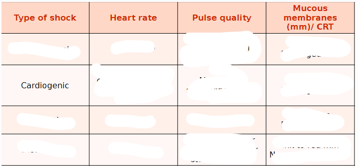

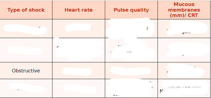


cardiogenic shock
Cardiogenic shock occurs when cardiac output is reduced.
Systolic and diastolic dysfunction and cardiac arrhythmias may cause decreased stroke volume and/or abnormal heart rates. It is important
to note that not all patients in congestive heart failure are in cardiogenic shock, while most animals in cardiogenic shock will concurrently be in congestive heart failure.
The prognosis is typically guarded for patients in
cardiogenic shock, except when it is caused by a reversible underlying disease such as sepsis-induced myocardial dysfunction.
Cardiogenic shock is usually treated with medications
hypovolaemic shock
shock resulting from a decrease in blood volume and is the most common form of shock seen in small animal practice
It may occur with volume loss due to:
Haemorrhage (internal or external)
Loss of other bodily fluids (e.g. vomiting, diarrhoea, polyuria)
Due to decreased intake (e.g. restricted access, regurgitation, vomiting, dysphagia)
hypovolaemia
used to refer to a fluid deficit in the intravascular space, while the term ‘dehydration’ is used to refer to a deficit in the interstitial space. It is very important to make a clinical distinction between a patient that demonstrates interstitial dehydration and one that is hypovolaemic. Hypovolaemia is life-threatening and must be treated immediately with acute resuscitative fluid therapy (e.g. boluses), while interstitial dehydration is not and can be corrected more slowly, over 24 hours
how to stabilise hypovolaemic shock
isotonic crystalloid boluses (via a large bore peripheral IV) – given as 10ml/kg boluses over 10 minutes (60-90ml/kg/hour)
If you have a very large patient (Great Dane etc.) you can use hypertonic saline – 5ml/kg as a single bolus (3ml/kg as a single bolus in cats with raised ICP)
dehydration IVFT
Maintenance requirement (fluid lost via normal mechanisms) - 2ml/kg/hr or 50ml/kg/24hr
Deficit: Volume in ml = % dehydration (0.05-0.12) x bodyweight (kg) x 1000
Ongoing losses:
Estimate volume lost (e.g. though V+ or D+) each time = 4ml/episode x weight (kg)
Estimate frequency of loss over 24-hour period e.g. vomiting four times a day
Volume in ml = volume of loss per episode (ml) x frequency of loss per 24 hours
From this you can then calculate the volume of fluid per minute and the drip rate
obstructive shock
results from physical obstruction to blood flow, either to or from the heart, or through the great vessels.
causes of obstructive shock
GDV
pericardial tamponade
tension pneumothorax
pulmonary or aortic thromboembolism
how to stabalise obstructive shock
removing the obstruction, e.g. remove gas from stomach in a GDV and give shock rate fluids as for hypovolaemia.
Always cannulate the front legs in GDV patients. If you
cannulate the back legs, none of the fluids will get to the heart because of the obstruction caused by the dilated stomach
distributive shock
defined as a maldistribution of blood flow, most commonly inappropriate vasodilatation due to
sepsis or the SIRS response.
It is clinically distinct from other forms of shock in that it
initially results in a hyperdynamic state, which in dogs is characterised by increased cardiac output and peripheral vasodilatation. As distributive shock worsens it usually progresses from a hyper- to hypodynamic state.
Non-infectious SIRS may be caused by insults such as
trauma, pancreatitis, shock, surgery or burns.
To stabilise treat the cause if possible:
Steroids and chlorphenamine for anaphylaxis
Address overwhelming infections with IV antibiotics (e.g. cefuroxime)
Reduce inflammatory drive (remove necrotic tissue if possible)
Use vasoactive drugs if available and shock rate fluids as for hypovolaemia