Quiz 4 A&P Lab
 Call Kai
 Learn
 Practice Test
 Spaced Repetition
 Match
 Flashcards
 Knowt Play
1/56

There's no tags or description
Looks like no tags are added yet.
57 Terms

Buccinator
Function: mastication, blowing, and whistling.

Depressor anguli oris
A muscle of facial expression originating from the mandible that inserts into the angle of the mouth, responsible for depressing the corners of the mouth downward (associated with frowning).
Biceps Brachii


Lateral vs. medial pterygoid muscles
Two muscles of mastication innervated by the mandibular nerve (V3). The lateral pterygoid depresses (opens) and protracts the mandible while facilitating side-to-side movements, whereas the medial pterygoid elevates (closes) and protracts the mandible while assisting in side-to-side chewing excursion.

Masseter
A primary muscle of mastication that originates from the zygomatic arch and inserts into the mandible, acting to elevate and close the jaw.

Orbicularis oculi
A ring-like facial muscle surrounding each eye orbit that functions to close the eyelids, blink, and squint.

Orbicularis oris
A circular muscle surrounding the mouth that closes, compresses, and protrudes the lips.

Occipitofrontalis
A broad, two-bellied scalp muscle consisting of frontal and occipital bellies connected by the epicranial aponeurosis, responsible for elevating the eyebrows and retracting the scalp.

Risorius
Function: grin or smirk and is innervated by the facial nerve

Temporalis muscle
A broad, fan-shaped muscle of mastication situated in the temporal fossa that elevates and retracts the mandible to close the jaw.

Zygomaticus major and minor
Pairs of facial expression muscles originating from the cheekbone that elevate and draw the angle of the mouth upward and backward to produce a smile.

Digastric
Function: depress the mandible and elevate the hyoid bone.

Levator scapulae
A skeletal muscle situated at the back and side of the neck that originates from the transverse processes of C1-C4 and inserts into the superior angle and medial border of the scapula, primarily acting to elevate the scapula.

Mylohyoid
A paired suprahyoid muscle originating from the inner aspect of the mandible and inserting into the hyoid bone, which forms the muscular floor of the oral cavity and elevates the hyoid and tongue during swallowing.

Platysma
A broad, superficial sheet of muscle in the anterior neck that tenses the neck skin, depresses the mandible and lower lip, and is innervated by the cervical branch of the facial nerve (CN VII).

Scalenes
A group of three cervical muscle pairs (anterior, middle, and posterior) located in the lateral neck that elevate the first two ribs during forced inhalation and assist in lateral flexion of the cervical spine.

Sternocleidomastoid
A major superficial neck muscle originating at the manubrium and clavicle and inserting into the mastoid process; it flexes the neck and rotates the head to the opposite side.

Sternohyoid
An infrahyoid neck muscle that originates at the manubrium of the sternum and medial clavicle, inserts on the body of the hyoid bone, depresses the hyoid bone during swallowing and speaking, and is innervated by the ansa cervicalis.

Stylohyoid
A slender suprahyoid muscle originating from the styloid process of the temporal bone and inserting onto the hyoid bone, functioning to elevate and retract the hyoid during swallowing; innervated by the facial nerve (CN VII).

Diaphragm
A dome-shaped sheet of muscle separating the thoracic cavity from the abdominal cavity that contracts and flattens during inhalation to facilitate breathing.

External and internal intercostals
Muscles situated between the ribs with opposing respiratory functions: external intercostals lift the ribs during inhalation, while internal intercostals depress the ribs during forced expiration.

External oblique
Paired superficial anterolateral abdominal wall muscle that bilaterally flexes the trunk and compresses abdominal viscera, and unilaterally facilitates lateral flexion and contralateral rotation of the torso.

Erector spinae
A deep muscle group composed of the iliocostalis, longissimus, and spinalis that extends vertically along the vertebral column to extend and laterally flex the spine while maintaining an upright posture.

Latissimus dorsi
A broad, flat muscle covering the middle and lower back that functions primarily in extension, adduction, and medial rotation of the arm at the glenohumeral joint.

Pectoralis major
A large, fan-shaped muscle located on the anterior chest wall that primarily functions to adduct, medially rotate, and flex the humerus at the shoulder joint.

Pectoralis Minor
A thin, triangular muscle of the anterior chest wall situated deep to the larger chest musculature; it originates from the third through fifth ribs, inserts into the coracoid process of the scapula, and functions to depress, protract, and downwardly rotate the scapula while aiding in forced inspiration.

Serratus anterior
Fan-shaped muscle on the lateral chest wall originating from ribs 1–8/9 and inserting on the medial border of the scapula; functions primarily to protract and stabilize the scapula, innervated by the long thoracic nerve.
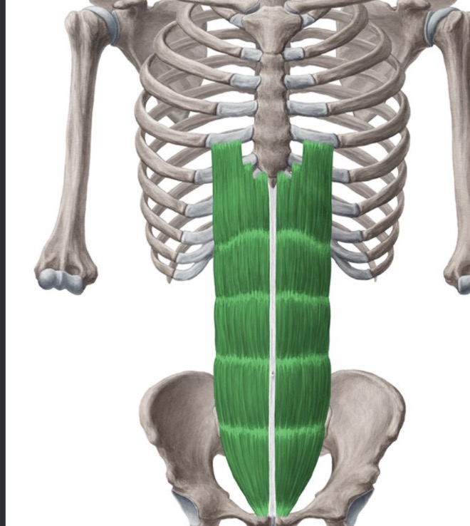
Rectus abdominis
(6 PACK) A paired muscle running vertically along the anterior abdominal wall, responsible for flexing the lumbar spine, depressing the rib cage, and compressing the abdominal viscera.

Rhomboid major
A back muscle originating from the spinous processes of T2–T5 and inserting onto the medial border of the scapula; acts to retract, elevate, and downwardly rotate the scapula, innervated by the dorsal scapular nerve.

Rhomboid minor
A small back muscle that originates from the nuchal ligament and spinous processes of C7 and T1, inserts onto the medial border of the scapula at the base of the scapular spine, and acts to retract and elevate the scapula.

Transversus abdominis
The deepest flat muscle layer of the anterolateral abdominal wall, with horizontally running fibers that compress abdominal contents and stabilize the lumbar spine and pelvis.

Trapezius
A large, superficial back muscle extending from the occipital bone to the lower thoracic vertebrae and scapula, primarily responsible for stabilizing, elevating, retracting, and rotating the scapula.

Internal obliques
An intermediate muscle layer of the anterolateral abdominal wall located deep to the external layer; it functions to compress the abdomen, assist in spinal flexion, and rotate the torso toward the same side.
Brachialis

Coracobrachialis

Deltoid


Infraspinatus
Subscapularis

Teres major

Teres minor

Triceps Brachii

Abductor pollicis longus

Brachioradialis

Extensor carpi radialis longus

Extensor carpi radialis brevis

Extensor digitorum

Extensor pollicis longus

Flexor carpi radialis

Flexor carpi ulnaris

Flexor digitorum superficials

Flexor digitorum profundus

Flexor pollicis longus

Palmaris longus

Pronator teres

Supinator

Adductor longus

Adductor magnus
