Digestive System
 Call Kai
 Learn
Learn Practice Test
 Spaced Repetition
 Match
Match Flashcards
 Knowt Play
1/212

Earn XP
Description and Tags
Bio 211
Name | Mastery | Learn | Test | Matching | Spaced | Call with Kai | Chat |
|---|
No analytics yet
Send a link to your students to track their progress
213 Terms
Functions of digestive system
Breakdown foods, Release their nutrients, Absorb those nutrients, Eliminate wastes
2 parts of digestive system
Alimentary canal/Gastrointestinal Tract, and Accessory digestive organs
Alimentary canal/Gastrointestinal Tract
mouth, pharynx, esophagus, stomach, small and large intestines, and anus
Accessory digestive organs
critical to the function of the digestive system. Includes the teeth, tongue, salivary glands, the gallbladder, liver, and pancreas are all accessory organs
layers of Alimentary canal/Gastrointestinal Tract
mucosa, submucosa, muscularis, and serosa
mucosa of alimenatry canal
Epithelial tissue lining of the tract, Produces mucus for lubrication and protection, Contains a very thin smooth muscle layer called the *muscularis mucosa* (which is not the same as the muscularis layer) Is in direct contact with ingested food
submucosa of alimentary canal
Dense connective tissue serves as the location for blood vessels, lymphatic vessels, and glands. Also contains a dense branching network of nerves.
muscularis of alimenatry canal
made up of layers of smooth muscle (circular and longitudinal except where otherwise indicated), the contractions of which promote mechanical digestion and propulsion. contains sphincters, skeletal muscle curved fibers located at certain locations along the length of the canal that give voluntary control over swallowing and defecation. Contractions of the muscularis promote deglutition, churning,
peristalsis, and segmentation
serosa of the alimentary canal
is the outermost layer. terminology: instead of serosa, the mouth, pharynx, and esophagus have a dense sheath of collagen fibers called the adventitia. adventitia serves to hold the alimentary canal in place near the ventral surface of the vertebral column since these organs aren’t as moveable
as the latter organs
histology of mucosa (epithelium)
Of the mouth, pharynx, esophagus, and anus: stratified squamous (structure complements function!) Of the stomach, small intestines, and large intestines: simple columnar (structure complements function!)
histology of mucosa (lamina propria)
Made of areolar connective tissue, blood vessels, nerves. In the small intestine, contains MALT called Peyer’s Patches (review Chapter 21)
histology of mucosa (muscularis mucosa)
Smooth muscle creates constant tension on mucosa, creating folds. (What are these folds for?)
ENTERIC NERVOUS SYSTEM
a nervous system within another system
What is food detected by?
Food is detected by receptors from the ENTERIC NERVOUS SYSTEM – a nervous system within another system
What 2 plexuses are Enteric neurons grouped into?
The myenteric plexus and The submucosal plexus
nerve supply
Extrinsic innervation (from without) is provided by the autonomic nervous system: sympathetic activation restricts/inhibits the activity of enteric neurons (reduces digestion) parasympathetic activation activates/stimulates those neurons (promotes digestion)
The functions of blood vessels in digestive system
transport absorbed the protein and carbohydrate nutrients – (recall that lipids are absorbed via lacteals) supply the organs with the nutrients and oxygen needed to drive their cellular processes
BLOOD SUPPLY TO/FROM THE DIGESTIVE SYSTEM
The supply comes FROM: thoracic aorta►abdominal aorta►and celiac trunk, superior, and/or inferior mesenteric arteries. Nutrient-filled blood goes TO: the hepatic portal system from digestive
organs about one-fourth of the blood pumped with each heartbeat enters arteries serving the intestines.

Peritoneum
Squamous epithelial issue making up the membranes of the abdominal cavity. Parietal peritoneum
adheres to abdominal cavity. Visceral peritoneum adheres to the abdominal organs. Visceral peritoneum is folded into a number of specific structures (next slide)
Parts of the visceral peritoneum
Greater omentum, Falciform ligament, Lesser omentum, Mesentery, Mesocolon
Greater omentum
Apron-like fatty structure that lies superficial to the stomach, small intestine and transverse colon
Falciform ligament
Anchors the liver to the anterior abdominal wall and inferior border of the diaphragm
Lesser omentum
Connects the stomach to the liver
Mesentary
Vertical band of tissue anterior to the lumbar vertebrae and anchoring all of the small intestine except the initial portion (the duodenum) – Like spokes on a bicycle wheel
Mesocolon
Attaches two portions of the large intestine (the transverse and sigmoid colon) to the posterior abdominal wall
Mouth
Ingestion; with salivary glands, begins breakdown of carbohydrates
Pharynx
Propulsion
Esophagus
Peristalsis
Stomach
Churning, begins breakdown of proteins, secretes intrinsic facto
Small intestine
Peristalsis, Segmentation, Major site of absorption
Large intestine
Peristalsis, Segmentation, Absorption of water/vitamins, concentration of chyme
salivary glands
Secretion of amylase and lingual lipase, lubrication
Liver
Produces bile, detoxification, metabolism, storage
Pancreas
Secretion of digestive enzymes, insulin and digestive enzyme
Gall Bladder
Stores, concentrates and secretes bile
6 digestive processes in alimentary canal
Ingestion, Propulsion (2 types), Mechanical digestion (3 types), Chemical digestion, Absorption, Defecation
Ingestion
entry of food into the alimentary canal through the mouth, mastication and mixing with saliva occur just after via the teeth, tongue, and salivary glands
Propulsion
forward movement of food through the digestive tract, includes deglutition (voluntary and involuntary), includes peristalsis (involuntary), is NOT considered mechanical digestion as nothing is physically being broken apart by the process
Mechanical Digestion
is purely physical, makes the food smaller to increase both surface area and mobility, involves mastication (voluntary), Involves churning (involuntary), involves segmentation (involuntary)
Chemical Digestion
takes place in the mouth, stomach, and small intestine, digestive secretions break down complex food molecules into their chemical building blocks using hydrolysis, the secretions contain water, various enzymes, acids, and salts
Absorbtion
Majority occurs in the small intestine It is different for carbohydrates, proteins, and nucleic acids, versus lipids
Defecation
Undigested materials are removed from the body as feces
Regulatory mechanisms
There are two ways that the digestive system is controlled (regulated) Nerves, Hormones. These two ways alter the responses to stimuli (or lack of) to change mechanical and chemical digestion
What are the receptors located on the walls of GI tract?
baroreceptors, chemoreceptors, and osmoreceptors detect pressure/stretch, chemicals, and hydration concentration
Two types of reflexes serve the digestive system
long and short
Extrinsic nerve plexuses create “long reflexes”
central and autonomic nervous systems, respond to stimuli from OUTSIDE (external to) the digestive system
Intrinsic nerve plexuses create “short reflexes”
nerves found within the walls of the alimentary canal, regulate activities in the specific area of the digestive tract where they are found
What produces gastrin?
G- cells in stomach
What produces secretin?
Duodenum
What producesCholecystokinin (CCK)?
Duodenum
What produces Glucose-Dependent Insulinotropic Peptide (GIP)?
Duodenum
What produces Gastric Inhibitory Peptide (GIP)*?
Duodenum
Gastrin function
Stimulates gastric acid secretion and motility
secretin function
Stimulates release of bicarbonate from the pancreas
Cholecystokinin (CCK) function
Stimulates secretion of pancreatic enzymes, bile from the liver, concentrated bile from the gallbladder
Glucose-Dependent Insulinotropic Peptide (GIP) function
Stimulates insulin secretion
Gastric Inhibitory Peptide (GIP) function
Inhibits gastric acid secretion, slows gastric motility and emptying
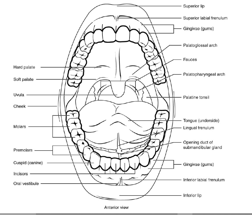
parts of the mouth
Oral cavity (also called the buccal cavity), Labia (also called the lips), Labial frenulum, Oral vestibule, Fauces, Hard Palate, Soft Palate, Uvula

the tongue
facilitates ingestion, facilitates mastication, facilitates chemical digestion by producing lingual lipase (very minor aid for lipid digestion beginning in the stomach), contains papillae under which are receptors that aid in sensation of taste, texture, and temperature of food aid in swallowing aid in vocalization

What is number 1?
Lips

What is 2?
Hard palate

What is 3?
Soft palate

What is 4?
uvula
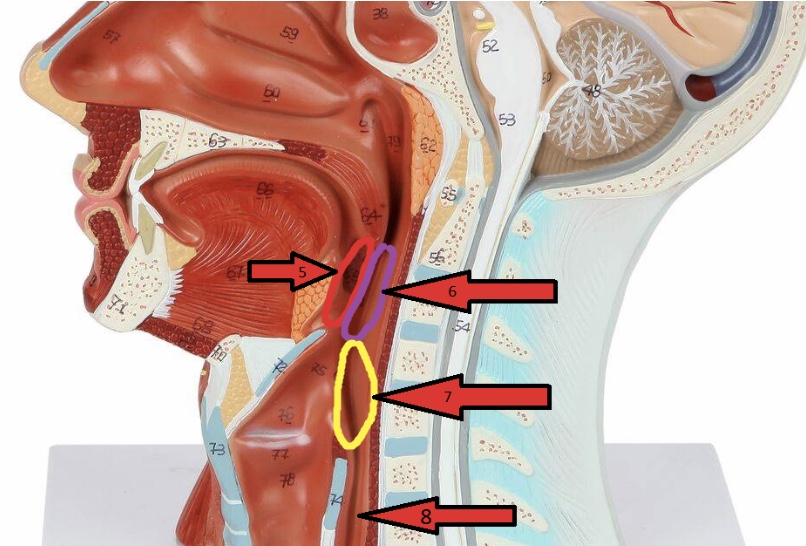
What is 5(opening)?
Fauces

What is 6?
Oropharynx

What is 7?
Laryngopharynx
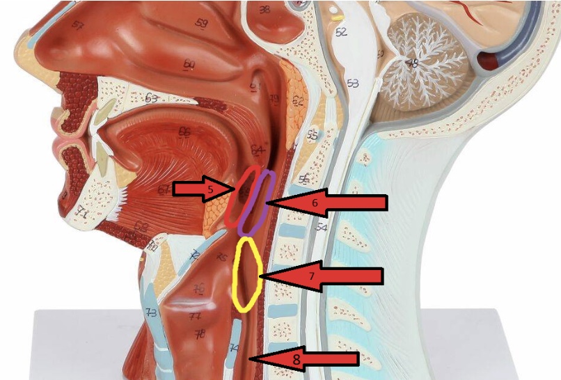
What is 8?
Esophagus
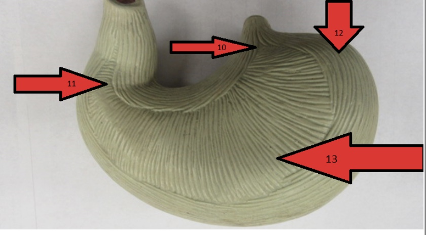
What organ is this?
Stomach

What is 10?
Cardia

What is 11?
Pylorus

What is 12?
Fundus

What is 13?
Body

What is 14?
Pyloric sphincter

What is 15?
Rugae

What is 16?
Greater curvature

What is 17?
Lesser curvature

What is 18?
Cardiac sphincter
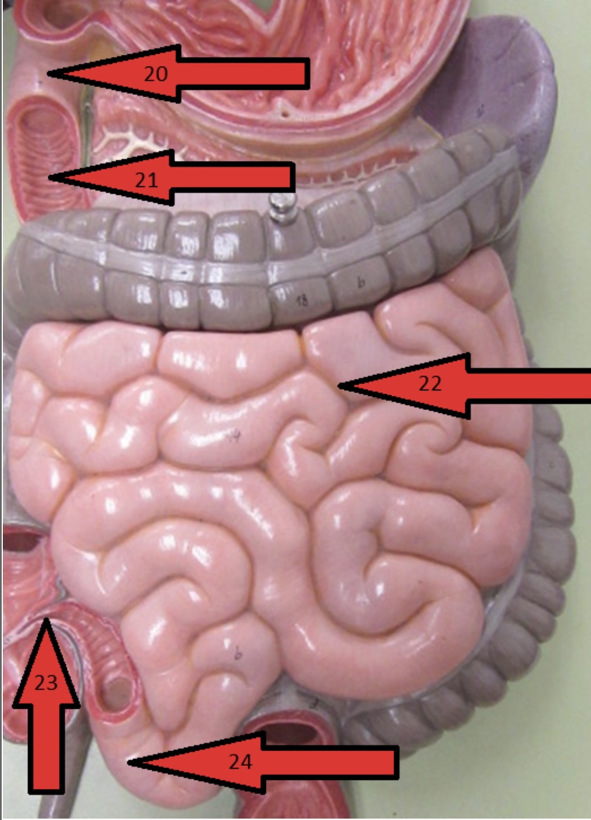
What organ are all the arrows pointing to?
Small intestine

What is 20?
Duodenum

What is 21(ridges)?
Plicae circulares

What is 22?
Jejunum

What is 23(valve)?
Ileocecal valve

What is 24?
Ileum
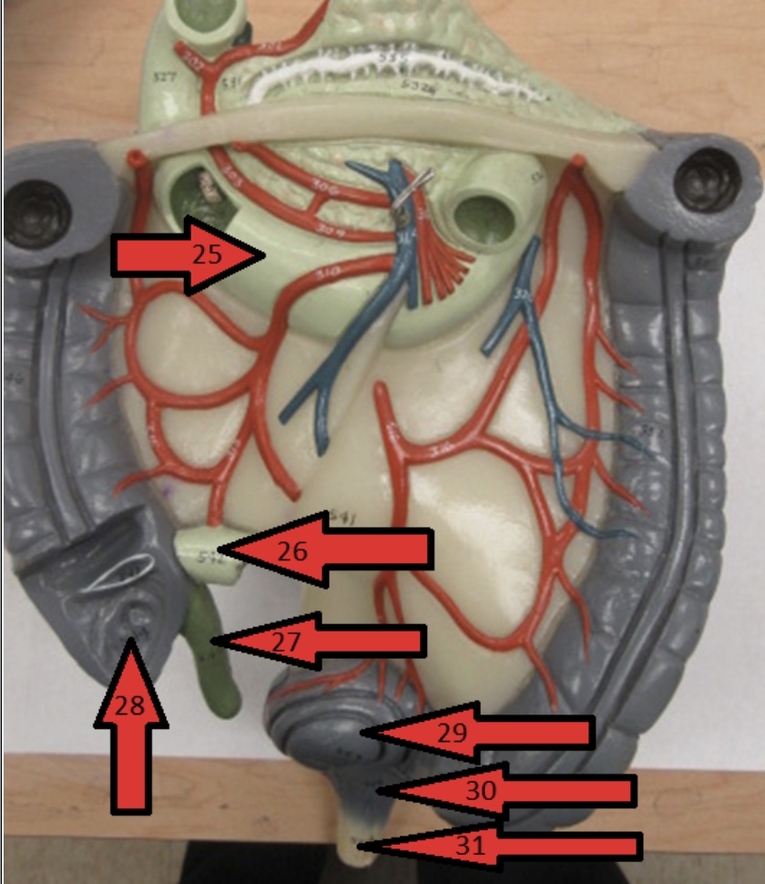
What is 25?
Duodenum

What is 26?
Ileum

What is 27?
Appendix

What is 28?
Cecum

What is 29?
Sigmoid Colon

What is 30?
Rectum

What is 31?
Anus

What is 32?
Ascending colon

What is 33?
Right (hepatic) colic flexure
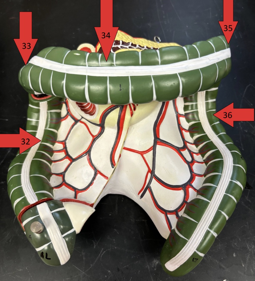
What is 34?
Transverse colon

What is 35?
Left (hepatic) colic flexure

What is 36?
Descending colon

What is 37?
Haustra

What is 38?
Taenia coli

What is 39?
Tongue tip

What is 40?
Tongue body

What is 41?
Tongue root