Chapter 12 UGI + Biliary
 Call Kai
 Learn
 Practice Test
 Spaced Repetition
 Match
 Flashcards
 Knowt Play
1/68

There's no tags or description
Looks like no tags are added yet.
69 Terms
Liver’s primary function
Produces large amounts of bile (about 1 quart/day)
Bile stored in gallbladder, used to break down and digest fats
Anatomy of the liver
Two larger lobes (larger right + smaller left)
Two smaller lobes only visible posteriorly (quadrate + caudate)
Separated by falciform ligament
Difference between caudate and quadrate lobes
Caudate - Superior + posterior to quadrate
Quadrate - On inferior surface of right lobe between gallbladder and falciform ligament
Where is the IVC located in terms of the liver?
Contours over surface of the caudate lobe
Pathway of bile
Small lobules of liver → small ducts → left and right hepatic ducts → common hepatic duct → cystic duct to gallbladder (for storage) OR common bile duct → hepatopancreatic sphincter → descending duodenum
Anatomy of the gallbladder
About 4” in length
Neck
Body
Fundus
Functions of gallbladder
Store bile for future use (2-2.5 TBSP)
Concentrate bile using hydrolysis
Contract when cholecystokinin releases
Anatomy of common bile duct
3” in length, diameter of a straw
Descends behind superior portion of duodenum and head of pancreas to enter descending duodenum
40% of people - CBD + pancreatic duct do NOT combine
60% of people - CBD + pancreatic duct combine (creating hepatopancreatic ampulla), common site of impaction of gallstones
Lateral view of gallbladder
More anterior to midcoronal
Duct system closer to midline
Necessary to place GB closest to IR (prone)
To drain GB, pt is placed supine
What does a LAO position demonstrate of the gallbladder?
Cystic duct
Neck, body, and fundus
What symptoms do gallbladder issues result in?
Nausea, vomiting, RUQ pain, heartburn, and feeling full
What’s the fancy word for gallstones
Choleliths
Choledocholithiasis
Presence of stones
Visualized using ERCP or cholangiography
Cholelithiasis
Condition of abnormal calcifications, caused by an increase in bilirubin, calcium, or cholesterol
Visualized using Cholescintigraphy
What are the two types of gallstones?
Cholesterol - 75%, highly radiolucent (unable to visualize)
Pigment
What are the only types of gallstones visible without contrast?
Crystalline calcium salt (about 20%)
What is milk calcium bile?
Emulsion buildup of gallstones, visualized as sandlike calcifications
Usually only helps to remove gallbladder (cholestectomy)
Cholecystitis
Inflammation of gallbladder typically due to a blockage in the cystic duct
Acute - due to blockages, infections, or ischemia
Chronic - due to gallstones, pancreatitis, or a carcinoma
Neoplasms
Rare growths, either malignant or benign
Malignant - 85% adenocarcinoma, 15% squamous cell carcinoma, likely to spread to liver, pancreas, or GI tract
Biliary Stenosis
Narrowing of a duct causing restriction of bile
May prevent passing of small gallstones leading to obstruction or inflammation
What is the pathway of the alimentary canal?
Oral cavity → pharynx → esophagus → stomach → small intestine → large intestine → anus
What are the accessory organs of the GI system?
Pancreas, liver, gallbladder, and salivary glands
What are the 3 salivary glands?
Parotid - largest, near the ear
Submandibular - below mandible
Sublingual - below tongue
Opening between the esophagus + stomach
Esophagogastric Junction
What allows food/fluid through the esophagogastric junction?
Cardiac sphincter
What is the opening between the stomach and duodenum?
Pyloric Sphincter
Fundus of the stomach
Top portion, looks like a balloon
Lies lateral + superior to cardiac orifice
Usually filled with air “gastric bubble”
Tends to move with diaphragm
Body of stomach
Main part
Lower end has partially constricted area to separate body from pyloric area called angular notch
Pylorus of stomach
Divided in two parts
Antrum - slight dilation distal to angular notch
Canal - ends at pyloric sphincter
The fundus + pyloric region of the stomach are slightly _____ to the stomach body
Posterior
Where does air + barium settle in the supine position?
Heavy barium settles in fundus
Gas settles in body and pylorus
Where does air + barium settle in the RAO prone position?
Gas fills fundus of stomach
Barium settles in anterior body/pylorus
Where does air + barium settle in the erect position?
Gas rises to fill fundus
Barium settles to fill pyloric region
Air fluid levels
Anatomy of the duodenum
7-9” long, shortest and widest of the small bowels
Close to head of pancreas, nestled in C-loop of duodenum
What are the four parts of the duodenum?
Superior - starts at pylorus, area of duodenal bulb, common site of ulcers (intraperitoneal)
Descending - largest segment, posses duodenal papilla where common bile + pancreatic duct open
Horizontal - curves to left to join 4th part
Ascending - ends to create duodenal flexure (junction of duodenum + jejunum, held in place by ligament of Treitz
GI system in hypersthenic
Short lungs
High diaphragm
Large intestine extends entire abdominal cavity
Gallbladder and stomach are high and transverse (T9-T12)
Duodenal bulb is @ T11-T12 right to midline
GI system in hyposthenic/asthenic
Long/narrow lungs
Low diaphragm
Large intestine pushed down low
J-shaped stomach (T11-L5)
Gallbladder + duodenal bulb (L3-L4)
GI system in sthenic
J-shaped stomach (T11-L2)
Duodenal bulb (L1-L2)
Gallbladder less transverse midway from lat to midline
Splenic flexure often quite high
What happens to abdominal organs when a patient goes from supine to erect
They drop about 1-2”
What parts of the GI system are visible without contrast?
Fundus of stomach and parts of the large bowel
What is negative/radiolucent contrast?
Swallowed air, CO2, and gas crystals (visualized as black)
What is positive/radiopaque contrast?
Barium and iodine
Characteristics of Barium Sulfate
Extremely insolubility in water or aqueous liquids
Must be chemically pure or it’s considered poisonous
Expelled rectally
Forms colloidal suspension when mixed with water (never fully dissolves)
Thin Barium
1 part to 1 part
Consistency of a thin milkshake
Used to study entire GI tract
Thick Barium
3-4 parts barium to 1 part water
Consistency of cooked cereal
Used to study esophagus
When should barium not be used?
If there’s any risk of it escaping into the peritoneal cavity (it can NOT be absorbed)
What is gastrografin
Iodinated contrast, 37% organically bound iodine
Body can absorb it
Travels faster than barium
Should not be used in severely dehydrated patients
What does double contrast mean?
Using both radiolucent + radiopaque contrast
Barium + room air/CO2 gas
Gas crystals like calcium + magnesium can be used to create CO2 gas
What is an esophagography?
The use of radiopaque contrast (typically thin + thick barium) to visualize the pharynx and esophagus during swallowing
What are the clinical indications for an esophagography?
Achalasia
Anatomic anomalies
Barrett Esophagus
Dysphagia
Esophageal varices
Zenker diverticulum
What is an upper GI series?
Studying the function of lower esophagus, stomach, and duodenum typically using thin barium
Clinical Indications for an upper GI series
Bezoar
Diverticula
Emesis
Gastric carcinoma
Gastritis
Hiatal hernia
Hypertrophic pyloric stenosis
Ulcers
What should the kVp be for barium, double-contrast, and iodinated contrast studies?
Barium - 110-125
Double - 90-100
Iodine - 80-90
Why is a RAO better for an esophagography than an LAO?
Provides better visibility of esophagus
Zencker’s Diverticula
Found at the pharyngoesophageal junction at upper end of esophagus
Diverticula
Pouch of variable size occurring normally or created by herniation of a mucous membrane through a defect in its muscular coat
Traction Diverticulum
Involves all layers of the esophagus and results from adjacent scar tissue that pulls the esophagus toward the area of involvement, more frequently in the middle third of the esophagus
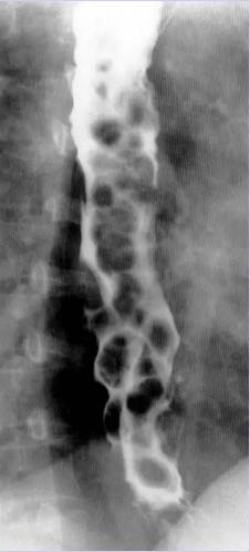
Esophageal Varices
Abnormally diluted veins in the distal esophagus often seen with acute liver disease, best demonstrated recumbent for more complete filling of the veins
Esophageal Reflux
Gastric contents return back through the gastric orifice and irritate the lining of the esophagus
Cleft Palate
Congenital anomaly where the roof of the mouth is open which allows food and fluid to pass into the nose
Esophageal Atresia
Congenital abnormality where esophagus fails to fully develop, causing choking/gagging/dyspnea/cyanosis
Hiatal Hernia
Protrusion of a structure into the thoracic cavity through the esophageal hiatus of diaphragm
Gastric/Peptic Ulcer
Erosion of the mucosal lining on stomach, could cause perforation if severe
Bezoar
Mass of undigested material that gets trapped in the stomach, could cause an obstruction
Polyp
Small mass growing from mucosal wall, could be malignant or benign
Achalasia
Neuromuscular disorder which results in failure of lower esophageal sphincter to relax causing dilation of the esophagus

Schatzke’s Ring
Type of hiatal hernia where esophagogastric junction has herniated through the diaphragmatic opening

Porcelain GB
Extensive calcification in the wall of the GB which forms an oval density that corresponds to the shape of GB. Blue discoloration and brittle consistency of wall.

Difference between cholecystogogue and cholecystopaque
Cholecystogogue - Agent that promotes contraction of GB (not contrast)
Cholecystopaque - Contrast media used for GB visualization