knee joint + surface anatomy
 Call Kai
 Learn
Learn Practice Test
 Spaced Repetition
 Match
Match Flashcards
 Knowt Play
1/59

There's no tags or description
Looks like no tags are added yet.
Name | Mastery | Learn | Test | Matching | Spaced | Call with Kai | Chat |
|---|
No analytics yet
Send a link to your students to track their progress
60 Terms

base of patella
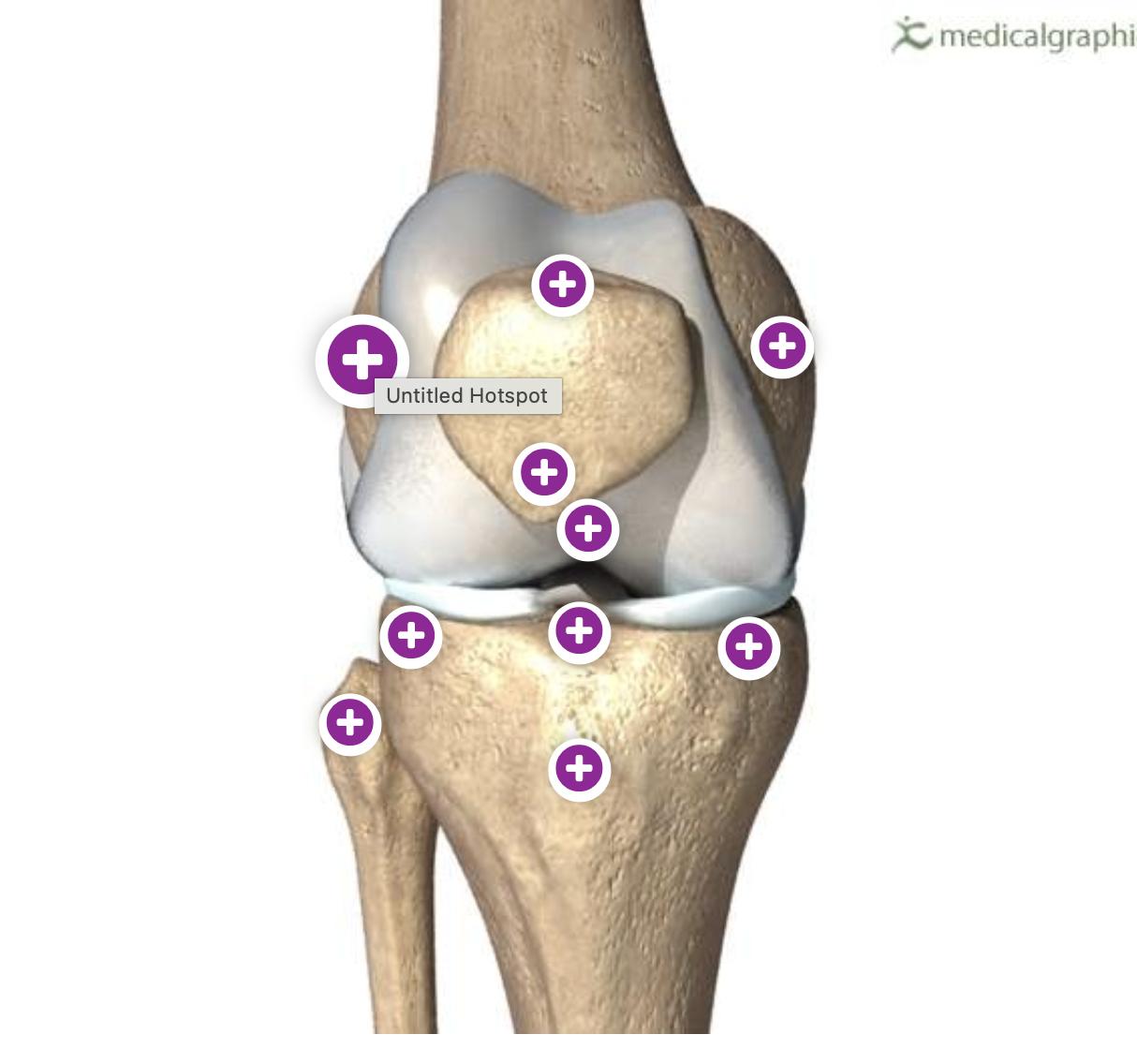
lateral condyle of femur
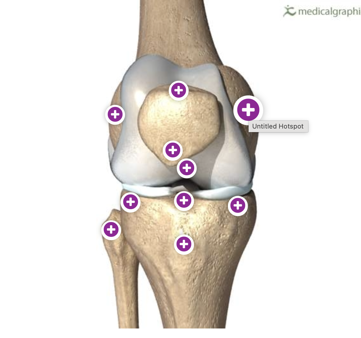
medial condyle of femur

apex of patella

intercondylar notch of femur

anterior intercondylar fossa of tibia
where is the attachment site of the anterior cruciate ligament
anterior intercondylar fossa of tibia

what is this
anterior cruciate ligament tear

medial tibial condyle

lateral tibial condyle
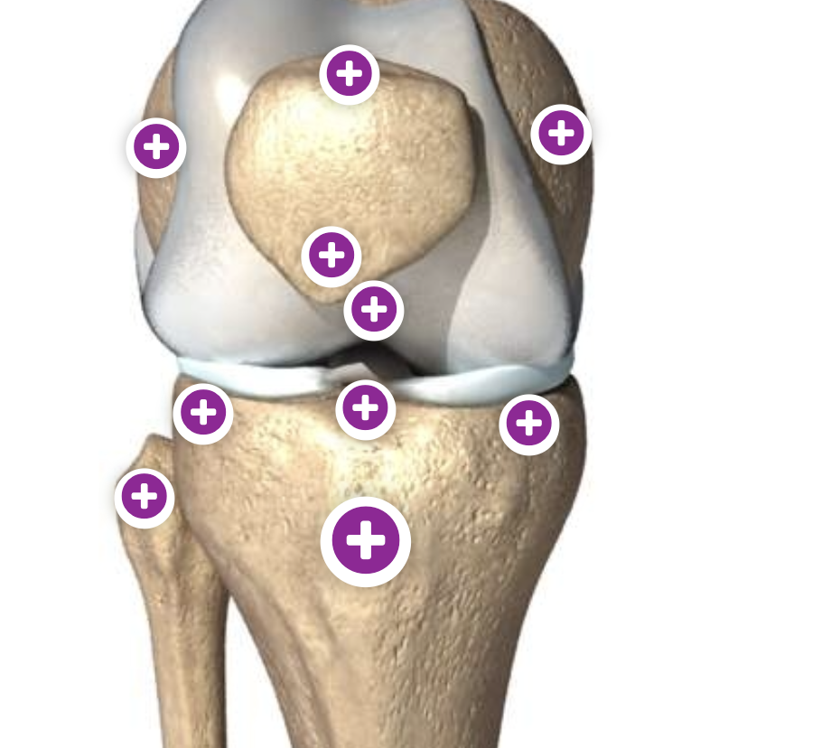
tibial tuberosity

head of fibula

greens
medial and lateral collateral ligament

blue
anterior cruciate ligament
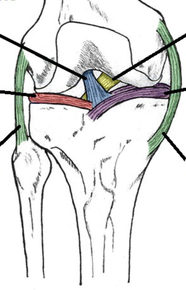
yellow
posterior cruciate ligament

red
lateral meniscus

purple
medial meniscus

patella ligament

what are the bursae of knee joint
suprapatella bursa, infrapatellar bursae (deep and superficial), prepatellar bursa
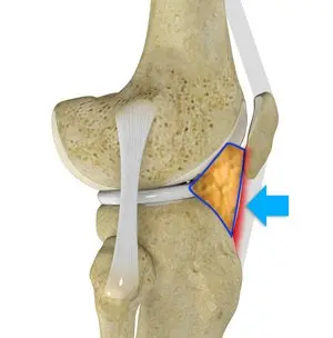
infrapatellar fat pad
what are the neurovascular bundles in anterior compartment of leg
anterior tibial artery and its venae comitantes, deep fibular/personeal nerve
what are the neurovascular bundles of lateral compartment of leg
perforating vessels of fibular artery, superficial fibular/peroneal nerve
what are the neurovascular bundles of posterior compartment of leg
posterior tibial artery and its venae comitantes, tibial nerve
what are the neurovascular bundles of dorsal compartment of foot
dorsalis pedis and its venae comitantes, deep fibular/peroneal nerve
what are the neurovascular bundles of medial bundle of foot
medial plantar artery and its venae comitantes, medial plantar nerve
what are the neurovascular bundles of lateral bundle of foot
lateral plantar artery and its venae comitantes, lateral plantar nerve
what are the contents of popliteal fossa
popliteal artery and vein, termination of the small saphenous vein, the tibial and common fibular nerve, popliteal lymph nodes and fat.
what are the two major superficial veins in the lower limb
short/small saphenous vein and long/great saphenous vein

small saphenous vein
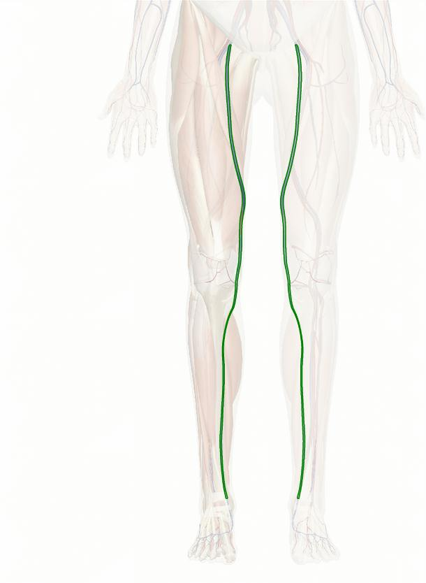
great saphenous vein
what are the bones in the foot
calcaneus, talus, cuboid, navicular, 3 cuneiforms, metatarsals, phalanges
what forms the ankle joint
distal tibia, distal fibula, talus
what are the lateral ligaments of ankle
anterior talogibular ligament, posterior talofibular ligament, calcaneofibular ligament
what are the medial collateral ligaments of ankle
posterior tibiotalar, tibiocalcaneal, tibionavicular, anterior tibiotalar

tibial nerve
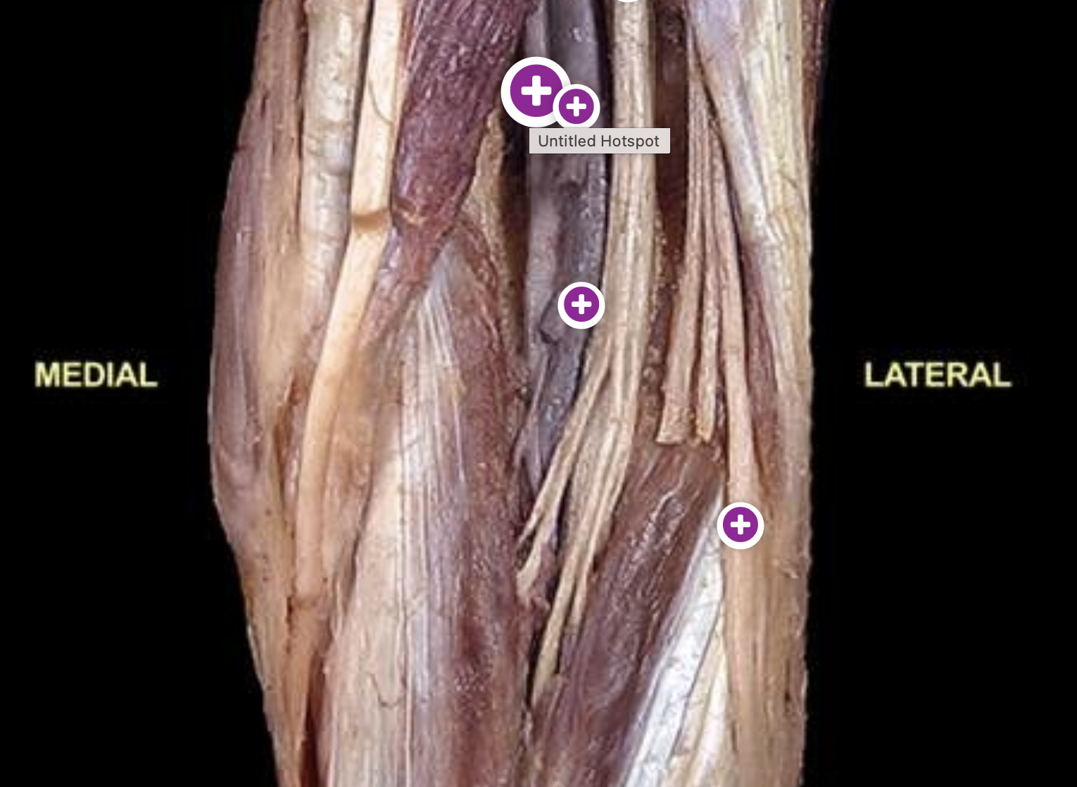
popliteal artery

termination of the small saphenous vein
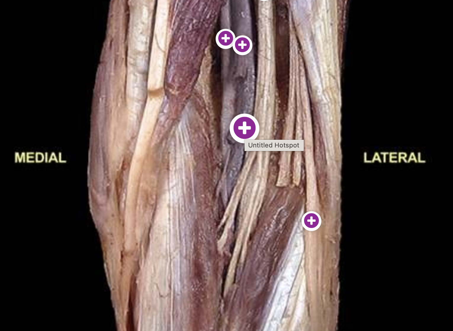
popliteal vein

common fibular nerve

obturator nerve

femoral nerve
the obturator and femoral nerve arise from the _____ which is at the level of ___
lumbar plexus, L1-4

sciatic nerve

tibial nerve

deep fibular nerve

common fibular nerve
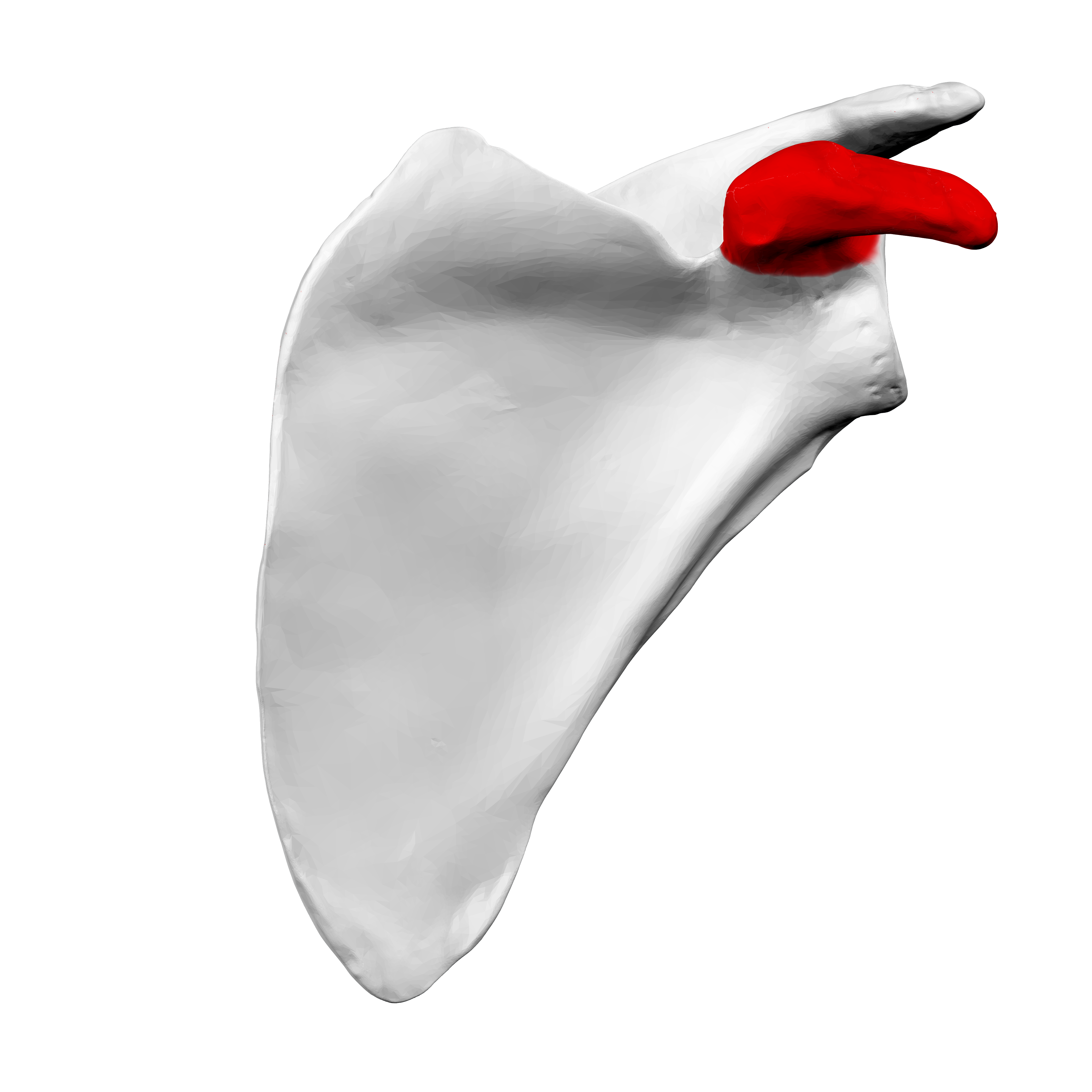
coracoid prcess of scapula

clavipectoral triangle
what does the anterior axilllary fold contain
pectoralis major muscle
what does the posterior axillary fold contain
latissimus dorsi and teres major muscles
what is located between the anterior and posterior axillary folds
axillary fossa

posterior superior iliac spines

anterior superior iliac spine

pubic tubercle
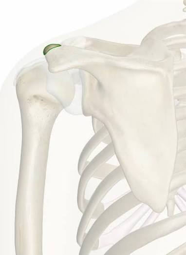
acromion of scapula
why is the medial meniscus more often torn than the lateral meniscus
it is less mobile due to its attachment to the medial collateral ligament and joint capsule, making itmore vulnerable to rotational forces
what is the shape of menisci in most sagittal sectional views of the knee joint
triangular
name the major extracapsular and intracapsular ligaments of the knee joint
extrascapular: patellar ligament, tibial collateral ligament, fibular collateral ligament, oblique popliteal ligament, arcuate popliteal ligament
Infrascapular: anterior cruciate ligament, posterior cruciate ligament

fabella