Genetics Exam 2
 Call Kai
 Learn
 Practice Test
 Spaced Repetition
 Match
 Flashcards
 Knowt Play
1/193

There's no tags or description
Looks like no tags are added yet.
194 Terms
genetic material
contains information
expression determines all traits and functions of organisms
this is what is leading to the traits that we observe, phenotypes
is stable, but must also have the ability to change
variation in many traits without lethality to organism
being passed to one generation to the next and being passed to one cell to 2 daughter cells during cell division
is carried on chromosomes
contain proteins and DNA (deoxyribonucleic acid)
meiosis where the chromosomes are organized at the metaphase plate so we wend up dividing the chromosomes up equally into the daughter cells that eventually become the gametes
helicase
unwinds DNA double helix
single-stranded binding protein
prevents the reannealing of DNA strands
DNA replication order
each genomic origin of replication assembles a prereplication complex
the initiation complex creates an active replication fork as helicase unwinds DNA
RNA primers are added to provide a 3’ end for elongation
DNA polymerase synthesizes both the leading and lagging strands from 5’ to 3’
RNA is replaced with DNA, and lagging strands are joined
active telomerase can extend the lost telomere region
Hershey-Chase experiment sequence
radiolabel phage DNA and phage protein
mix each type of phage with separate bacteria
agitate to separate cells from phage bodies
centrifuge to separate bacteria into a pellet
measure pellet and liquid for radioactivity
Initiator protein
bind to the oriC and unwind the DNA, allowing for the binding of
DNA helicase and single-stranded binding proteins to the DNA.
If these proteins are unable to bind, then DNA replication initiation will not occur, so DNA synthesis will not take place
Griffith’s Experiment
Smooth (S)
colonies are smooth and shiny
cells are enclosed in polysaccharide capsule
virulent (infectious)
wild-type
Rough (R)
colonies are rough with jagged edges because there is no polysaccharide capsule
nonvirulent (easy to engulf and destroy)
mutant
experiment
S (virulent) → dead mouse → S bacteria was recovered
R (nonvirulent) → alive mouse → no bacteria recovered
boiled S → alive mouse → no bacteria recovered
boiled S and living R → dead mouse → S bacteria was recovered
protein denatured when boiled
Conclusion
living cells are necessary for virulence
transformation
conversion of rough → smooth
inject living R strain into an S strain
Avery, MacCleod, McCarty
experiment
boiled S → split into 3 samples
add RNase (destroys RNA) → add to R → transformation principle destroyed
add protease (destroys proteins) → add to R → transformation principle destroyed
add DNase (destroys DNA) → add to R → transformation principle was NOT destroyed
conclusion
transforming principle is made of DNA
transformed bacteria remained virulent in subsequent generations → transformed traits are heritable (passed to one gen to the next)
Hershey and Chase
studied Bacteriophage T2
virus that infects bacteria
DNA core surrounded by protein coat
protects core that has DNA
structure
phage = attaches to the outside of cell, take over machinery of host and use machinery to make more copies of itself
protein coat remains on the outside of the bacterial cell
knew T2 is ~50% protein, ~50% DNA
infection steps
adsorption
phage attaches to bacteria via tail fibers
phage material enters bacteria
production of new viruses within bacterial cell
this must be the genetic material of the phage … it is necessary to make new phage
because this material is allowing the phage to make more copies of itself
bacterial lysis
new phage progeny released
radioactive labeling is used to “see” molecules
sulfur (35S) marks proteins
phosphorus (32P) marks DNA
experiment
phage with labeled protein or DNA → infect with E. coli (genetic material must enter cells here) → blend (shears off what’s left on outside of bacteria because want to get rid of any phage that is left) → spin (infected bacteria will pellet)
protein (35S) radioactive in liquid
DNA (32P) radioactive in pellet
conclusion
DNA is genetic material
purines
adenine (A)
guanine (G)
nitrogenous base has 2 rings
pyrimidines
cytosine (C)
thymine (T)
nitrogenous base has 1 ring
nucleotides
made up of phosphate, sugar, and a base
they are bonded covalently
basic building block of DNA
deoxyribose structure
C1’ has N-base attached
C2’ has H in DNA OH in RNA
C3’ has OH group
C5’ has phosphate attached
Erwin Chargaff
studied composition of DNA from many organisms
total pyrimidine (T+C) = total purine (A+G)
T = A and G = C but A+T ≠ G+C
Rosalind Franklin and Maurice Wilkins
studied X-ray diffraction of DNA
conclusion
DNA is made of 2 parallel parts
DNA is helical
helix contains 10 nucleotides (nt) per turn
measured helix diameter
Watson and Crick
DNA is a double helix
2 strand of DNA wound around each other
each strand is one chain of nucleotides
5’ phosphate of one nucleotide is linked to the 3’ OH of another
covalent bonds that are joining each nucleotide to its neighbor
complementary bases
A pairs with T
2 hydrogen bonds holding it together
C pairs with G
3 hydrogen bonds holding it together
2 strains are antiparallel
runs in opposite directions
5’→3’ and 3’ → 5’
DNA structure is likely important for replication
complementary strands → each strand could be template for the other
specific base-paring → only one sequence is possible from a template
DNA Structure
two strands of DNA form one helix
antiparallel
sugar-phosphate backbones of each strand are on the outside of helix
covalent bonds between sugar and phosphate hold nucleotides together in a single chain
Bases (A, G, C, T) are stacked inside
hydrogen bonding between complementary bases keeps 2 DNA strands together
major and minor grooves are formed
important for protein binding
minor = distance between two backbones
major = area between turns of double helix
A form DNA structure
less hydrated
more compact (10.9 bp/turn)
right-handed
seen under high salt concentrations
in test tube → in vitro
B form DNA Structure
more hydrated
less compact (10bp/turn)
right-handed
typically in vivo (in cell)
proposed by Watson and Crick
Z form DNA Structures
left-handed
zig-zag sugar phosphate backbone
seen in short DNA pieces with alternating purine/pyrimidine
oligonucleotide
short chain of nucleotides
single-stranded DNA
denaturing
separating 2 strands of DNA
also called “melting”
make single stranded
can be done by heating DNA, which breaks H-bonds between strands
G/C rich DNA has higher melting temp than A/T-rich DNA
the H bonds that are only hold two bonds are less stable sot they are easier to break when heating
annealing
when 2 complementary single strands of DNA H-bond back together
DNA
sugar: deoxyribose
bases: A, G, C, T
structure: double-stranded
relative stability: more
RNA
sugar: ribose
Bases: A, G, C, U
structure: single-stranded
relative stability: less
synthesized as single stranded nucleic acid
hairpins, bulges, stems, loops, etc fold on each other
create binding sites for proteins
overall molecule will be most stable if complement base pairing is allowed to happen
fold into complex secondary and tertiary structures
hairpin
sequences on same strand are inverted complements
sequence between inverted repeats forms loops
found in single-stranded nucleic acid
more often in RNA than DNA
stem
hairpin with no loop
found in single-stranded nucleic acid
more often in RNA than DNA
conservative model of DNA replication
1st round of replication
2 strands original
2 strands new
2nd round of replication
1 helix original
3 helices new
old strands back together and new strands back together

semi-conservative model of DNA Replication
1st round of replication
1 strand original; 1 strand new
1 strand original; 1 strand new
2nd round of replication
2 helices new
2 helices old + new strands
the two experiments that proved this model
Meselson-Stahl experiment (in prokaryotes)
studied E. coli → 1 circular chromosome
used heavy isotope of Nitrogen (15N) to label DNA
“normal” Nitrogen is 14N
Taylor experiment (in eukaryotes)
all DNA in the sample consists of one heavy strand and one light strand

dispersive model of DNA replication
1st round of replication
2 strands mixed
2 strands mixed
2nd round of replication
4 helices mixed
here long chromosomes gets chopped up into smaller pieces and each piece has 2 strands separate so that each strand acts as a templet and then somehow the whole long chromosome gets reassembled
the chopped up pieces come back together

Meselson-Stahl experiment (in prokaryotes)
studied E. coli → 1 circular chromosome
used heavy isotope of Nitrogen (15N) to label DNA
“normal” Nitrogen is 14N
used density gradient centrifugation
take their cell samples at various time points during the process of DNA replication
chop DNA up so they can analyze it and they are going to put it onto a gradient
basically the light DNA sits on the top and the heavy DNA sits on the bottom
consistent with the semi-conservative model because 2 different populations of DNA present
density gradient centrifugation
DNA is extracted from cells cut into pieces and added to a gradient of Cesium Chloride (CsCl)
samples are centrifuged at high speeds
at equilibrium, DNA concentrates within the gradient where its density is equivalent to that of CsCl
see DNA by straining with ethidium bromide and shining uv light
binds to DNA and can see where etBr is for where the DNA is located
method
take from their cell samples at various time points during the process of DNA replication
chop DNA up so they can analyze it and they are going to put in onto a gradient
DNA moves so it matches density with the CsCl
lighter DNA sits at the top while the heavier DNA sits on the bottom
conservative model
1st round: will have a band at 14N and 15N
semi-conservative model
1st round: one at the intermediate
2nd round: will have a band at 14N and one at the intermediate
dispersive model
will have one band at the intermediate that progressively shifts towards the light
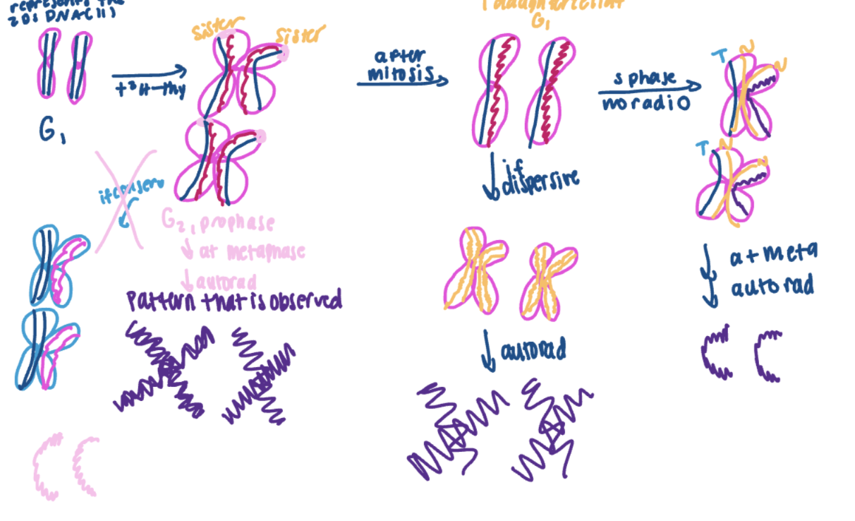
Taylor Experiment
studies bean root tups (eukaryotic cells)
followed cells through mitosis (2 rounds)
labeled DNA with tritiated (3H) thymidine → radioactive
the conservative model would suggest only one chromatid containing 2 newly synthesized strands would be radiolabeled
meiosis 1
G1 → (goes through S, G2, prophase)
Metaphase (DNA is most condense, stop cells from growing here)
anaphase (all chromosomes are lined up and sisters are separating from each other)
chromatids migrate into separate cells
both sister chromatids are radioactive
meiosis 2
G1 unlabeled thymidine and chromosome goes through S, G2, prophase
metphase
only 1 chromatid is labeled
autoradiography
exposed DNA to film dark areas = radioactivity
DNA polymerase
enzyme that synthesizes DNA
in E coli Pol 1 and Pol 3 do most work during DNA replication
more types in eukaryotes
makes new DNA strand 5’ → 3’
only works in one direction
needs 3’-OH group to begin
cant do it by itself
DNA synthesis
is synthesized in 5’ → 3’
polymerase moves down template in 3’ → 5’ direction
anti-parallel
theta replication
circular chromosome
autoradiographs
you radiolabeled the DNA and then put piece of film on that, develop the film, and where you see the dark signal that is where the DNA is present
in prokaryotes
rolling circle replication
plasmids
extrachromosamal, circular piece of DNA that is present in one bacterial cell
if present, bacterial cell will replicate it at the same time it replicates circular chromosomes before cell division
origin of replication
where replication begins
eukaryotic chromosomes have many
only 1 in e. coli → OriC
A/T rich
important because at ORiC, the two strands have to begin synthesizing the new DNA
easier to do it lots of A’s and T’s becase A and T only have 2 hydrogen bonds
required less energy to do this
sequence is recognized by initiator proteins
recognize tha there is a group of proteins that bind to the OriC and begin this process by melting the DNA
theta model in prokaryotes
starting at oriC, DNA is melted so single stands can act as templates
DNA synthesis happens simultaneously in both directions (bidirectional)
1 replication bubble made of 2 replication forks
product = 2 circular chromosomes with 1 old + 1 new DNA stand
theta replication in eukaryotes
many replicons on eukaryotic chromosomes
replicons = point of origin + area of DNA replicated
1 bubble (2 forks) from each origin
bidirectional DNA synthesis
final product = 2 linear chromosomes
1 old and 1 new DNA strand
contains
leading strand
each fork will have only 1 strand made continuously
lagging stand
made discontinuously
contains ozaki fragments
leading strand
since DNA polymerase makes DNA 5’ → 3’, each fork will only have 1 strand made continuously
lagging strand
made discontinuously
short pieces are called the Okazaki Fragments
DNA Replication
overall sequence
initiator proteins bind oriC
DNA melts at A/T rich regions
DNA Helicases bind and continue denaturing
single-strand-binding proteins (SSBs) keep DNA melted
RNA Primer Removal and DNA Ligase
RNA primers are removed by DNA Pol 1 in prokaryotes
has 5’ → 3’ exonuclease activity
has 5’ → 3’ polymerase activity to replace RNA primer with DNA
has 3’ → 5’ exonuclease activity for proofreading
DNA Ligase
seals final hole (nick) in sugar/phosphate backbone
DNA gyrase
important for initiation of replication
its a specific topoisomerase (enzyme)
works ahead of replication fork to reduce strain due to helix unwinding
prevents supercoiling
functions upstream of replication fork
primase
RNA polymerase
needs no free 3’-OH group to work
makes an RNA primer using ssDNA as template
RNA primer
short oligonucleotide
provides free 3’-OH for DNA polymerase to add onto
DNA Polymerase III
prokaryotic enzyme
does most of DNA synthesis
elongates RNA primer by adding new DNA
very fast (~1,000 nucleotides/sec)
has 3’ → 5’ exonuclease activity to correct mistakes
3’ → 5’ exonuclease
enzymatic activity that destroys nucleic acid such as DNA
destruction from end and not middle
start at 3’ end and destory it
catch some mistake right after the fact but will not catch all mistakes
only works for nucleotides just added
called proof-reading
base pairing not correct, will realize it and step backward to remove it (3’ → 5’)
DNA Polymerase 1
5′ → 3′ exonuclease activity = Removes RNA primers
destroys RNA primer
5′ → 3′ polymerase activity = Adds DNA nucleotides
3′ → 5′ exonuclease activity = Proofreading
5′ → 3′ exonuclease activity
remove RNA primer
if mutant, RNA primers cannot be removed
3′ → 5′ exonuclease activity
proofreading, step back and take away
if mutant, more mistakes will be in the newly made DNA
5′ → 3′ polymerase activity
used to synthesize new DNA to replace RNA primers
if mutant, RNA primers will not be replaced by DNA Pol 1 with DNA
DNA ligase
seals final hole (nick) in sugar/phosphate backbone
creates final covalent bond that joins the two Ozakai fragments together so RNA fragments has been replaced by DNA primer
creating covalent bond between 5’ phosphate and 3’ OH to join together 2 nucleotides in their newly synthesized DNA strands
DNA replication eukaryotes
these chromosomes have many DNA packing proteins bound to DNA
histones
making more of DNA packing proteins so DNA can be packaged properly
must be synthesized during this
have many replicons/chromosome
have many DNA polymerases
different DNAPs function at specific times, with varying speeds, error rates and ability to handle mismatches and distortions in DNA
use replication licensing factor (RLF) to control timing of replication initiation
RLF help keep track where DNA replication has happened and where it needs to happen still
contains several proteins that bind to OriC during G1 phase of cell cycle
marks an OriC as ready for DNA replication
once replication begins, different proteins will bind to maker that OriC as already replicated
have linear chromosomes with telomeres (contain repetitive sequence that doesn’t code for anything) on the ends
difficult to replicate the end of a lagging strand
length is linked to aging and disease
chromosomes shortening
DNA gets shorter and shorter by RNA primer length
DNA replication prokaryotes
have 5 DNA polymerase
replication licensing factor (RLF)
helps keep track where DNA replication has happened and where it needs to happen
telomeres
contain highly repetitive DNA
contains repetitive sequence of DNA that doesn’t code for anything
end-replication of linear chromosomes
when RNA primer on 5’ end is removed, there’s no 3’-OH for DNA Polymerase to use
very end of chromosome can’t be replicated
chromosome should get shorter after every round of replication
there is a cap left by removal of primer
cant add to it because DNA primer needs a free 3’-OH, cant go in opposite direction, must go in 5’ → 3’ direction
happens in DNA replication of eukaryotes
single-stranded DNA overhang
happens when there is __ on the 3’ side
to fix this problem
telomerase enzyme extends DNA at chromosome ends and has a protein and RNA component
protein part synthesizes new DNA
RNA part acts as template for DNA synthesis
telomerase moves down and continues extending 3’ overhang
primase makes primer
DNA Polymerase fills in
primer removed
telomerase solves the problem of the chromosome shortening after every round of DNA replication
telomerase enzyme
extends DNA at chromosome ends to help with DNA overhang
has protein and RNA components
protein
synthesizes new DNA
RNA
acts as template for DNA synthesis
expressed in
germ cells
gives rise to gametes
stem cells
immortal cells that aid in such things as wound healing
telomerase protein part
synthesizes new DNA to help with DNA overhang
telomerase rna part
acts as template for DNA synthesis
helps with DNA overhang
why does our body cells have finite life span?
because telomerase doesn’t work on them so once it gets too short, the self-death of cell is activated
is useful because can help get rid of mutations that have occurred in our DNA
DNA packing in prokaryotes
formation of loop domains
each loop is held in place by DNA binding proteins
supercoiling
happens within loop domains
allow DNA to be more condense so more can get added
superpacked ≠ gene expression
relaxed DNA ←(topoisomerases)→ supercoiled DNA

topoisomerases
enzymes that add or remove rotations from DNA helix
super coiling from looped domains
relaxes supercoiled DNA
chromatin
DNA + proteins
DNA is always existing as chromatin, bound by protein
example is nucleosomes by histones
DNA is never really by itself
chromosome
chromatin that is highly condensed
DNA complex during G1, S, G2, prophase → chromatins
heterochromatin
remains highly condensed (tightly packed) throughout cell cycle
no gene expression here because chromatin is tightly packed in these regions so its difficult for all machinery to come in and access the DNA in order for gene expression to happen
can be
constitutive
always condensed
examples: centromeres and telomeres
facultative
condensed at certain times
example:
inactivation X chromosome
happens early on in development
½ moms inactivated and ½ dads inactivated
euchromatin
packaging changes during cell cycle
contains expressed genes
more losely packed when gene expression happens
centromeres are made out of
heterochromatin
part of chromosome that attaches to spindle fibers
required for proper segregation of chromatids
no genes present here; DNA sequence is highly repetitive
# of repeats on each chromosome varies
DNA remains tightly packaged throughout cell cycle
telomeres are made up of heterochromatin
POT (Protection of Telomere) proteins bind single stranded overhang
provide stability
prevent ends from degrading and/or sticking together
sometimes ends fold over to form triple-stranded structure
DNA remains tightly packaged throughout cell cycle

histones
“beads on a string”
simplest level of chromatin structure
visible under low salt conditions
small, positively charged (R groups)
Arg and Lys amino acids
bind tightly to DNA
bind tightly to DNA
where is the tightest packaging seen?
at the metaphase plate
and anaphase so that DNA or chromatin can go to daughter cells
chromosomes
nucleosome
simplest level of chromatin structure
contains histone proteins
small, positively charged
Arg and Lys amino acids
bind tightly to DNA
Five types:
H1
linker histone
H2A
core histone
H2B
core histone
H3
core histone
H4
core histone
need 2 of each of the core histones since 8 histones make one bead

net charge of DNA is
negative because of the phosphate groups in the backbone
epigenetics
study of phenotypic variation caused by variation in gene expression … NOT caused by variation in nucleotide sequence
changes in gene expression due to changes in chromatin structure
can be stable through cell divisions (mitosis)
can be stable over generations (meiosis)
can be influenced by environment
chromatin structure is always changing and are reversible but can be maintained because they are heritable
chromatin structure is altered by
chemical modification on DNA and histones
reversible
important for regulating gene expression
modified by
DNA modification
histone modification
DNA modification
one type: methylation of nucleotide base (mostly Cytosine)
frequently CpG dinucleotides
both strands of DNA are modified
associated with lower gene expression
happens where there is repeat of C’s and G’s
histone modification
several types: phosphorylation, methylation, acetylation, ubiquitination
affects binding to DNA
often happens in positively charged “tails”
groups of amino acids at end of histone proteins
wrap around DNA and interact with phosphates in backbone
tail becomes a littler arm
can interact tightly with backbone → giving it a little hug essentially
can act strongly with the negative charge in the sugar phosphate backbone of DNA
acetylation can mask positive charges
often stimulates gene expression
methylation can create binding sites for transcription factors
stimulates gene expression
it can mark on area for increased or decreased gene expression
histone acetylation
can mask positive charges
makes binding to DNA weaker
not hugging it as tightly
makes chromatin structure more open
losely packed
often stimulates gene expression
remember: open chromatid structure and increased gene expression
histone methylation
can create binding sites for transcription factors
stimulates gene expression
can “mark” an area for increased or decreased gene expression
specific amino acid in certain histones
this is heritable
enzymes are recruited by transcription factors, pre-existing epigenetic marks, noncoding RNAs
are recruited during S-phase
new histones being made
mechanisms for maintenance during cell division are not well understood
how is level of DNA Methylation maintained after DNA replication?
DNA replication
melt DNA
2 olds strands act as template
both DNA molecules are hemimethylated
methylated on 1 DNA strand
DNA repair machinery can distinguish old and new DNA strands at this point
helps DNA repair machinery because if they are scanning and see base pairs not proper, strand not methylated is the new strand so that must be where the mistake is
G2 cell cycle
enzymes that will check DNA and correct any mistakes that were made during DNA replication
special methyltransferase enzymes recognize hemimethylated DNA
can see where methyl group is supposed to be by looking at the old strand and add methyl groups on the appropriate place on the new strand
chemical modifications are
heritable
can be passed on to one cell to the daughter cell during mitosis and can be passed on to parent to progeny when we create gametes
DNA methylation and gene expression
it down-regulates gene expression
how it does it
methyl group in major groove prevents binding of proteins that stimulate transcription (transcription factors → important for turning on gene expression, which increases gene expression)
creates binding site for histone deacetylase enzymes
removes acetyl groups from histone proteins
it will ALWAYs lead to down regulation of gene expression by preventing transcription factors from binding because methylation of DNA
histone deacetylase
remove acetyl groups from histone proteins
histones bind DNA more tightly
chromatin structure is more condensed
gene expression downregulated
harder for machinery to get in and read DNA in order to start the process of gene expression
DNA methylation in honeybees
queen and work bees develop from genetically identical eggs
queen
female
larger body
functional ovaries (just means fertile)
behavior includes mating flight and egg production
worker bees
female
smaller body
sterile
behavior includes collecting nectar and pollen, tending the queen, raising babies
differences between the two are caused by changes in gene expression and diet
queen’s diet
queen is the only one that eats royal jelly or siRNAs
is an RNA silencer
Dnmt3 gene repressed
gene codes for methyltransferase enzyme
less methylation in genome, increased gene expression
this is an example of epigenetics
X-inactivation epigenetics
random even that happens early in development
either moms or dads X is inactivated
leads to stable change in gene expression
passed on to daughter cells after mitosis
will have the same x-inactivation gene
histone modification is important
x-inactivation center
segment of X-chromosome where inactivation begins
contains several important genes:
X-inactivation-specific transcript (X-ist)
Tsix
important for keeping one X-chromosome activated
Jpx
important for keeping one X-chromosome activated
Xite
important for keeping one X-chromosome activated

X-ist gene
encodes long RNA that does not code for protein that doesn’t get translated
bind X-chromosome from which it was made
attracts protein complexes (PRC2; PRC1)
methylate histones to turn off gene expression
create “epigenetic mark” for repressed gene expression
because of long non-coding RNA binding
recruit proteins that will modify histones
methylated histones create the signal (the epigenetic mark) that results in a decrease in gene expression
the long noncoded RNA will bind to chromosome where it wasn’t synthesized
when RNA binds, it will attract protein complexes
Tsix is complement to this gene
meaning they are 2 genes that are opposite from one another
part of x-inactivation center
Tsix gene
keeps active x chromosome from being inactivated
encodes long RNA that does not code for protein
anti-sense to X-ist RNA
reverse complement
complementary antiparallel sequence
the interaction prevents the Xist chromosome from binding to one x-chromosome
can hydrogen bond with one another
prevents X-ist expression/function on activated X
part of x-inactivation center
Jpx gene
encodes lon RNA that does not code for protein
stimulates (increases) synthesis of Xist on inactive chromosome
part of x-inactivation center
Xite gene
encodes long RNA that does not code for protein
sustains Tsix expression on active X chromosome
part of x-inactivation center
genomic imprinting
gene expression affected by whether gene comes from Mom or Dad
results in autosomal traits that show different results for reciprocal crosses
genes often in clusters of 3-12
encode proteins and noncoding RNAs
key to process basically
chromatin modification differ between alleles
tightly or more loosely packed
genetic conflict hypothesis attempts to explain
dad might benefit more when certain genes are turned on than mom would benefit if it was turned on
example
growth factor of baby
dad wants big baby while mom wants a small baby for giving an easier birth
it matters which parent supplies gene to progeny
mom and dad each give one and one is turned on while the other is off
example
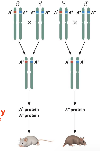
A1 allele (reciprocal cross)
A1 allele only expressed if from Dad
moms A1 gene is not expression (as shown on the right side of the picture
reciprocal crosses between donkey and horse produce hybrid animals with very different phenotypes
female donkey x male horse → hinny
more like a horse than a donkey
male donkey x female horse → mule
more like a donkey than a horse
THIS IS AN EXAMPLE OF EPIGENETICS
behavioral epigenetics
The study of how environmental experiences influence gene expression through epigenetic modifications without changing the DNA sequence, potentially affecting behavior.
Example: Maternal care in rats can alter DNA methylation and stress responses.
variations in chromosome structure
chromosomal rearrangements
changes in parts of chromosomes
involve 1 or more breaks in chromosomes
means that the free ends of DNA are unable
can be degraded by exonucleases (enzymes that destroy nucleic acid from the end)
can bond with other free ends (sometimes telomers)
can be repaired by cellular DNA repair machinery
mistakes here can create rearrangements
may also be caused by unequal genetic recombination
prophase 1 of meiosis
includes
deletion
loss of chromosomal segment
break, then degradation
duplication
inversion
translocation
chromosomal deletion
loss of chromosomal segment
break, then degradation
effects
phenotype depends on which genes/gene parts are deleted
in het (diploid)
__ dominat allele → expression of recessive allele
this is called psuedodominance
gene dosage effects may occur
haploinsufficiency
looped out structure forms during meiosis prophase 1
if its only one region and that region does not influence genome, could see no phenotypic variation
deletion of one or more genes depending on what those genes code for, you will see a resulting phenotype

intercalary deletion
deletion of an internal region of the chromosome
example
wt: A B C · D E F G
mutation: A B C · D G
terminal deletion
deletion of the end of the chromosome
example
wt: A B C · D E F G
mutation: A B C · D E