neurology part 1 (Essay 1,2,3)
 Call Kai
 Learn
 Practice Test
 Spaced Repetition
 Match
 Flashcards
 Knowt Play
1/32

There's no tags or description
Looks like no tags are added yet.
33 Terms
Definition of a reflex
A reflex is an involuntary, rapid and stereotyped response of the body to a particular stimulus, mediated through a neural pathway called a reflex arc
What makes up the reflex arc
1.Receptor Receives the stimulus
2. Afferent neuron Carries sensory impulses towards the CNS
3. Integration centre Processes the impulse in the spinal cord or brainstem
4. Efferent neuron Carries motor impulses away from the CNS
5. Effector Muscle or gland that produces the response
Define deep tendon reflexes
Deep tendon reflexes are involuntary muscle contractions produced when a tendon is tapped
What are the Important characteristics of deep tendon reflexes
They are predominantly monosynaptic.
They are influenced by descending inhibitory pathways from the brain.
The reflex arc of the mandibular reflex as well as the clinical significance
Afferent :Mandibular division of CN V
Centre :Pons
Efferent: Motor fibres of CN V
Effector :Masseter and other jaw-closing muscles
Clinical significance : corticobulbar dysfunction
Reflex arc of triceps
Afferent Radial nerve
Centre C7–C8 (predominantly C7)
Efferent Radial nerve
Effector Triceps brachii
Reflex arc of biceps
Afferent Musculocutaneous nerve
Centre C5–C6
Efferent Musculocutaneous nerve
Effector Biceps brachii
Reflex arc of brachioradialis
Afferent Radial nerve
Centre C5–C6
Efferent Radial nerve, median, musculocutaneous
Effector Brachioradialis
Reflex arc for patellar reflex
Afferent Femoral nerve
Centre L3–L4 (predominantly L4)
Efferent Femoral nerve
Effector Quadriceps femoris
What does increased & decreased patellar reflexes mean
Reduced or absent response may suggest femoral neuropathy, L3–L4 root disease or peripheral neuropathy.
Increased response may suggest an upper motor neuron lesion above the relevant spinal segments.
Reflex arc for Achilles Tendon
Afferent Tibial nerve
Centre S1–S2 (predominantly S1)
Efferent Tibial nerve
Effector Gastrocnemius and soleus
What does increased and decreased reflexes of Achilles mean
Reduced or absent Achilles reflex is common in S1 radiculopathy and peripheral neuropathy.
Increased Achilles reflex may indicate an upper motor neuron lesion.
It is particularly useful when assessing suspected peripheral neuropathy.
What are the deep reflexes
Superficial reflexes are involuntary responses produced by stimulation of the skin or mucous membranes.
According to your notes, they are mostly nociceptive reflexes and are generally polysynaptic.
Define the characteristics
Elicited by stimulation of the skin or mucous membranes.
Mostly polysynaptic.
Their reflex arcs involve sensory and motor pathways.
They can be absent in corticospinal tract lesions.
What is the reflex arc of the corneal reflex/ conjunctival
Reflex arc Structure
Afferent sensory fibers ofOphthalmic division of CN V (V1)
Centre trigeminal spinal nucleus of trigeminal nerve and nucleus of facial nerve
Efferent motor fibers Facial nerve (CN VII)
Effector Orbicularis oculi
The reflex arc of the pharyngeal reflex
Afferent Glossopharyngeal nerve (CN IX)
Centre Medulla oblongata( nucleus ambigus, nucleus spinal trigeminal)
Efferent Vagus nerve (CN X), with contributions to the pharyngeal motor response
Effector Pharyngeal muscles
What is the reflex arc of the abdominal reflex
Reflex Spinal segments
Upper abdominal T5–T9
Middle abdominal T7–T10
Lower abdominal T8–T12
Afferent & efferent: sensory & motor fibers of intercostal nerve
When can there be absence in the abdominal reflex
Unilateral absence may be an early sign of a contralateral motor cortex or pyramidal tract lesion.
They may also be absent in lesions of the corresponding reflex arc.
They can be absent physiologically in elderly people, obese patients and patients who have undergone abdominal surgery.
What is the reflex arc for the cremasteric reflex
Reflex arc Structure
Afferent Ilioinguinal nerve (L1)
Centre L1–L2 spinal segments
Efferent Genital branch of genitofemoral nerve (L1–L2)
Effector Cremaster muscle
What is the reflex arc for the plantar reflex
Reflex arc Structure
Reflex arc Structure
Afferent Tibial nerve
Centre Spinal cord, predominantly L5–S1
Efferent Tibial nerve
Response Plantar flexion of toes
Explain pathological reflexes
Pathological reflexes are abnormal reflex responses that appear due to dysfunction or damage of the nervous system, particularly the corticospinal (pyramidal) tract or other descending inhibitory pathways.
Why is Babinski sign normal in children
Babinski sign is normal in young infants because the corticospinal system is still developing
What are the extensor plantar reflexes
These four signs belong to the Babinski group (extensor plantar reflexes). They are used to detect an upper motor neuron (UMN) lesion, particularly damage to the corticospinal (pyramidal) tract.
1. Oppenheim's sign
How: Firmly stroke down the anterior surface of the tibia (shin) using your knuckles or fingers.
Positive: Big toe extends upwards, with possible fanning of the other toes.
2. Gordon's sign
How: Squeeze the calf muscles firmly.
Positive: Big toe extends upwards, with possible fanning of the other toes.
3. Chaddock's sign
How: Stroke the skin around the lateral malleolus (outer ankle), moving around the lateral side of the foot.
Positive: Big toe extends upwards, with possible fanning of the other toes.
4. Schaefer's sign
How: Squeeze the Achilles tendon.
Positive: Big toe extends upwards, with possible fanning of the other toes.
Explain primitive reflexes
Sucking Sweep a tongue depressor along the lips. Involuntary sucking movement
Snouting Tap gently around the mouth. Puckering of lips
Nasolabial Tap gently over the nose. Sucking movement of lips
Palmomental (Wartenberg) Scratch the thenar eminence of the palm. Contraction of the chin muscle on the same side
Explain the grasping reflexes
Grasping reflexes
Indicate contralateral frontal lobe disease.
They are normally seen in infants but disappear with maturation.
Their reappearance in adults is pathological.
There are 2 types:
A. Palmar grasp reflex
Place your finger or the handle of a reflex hammer in the patient's palm.
Stroke the palm.
The patient's fingers involuntarily close and grasp your finger.
B. Plantar grasp reflex
Stimulate the sole of the foot.
The toes involuntarily flex and adduct (move towards each other).
What are the rossolimo group
1. Rossolimo reflex
Rapidly tap the plantar surfaces of the distal phalanges of the toes.
Positive response: Sudden flexion of the toes.
Clinical significance: May indicate corticospinal tract dysfunction.
2. Zhukovsky reflex
Tap the palmar surface near the base of the middle fingers or metacarpal region.
Positive response: Flexion of the fingers.
Clinical significance: May indicate pyramidal tract dysfunction.
Mendel sign
Tap the dorsum of the foot over the region of the cuboid and lateral cuneiform bones.
Positive response: Flexion of the toes.
Clinical significance: Suggests pyramidal tract dysfunction.
hoffmann -:
Hold the patient's middle finger.
Support the distal phalanx.
Flick or suddenly release the distal phalanx of the middle finger.
Positive response:
Flexion of the thumb.
Flexion of the index finger, sometimes accompanied by flexion of other fingers.
Clinical significance:
May indicate increased corticospinal excitability, particularly in cervical spinal cord disease or other upper motor neuron disorders.
Tomner
Ask the patient to relax the hand.
Hold the middle finger.
Tap the palmar surface of its distal phalanx upwards.
Positive response: Flexion of the thumb and index finger.
Clinical significance: May indicate corticospinal tract dysfunction, particularly in cervical myelopathy.
List the qualitative reflexes
Quantitative
1. Areflexia
Definition: Complete absence of a reflex response.
Causes include:
Deep coma.
Spinal shock.
2. Hyporeflexia
Definition: A reduction in the strength of a reflex response.
Causes include:
Neuropathy.
Muscle or neuromuscular disorders affecting the reflex arc.
3. Hyperreflexia
Definition: Exaggerated or increased reflex responses.
Causes include:
Upper motor neuron lesions.
Corticospinal tract lesions.
It occurs because descending inhibitory control of spinal reflexes is reduced.
4. Clonus
Clonus is a series of rhythmic, involuntary muscle contractions produced by sudden sustained stretching of a muscle.
How to test ankle clonus:
Ask the patient to relax.
Support the lower leg.
Quickly dorsiflex the foot.
Maintain the dorsiflexion.
List the quantitative reflexes
Babinski sign.
Oppenheim sign.
Gordon sign.
Hoffmann sign.
Rossolimo sign.
Primitive reflexes.
Grasping reflexes.
What is anisoreflexia
Anisoreflexia refers to unequal reflex responses on the two sides of the body.
For example:
Right knee reflex is brisk.
Left knee reflex is normal
What is the UMN lesion
An upper motor neuron lesion occurs when there is damage to the motor pathways above the anterior horn cells of the spinal cord or the motor cranial nerve nuclei.
Examples:
Stroke.
Multiple sclerosis.
Reflex findings:
Increased deep tendon reflexes (hyperreflexia).
Positive Babinski sign.
Clonus.
Increased muscle tone (spasticity).
Superficial abdominal reflexes may be absent.
Loss of normal descending inhibition.
What is the LMN lesion?
A lower motor neuron lesion occurs when there is damage to the anterior horn cells, motor cranial nerve nuclei, nerve roots, plexuses or peripheral motor nerves.
Examples:
Peripheral neuropathy.
Poliomyelitis.
Reflex findings:
Reduced or absent deep tendon reflexes.
Hyporeflexia or areflexia.
Hypotonia.
Muscle atrophy.
Fasciculations may occur.
No Babinski sign from an isolated LMN lesion.
What is the grading used for deep tendon reflexes
Grade Meaning
0 Absent
1+ Reduced or diminished
2+ Normal
3+ Brisk, may be normal depending on context
4+ Very brisk, often with clonus
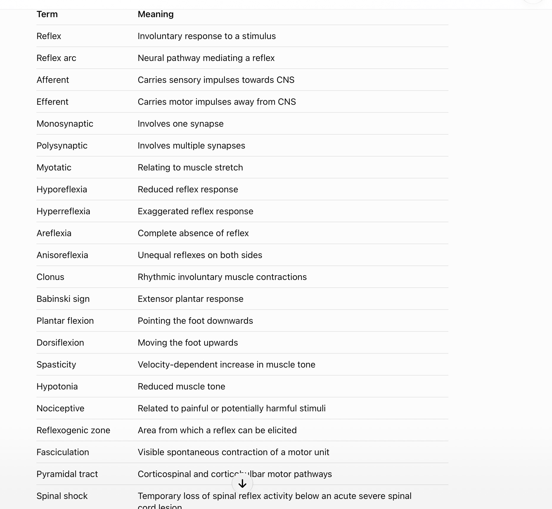
Explain the following terms