Congenital Heart Diseases in Dogs and Cats
 Call Kai
 Learn
 Practice Test
 Spaced Repetition
 Match
 Flashcards
 Knowt Play
1/30

There's no tags or description
Looks like no tags are added yet.
31 Terms
What are the two main categories of heart murmurs found in puppies and kittens?
• Pathological → Structural reason is present (i.e. abnormal valve, abnormal cardiac structure, ...)
• Nonpathological
Functional (physiological) murmur → There is a physiological reason for the murmur, heart is not the culprit
Innocent murmur

How can nonpathological heart murmurs and pathological be characterized?

What are some causes of functional heart murmurs?
• Changes in blood viscosity
Anemia, Hypoproteinemia
• Increased cardiac output state
Fever, Pregnancy, Hyperthyroidism
Athlete's heart syndrome

What are the prevalence of congenital heart diseases in canines?
• Dogs: < 1% (Estimated to be)
Pulmonic Stenosis (30%)
Patent Ductus Arteriosus (PDA, 17-25%)
Subaortic Stenosis (15%)
What are the prevalence of congenital heart diseases in felines?
• Cats: <1%
• Ventricular septal defect (VSD, 40-20%)
PDA (3-10%)
Tricuspid valve dysplasia (TVD, 5- 10%)
Atrioventricular septal defect (AVSD, ASD, 5-10%)
Mitral valve dysplasia (MVD, 9- 13%)
What are the common predispositions for congenital heart disease?
• Unknown factors: Genetic/Epigentic/Environmental?
• Breed predispositions
• Boxer: Pulmonic and subaortic stenosis.
• German Shepherd Dog: PDA, persistent right aortic arch
• Bulldogs: Pulmonic stenosis
• Labrador: Tricuspid valve dysplasja
• Bull Terrier: Mitral valve dysplasia
How are congenital heart diseases usually diagnosed?
• Presentation
• Breed, Age
Tend to be younger animals
• Auscultation: Heart murmur depends on disease
PDA: Continuous murmur
Mostly systolic
• Radiography, ECG can help...
• Echocardiography is the method of choice!
How does the heart respond to haemodynamic issues?
Cardiac remodeling
Based on pressure or volume overload - hypertrophy - what type? Is it concentric or eccentric and what side?
Volume: Sarcomeres in a series - in length
Pressure: Sarcomere in parallel - in width

Hypertrophy only applies to the….
Ventricles, not the atria, no matter the cause, the atria respond the same way
Eccentric or concentric hypertrophy refer to the architectural answer of the ventricles (left and right).
How do the atria respond to haemodynamic issues?
• The left atrium remodelling is "only about dilatation/enlargement" which may occur in response to pressure and volume overload.
What are the main features of pulmonic stenosis?
• Common CHD in dogs, rare in cats.
• Structural malformation that cause obstruction of the blood flow from the right ventricular outflow tract (RVOT) to the pulmonary artery (PA), right ventricle struggles to push blood out due to narrowing of valve
• Pulmonary valve stenosis
Pure valve problem or annulus problem
Leads to PRESSURE overload, think putting thumb over garden hose
Subvalvular PS
Supravalular PS
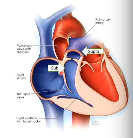
How does pulmonic stenosis affect the heart?
What are common history and clinical findings?
• Asymptomatic, "just a murmur"
Left cranial systolic crescendo-decrescendo murmur; evtl. right sided component (tricuspid regurgitation)
Weak pulse
• Exercise intolerance, syncope, sudden death, right sided heart failure (ascites)
Louder heart murmur = more severe disease

How might an ECG appear in an animal with a pulmonic stenosis?
• Right axis deviation - due to enlarged right side of heart
• Deep S waves in lead II, Ill with prolonged QRS duration: right bundle branch block (RBBB).
• Arrhythmias possible.
How can pulmonic stenosis be treated?
Balloon valvuloplasty for pulmonary stenosis - inflate balloon to allow valve to open properly
What are the main features of a patent ductus arteriosis.
• Part of fetal circulation
• At birth should close itself and regress to form the ligamentum arteriosus.
• If it does not close, stays patent and allows blood shunting from left to right (but could lead to flow right to left).
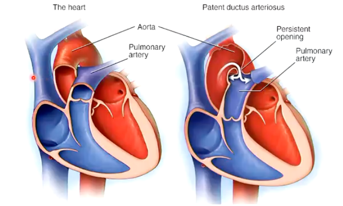
How does a PDA affect circulation and the heart?
Initially: shunt of blood from left side to the right side, systemic circulation is reentering pulmonary circulation → increased flow to lungs → volume overload to left side since left side is the end recipient of pulmonary circulation
Later: increased pressure in pulmonary artery, becoming less elastic, pressure overload, backs into the right ventricle, which can develop into R → L shunt (Reversal of shunt) and hypertrophy of the right ventricle
If the PDA is large and untreated:
Excessive blood flow repeatedly passes through the pulmonary circulation.
Pulmonary blood vessels undergo structural changes, including thickening and narrowing.
Pulmonary vascular resistance (PVR) increases.
Pulmonary arterial pressure rises.
Eventually, pulmonary arterial pressure can exceed systemic pressure.
Blood now flows from the pulmonary artery into the aorta: right-to-left shunting.
Blood entering brachycephalic trunk is normally oxygenated
Everything else is mixed
^^Differential, front oxygenated, back is not

What are the most common history and clinical findings of a PDA?
If Left to Right:
• Asymptomatic, loud continuous murmur on the left base (machinery murmur) - location of great vessels)
• Exercise intolerance, left sided congestive heart failure (CHF)
• Bounding pulse (water hammer pulse)
• If right to left:
No continuous murmur (evtl. systolic on the right secondary to tricuspid regurgitation)
Hindlimb weakness (differential cyanosis)
Describe the main features of a subaortic stenosis (SAS).
• Malformation affecting the left ventricular outflow tract (LVOT).
• Most common is the obstruction arising at subvalvular (subaortic) level.
• Valvular aortic stenosis is possible, but less common.

What are the consequences of a subaortic stenosis?

What are the common history and clinical findings of a subaortic stenosis?
• Asymptomatic, "just a murmur"
Left cranial systolic crescendo-decrescendo murmur
Weak pulse
• Exercise intolerance, syncope, sudden death, left sided heart failure
Can see more defined aortic arch, but ECG needed to identify, produces concentric hypertrophy
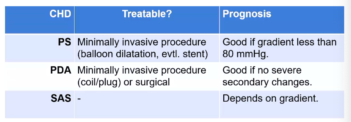
What are the main treatment options for congenital heart diseases?
PS
PDA
SAS
PDA surgical - find abnormal connection and ligate
SAS - long term management not good
What are the main features of atrioventricular septal defects?
• Different issues during the development can produce:
• Defect in the ventricular septum
• Defect in the atrial septum
• Combination of the two
• Affect the atrioventricular valves
**An atrioventricular septal defect (AVSD)—also called an atrioventricular canal defect or endocardial cushion defect—is a congenital heart condition (present at birth) where there are holes in the walls (septa) between the heart's chambers and abnormalities in the central heart valves.
How do AVSD arise?

What are the consequences of a ASD?
Atrial septal defects
Atrial septal defects (ASD) lead to a right ventricular eccentric hypertrophy (volume overload of the right).
Since right side is more elastic and more compliant - therefore more blood ends of here
What are the features of Ventricular Septal Defects (VSD)?
• Communication between the left and right ventricles.
Exception: Gerbode Defect Left Ventricle and Right Atrium connection.
Mostly common in cats
• In small animals majority is peri-membranous - usually in high area beneath the aorta
What are the consequences of Ventricular Septal Defects (VSD)?

What are the history and clinical findings associated with VSDs?
• Asymptomatic, "just a murmur"
Right systolic murmur (L → R)
Strong pulse
• Exercise intolerance, left sided congestive heart failure.
• Smaller Defect = Louder Murmur → Does not correlate with severity!
Does in SAS, PS, PDA
What is the tetralogy of fallot?
1. Overriding Aortic Arch - biggest issue
The body's main artery (the aorta) is shifted to the right and sits directly above the VSD, allowing it to receive low-oxygen blood from both ventricles.
2. Ventricular septal defect
A hole in the wall (septum) separating the two lower pumping chambers (ventricles). This allows oxygen-poor blood from the right ventricle to mix with oxygen-rich blood in the left ventricle.
3. Pulmonic stenosis
4. Right ventricular concentric hypertrophy
What is the main feature of the tricupsid valve dysplasia?
• Systolic murmur in the tricuspid area tricuspid valve is malformed
• Potential right sided congestive heart failure
• Labrador
What is the main feature of the double-chambered right ventricle?
The right ventricle is subdivided in two chambers:
High pressure chamber and low-pressure chamber
High pressure chamber below becomes hypertrophic
Similar to a subvalvular pulmonic stenosis.
• Clinically like a pulmonic stenosis
• Associated with VSD?
What is the main feature of a persistent right aortic arch?
• Regurgitation
• Resophraguy to identify changes in the
• German Shepherd Dogs
• Surgery!
Clinically not a big problem for the heart