Bio201 Axial skeleton
 Call Kai
 Learn
Learn Practice Test
 Spaced Repetition
 Match
Match Flashcards
 Knowt Play
1/100

There's no tags or description
Looks like no tags are added yet.
Name | Mastery | Learn | Test | Matching | Spaced | Call with Kai | Chat |
|---|
No analytics yet
Send a link to your students to track their progress
101 Terms
Sutures
Fibrous joints between bones of the skull
Coronal suture
Fibrous joint between parietal bones and frontal bone

Sagittal suture
Fibrous joint between two parietal bones

Lambdoid suture
Fibrous joint between occipital bone and parietal bones

Squamous suture
Fibrous joint between parietal bone and temporal bone

Frontal bone
Shell shaped bone forming the anterior cranium and the superior orbits

Supraorbital foramen/notch
Channel for supraorbital nerve and blood vessels

Glabella
Muscle attachment of the forehead

Parietal bone
Large bones forming most of the superior and lateral aspects of the cranium

Temporal bone
Bones that form the inferolateral aspects of the skull

External acoustic/auditory meatus
Passageway that guides sound waves to the tympanic membrane (ear drum)

Internal acoustic/auditory meatus
Passageway for the Vestibulocochlear nerve (VIII) carrying hearing and balance impulses
Zygomatic process of temporal bone
Site of muscle attachment (masseter)

Mastoid process
Enlargement posterior to earlobe; attachment site for muscles that move the skull

Styloid process
Attachment site for muscles that move the tongue, throat, and mandible

Stylomastoid foramen
Opening for nerves and blood vessels

Mandibular fossa
Depression on temporal bone articulating with mandible condyle (temporomandibular joint)

Carotid canal
Passageway for internal carotid artery supplying blood to the brain

Jugular foramen
Passageway for jugular vein draining blood from the brain

Occipital bone
Large bone forming most of posterior wall and base of skull

Occipital condyle
Rounded processes articulating with atlas (C1) for head movement

Foramen magnum
Large opening where brain stem extends to connect with spinal cord

Hypoglossal canal
Opening for nerve supplying the tongue muscle

Sphenoid bone
Butterfly shaped keystone bone forming posterior wall of orbits

Greater wing (Sphenoid)
Forms anterior/medial floor of middle cranial fossa

Lesser wing (Sphenoid)
Makes up part of the orbit
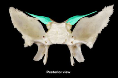
Sella turcica
Depression in sphenoid protecting and housing the pituitary gland

Optic canal
Round hole for optic nerve carrying visual information

Foramen rotundum
Sphenoid opening allowing nerves and arteries to pass

Foramen ovale
Sphenoid opening allowing nerves and arteries to pass

Foramen spinosum
Sphenoid opening allowing nerves and arteries to pass

Foramen lacerum
Sphenoid opening allowing nerves and arteries to pass

Ethmoid bone
Deepest skull bone lying between nasal bones and sphenoid

Perpendicular plate
Forms superior nasal septum with vomer and nasal cartilage

Superior nasal concha
Filters and humidifies inhaled air

Middle nasal concha
Filters and humidifies inhaled air

Crista galli
Midline projection holding brain membrane in place ("rooster crest")

Cribriform plate
Flat portion on either side of crista galli where olfactory bulbs lie

Olfactory foramina
Small holes in cribriform plate for olfactory nerves
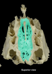
Frontal sinus
Air-filled cavity in frontal bone reducing skull weight and aiding resonance

Maxillary sinus
Air-filled cavity in maxilla reducing skull weight and aiding resonance

Ethmoid sinus
Air-filled cavity in ethmoid reducing skull weight and aiding resonance

Sphenoid sinus
Air-filled cavity in sphenoid reducing skull weight and aiding resonance

Vomer
Forms inferior nasal septum

Palatine bone (2)
Forms posterior 1/3 of hard palate

Inferior nasal concha (2)
Filters and humidifies inhaled air

Nasal bone (2)
Forms external surface of nose and protects nasal cavity

Mandible
Lower jaw bone enabling chewing and speaking

Coronoid process
Mandibular projection for muscle attachment moving mandible upward

Mandibular condyle/Condylar process
Rounded structure articulating with mandibular fossa of temporal bone

Alveolar process
Ridges on mandible and maxilla forming sockets for teeth

Mandibular foramen
Opening for inferior alveolar nerve supplying gums and teeth

Mental foramen
Opening for mental nerve supplying lower lip and chin

Ramus of Mandible
Vertical portion of mandible

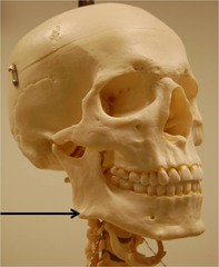
Mandibular angle
Posterior inferior corner of mandible
Maxilla (2)
Upper jaw bone enabling chewing and speaking

Infraorbital foramen
Opening for infraorbital nerve and blood vessels

Palatine process
Anterior 2/3 of hard palate

Lacrimal bone (2)
Supports structures of lacrimal apparatus (tear production)

Nasolacrimal canal
Drains tears into nasal cavity

Zygomatic bone (2)
Cheek bone providing shape and structure to face

Temporal process of zygomatic bone
Site of muscle attachment (masseter)

Zygomatic arch
Bridge formed by temporal and zygomatic processes for muscle/ligament attachment

Hyoid bone
Unattached neck bone inferior to mandible anchoring throat, larynx, and tongue

Cervical vertebrae count
7 (Cervical vertebrae in neck)

Thoracic vertebrae count
12 (thoracic vertebrae are in chest region)

Lumbar vertebrae count
5 (lumbar vertebrae associated with lower back)

Sacral vertebrae count
5 fused (base of spine between lumbar and coccyx)

Coccyx count
3-5 fused (end of vertebrae column)

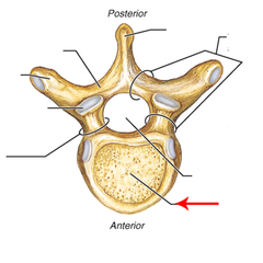
Body (centrum) of vertebrae (main part)
Thick, cylindrical weight-bearing part of vertebra
Vertebral foramen
Hole for spinal cord and cauda equina

Spinous process
Posterior projection for muscle/ligament attachment

Transverse process
Lateral projection for muscle attachment and thoracic rib articulation

Superior articular process/facet
Site of articulation with adjacent upper vertebra

Inferior articular process/facet
Site of articulation with adjacent lower vertebra
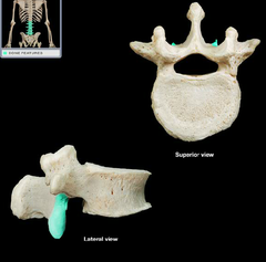
Pedicle
Cylinder projections joining front and back parts of vertebra

Lamina
Posterior roof segment protecting backside of spinal cord
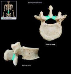
Intervertebral foramen
Opening between adjacent vertebrae for exiting spinal nerves

Atlas (C1)
First cervical vertebra articulating with occipital condyles for "yes" movement

Axis (C2)
Second cervical vertebra featuring dens for "no" rotational movement

Dens
Bony projection on axis around which atlas rotates

Transverse foramen
Openings in cervical vertebrae for vertebral arteries supplying brain

Cervical Vertebrae
has C1, C2, and C7 (most notable but has all C1-C7)

Thoracic vertebrae
Vertebrae articulating with ribs

Lumbar vertebrae
Low back vertebrae bearing most body weight

Sacrum
Five fused vertebrae forming posterior wall of pelvis

Coccyx
Tailbone formed by 3-5 fused vertebrae

Sternum
Breastbone serving as anchor point for clavicles and ribs

Manubrium
Superior bone of sternum attaching to clavicles

Jugular notch
Indentation on superior border of manubrium

Body of Sternum
protects the organs and muscles inside your chest from injury

Xiphoid process
Inferior sternal tip for muscle attachment and CPR landmark

True ribs
Ribs 1-7 (directly attach to sternum via costal cartilage)

False ribs
Ribs 8-12

Floating ribs
Ribs 11-12 (no anterior attachment)
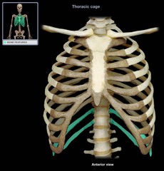
Costal cartilage
Hyaline cartilage attaching ribs to sternum

Head of rib
Articulates with costal facet on body of thoracic vertebra
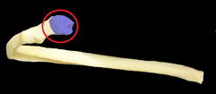
Tubercle of rib
Articulates with transverse process of thoracic vertebra

Neck of Rib
the flattened, narrow portion of a typical rib that extends laterally from the head to the tubercle
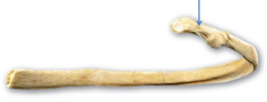
Costal end
Rib end attaching to costal cartilage
