Renal and Urologic Systems Disorders
 Call Kai
 Learn
Learn Practice Test
 Spaced Repetition
 Match
Match Flashcards
 Knowt Play
1/14

There's no tags or description
Looks like no tags are added yet.
Name | Mastery | Learn | Test | Matching | Spaced | Call with Kai | Chat |
|---|
No analytics yet
Send a link to your students to track their progress
15 Terms
Starling’s Forces
The Starling equation calculates:
Movement of edema fluid
Depends on hydrostatic and oncotic pressure differences between capillaries and interstitium
Glomerular filtration depends on same opposing forces that produce the exchange of fluids in every capillary in our body
Hydrostatic Pressure
Force within a fluid compartment
Major force that pushes water out of vascular system at capillary level
Pushes out “bag chair crew”: blood pressure.
Oncotic Pressure
Osmotic pressure exerted by colloids in solution
albumin is a major colloid.


Fluid Movement in Capillaries
Amount and direction of movement determined by
Capillary hydrostatic pressure – pushes H2O out of capillary
Plasma oncotic pressure- pulls H2O back into capillary
Interstitial hydrostatic pressure- opposes movement of H20 out of capillary
Interstitial oncotic pressure- pulls H20 out of capillary into interstitial spaces
Filtration: controls the outward movement of fluid from capillary
Reabsorption: pulls fluid back into capillary
WHY DOES IT MATTER?
LOW BP = drop in hydrostatic pressure > drop in filtration > acute kidney injury
LOW PROTEIN STATE = drop in oncotic pressure > fluid leaking out > edema
edema in relation to solutes from liver and ascites.
Fluid Shifts
Plasma-to-interstitial fluid shift results in edema.
__increase__ in hydrostatic pressure in capillaries
__decrease___ in plasma oncotic pressure
__increase___ in interstitial oncotic pressure
Interstitial fluid to plasma
Fluid drawn into plasma space with increase in plasma osmotic or oncotic pressure
Hypertonic solutions
Osmotic diuretic- mannitol
Colloids (albumin, dextran, or FFP)
___compression__ stockings decrease peripheral edema.
how does it work
increases interstitial hydrostatic pressure
A patient with low serum albumin is at risk for which complication related to Starling’s forces in the glomerulus?
A. Hypertension
B. Dehydration
C. Edema
D. Hyperkalemia
C. Edema


Fluid Distribution
Intracellular (ICF) = 2/3 of body fluid
Extracellular fluid (ECF) = 1/3 of body fluid
Intravascular (Plasma) – inside blood vessels
Interstitial- between cells
What is the difference between interstitial fluid and extracellular fluid?
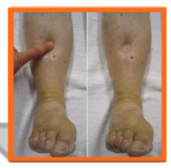
Edema - ↑ interstitial fluid volume
ELECTROLYTES AND OSMOTIC ACTIVE SOLUTES
ICF
• ↑ Potassium
• Moderate Magnesium
• ↓ Sodium, Chloride, Bicarbonate, Phosphorus
• Very little Calcium
ECF
• ↑ Sodium, Chloride
• Moderate Bicarbonate
• ↓ Potassium, Magnesium, Calcium, Phosphorus

Isotonic Fluid- used to increase fluid in vascular space
Isotonic Fluid- used to increase fluid in vascular space
• Cells stay the same
• And fluids stay in the intravascular space
Hypotonic fluid- uses hypovolemia with hypernatremia and/or dehydration
• Cells swell as fluid goes into the cells
.45
• Could cause cells to burst (lyse)
Hypertonic fluid- used for severe hyponatremia or hypoglycemia
3NaCL

A nurse is administering Normal Saline (0.9% sodium chloride) and knows that this fluid type:
A) Has a higher concentration of solutes than the fluid inside the body’s cells
B) Has the same concentration of solutes as the fluid inside the body’s cells
C) Has a lower concentration of solutes than the fluid inside the body’s cells
B) Has the same concentration of solutes as the fluid inside the body’s cells

RAAS
What sparks the RAAS to activate?
Dehydration
Hyponatremia
Hemorrhage
Decreased cardiac output
Decreased blood volume
Decreased blood pressure.
RAAS & HIGH SODIUM DIET
Consider how the RAAS responds to a high salt diet
Increased salt→ keep water in → increased blood pressure→ increased blood flow to kidneys → renin decreased→ decrease salt reabsorption from tubules → decreased extracellular fluid → decreased blood pressure

Antidiuretic hormone
ANTIDIURETIC HORMONE
• ADH -also called vasopressin.
• Helps kidneys control the amount of water your body loses through the urine
retains fluid
• Produced in the hypothalamus but released by the pituitary gland
• If too much is produced -Syndrome of Inappropriate Antidiuretic Hormone (SIADH))
Kidneys hold onto too much water
Hyponatremic because diluted by excess water
Fluid overload
Fluid Volume Excess
FLUID VOLUME EXCESS - HYPERVOLEMIA
Define: Too much fluid volume typically intravascular but can become interstitial.
Nurses call these patients WET.
Common causes:
Heart failure
Kidney disease- Chronic or acute renal failure or acute kidney injury
Liver disease- cirrhosis and ascites
Diabetes
Manifestations:
Edema
Bloating
↑ weight
Dyspnea
Hypertension
Crackles
Treatment
Remove fluid without changing electrolyte composition.
Give a diuretic!
Fluid Volume Deficit
FLUID VOLUME DEFICIT - HYPOVOLEMIA
Define: Extracellular fluid volume deficit, can’t perfuse the body.
Nurses call these patients “dry”. Think Hypovolemic Shock!
Causes:
Diarrhea
Hemorrhage (blood loss)
Excessive sweating
Manifestations:
Dizziness when standing
Dry skin and mucous membranes
Fatigue/weakness
Muscle cramps
Decreased urinary output, increase in urine specific gravity. (urine very concentrated)
Treatment
Replace water and electrolytes with balanced IV solutions.
Replace whatever they are missing

RENAL CALCULI: NEPHROLITHIASIS
Also known as “kidney stones”
Complex etiology
• Increase in stone components in blood and urine
• Anatomic changes
• Metabolic, endocrine & dietary influences
• Infection
Risk factors:
• Family/personal Hx
• Dehydration
• Diet high in protein, Na & sugar
• Obesity
• Digestive diseases/surgery
Signs & symptoms
Hematuria
Pain
Renal colic (spasms): acute, intermittent, excruciating pain
May radiate down into groin for men
Nausea/Vomiting
TREATMENT
Fluids- oral and IV
Pain management
Medications:
• Tamsulosin (Flomax) (releases sphincter action/quicker)
• Dutasteride and tamsulosin (Jalyn)
Procedures/surgeries
• Extracorporeal shock wave lithotripsy (ESWL)
• Percutaneous nephrolithotomy
• Ureteroscopy with or with/out stent placement
• Parathyroid gland surgery- if hyperparathyroidism caused


Types of Stones and causes
Calcium (most common)
• High doses of Vit D
• Intestinal bypass surgery (d/t metabolic changes)
• Metabolic disorders- renal tubular acidosis
Struvite- calcium, magnesium, ammonium and phosphate
• Usually caused by a UTI
Uric acid
• Chronic diarrhea or malabsorption
• High protein diet
• Diabetes
• Metabolic syndrome
Cystine (amino acid)
• Hereditary disorder - cystinuria
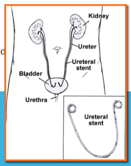
A 45-year-old male presents to the emergency department with sudden onset of severe flank pain radiating to the groin, accompanied by nausea and hematuria. A non-contrast CT scan reveals a 6 mm calculus in the left ureter.
Which of the following best explains the pathophysiological mechanism leading to his symptoms?
A. Inflammation of the glomerular basement membrane causing proteinuria
B. Obstruction of urinary flow causing increased intrarenal pressure and ureteral spasm
C. Autoimmune destruction of renal tubular cells causing acute tubular necrosis
D. Excess aldosterone secretion leading to sodium retention and hypokalemia
B. Obstruction of urinary flow causing increased intrarenal pressure and ureteral spasm