Muscles of Lower Extermities
 Call Kai
 Learn
Learn Practice Test
 Spaced Repetition
 Match
Match Flashcards
 Knowt Play
1/163

There's no tags or description
Looks like no tags are added yet.
Name | Mastery | Learn | Test | Matching | Spaced | Call with Kai | Chat |
|---|
No analytics yet
Send a link to your students to track their progress
164 Terms
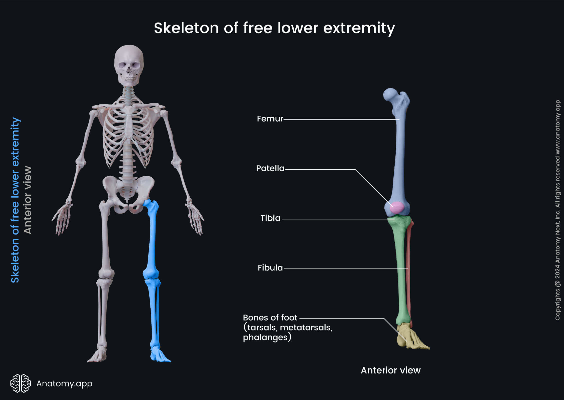
What is the Lower-Extrm
Thigh
Leg
Foot
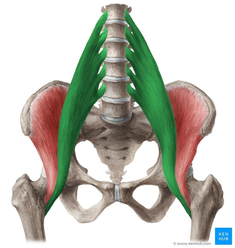
What multiarticular / biarticular muscle?
Psoas Major
Multi-articular; orginates from lumbar spine - acts on hip
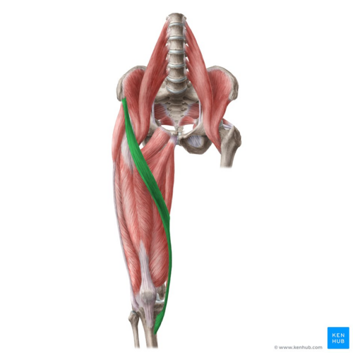
What multiarticular / biarticular muscle?
Sartorius
O: Anterior Superior Iliac Spine
I: Anteromedial Tibial Condyle (pes anserine)
A: Flexion, Abduction, External rotation of femur (femoracetabular joint); Weak Flexion / Internal Rotation (when flexed) of knee/ leg
IN; Femoral Nerve
Cross both the hip / knee joint
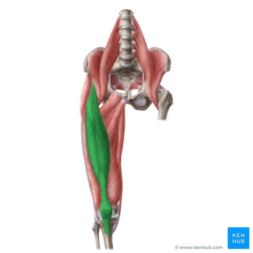
What multiarticular / biarticular muscle?
Rectus Femoris (Quad Group)
O: Anterior Inferior Iliac Spine (AIIS)
I: Patella (via quadriceps tendon) / tibial tuberosity; (via pateallar tendon/ligament)
A: Flexion of thigh/hip - extension of leg/knee (tibiofemoral joint)
IN Femoral Nerve
Crosses the hip/knee - contrib to hip flexion / knee extension
(BI-Articular quad muscle)
What is the Fascial / Tendinious Strcuture of Knee / Thigh
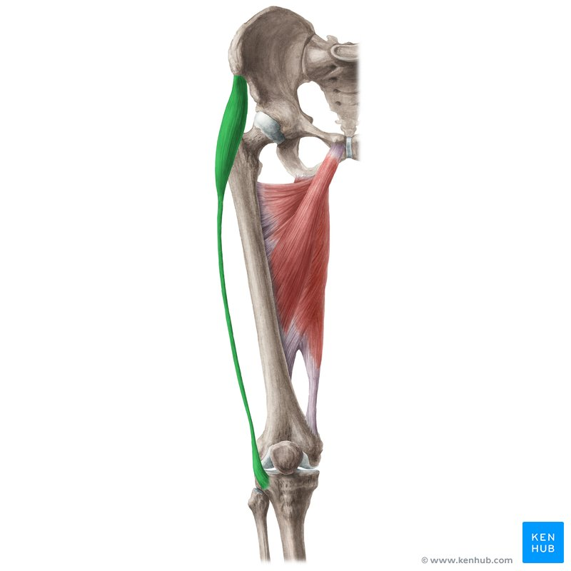
Iliotibial (IT) Band / Tract
Dense band of fascia running along the lateral thigh from hip to knee (superficial - vastus lateralis) contrb to lateral staibility of hip / knee
Attachments: Recivies fibres from Glutues Maximus / Tensor Fascia Lata - inserts distally at Gerdys Tubercle on ANterolateral Tibia
What is the Fascial / Tendinious Strcuture of Knee / Thigh
Attachment
What is the Fascial / Tendinious Strcuture of Knee / Thigh
Pes Anserine (‘gosse foot’)
Shared tendon on upper inner > anteromedial tibia formed by Sartorius / Gracilis / Semitendinosus Muscles
Assists in Medial Knee Stability (secondary restraint to valgus forceses)
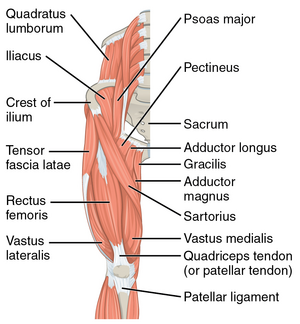
What Anterior Hip Muscle? (COVERED?)
Iliospoas
Sartorius’'
Pectineus
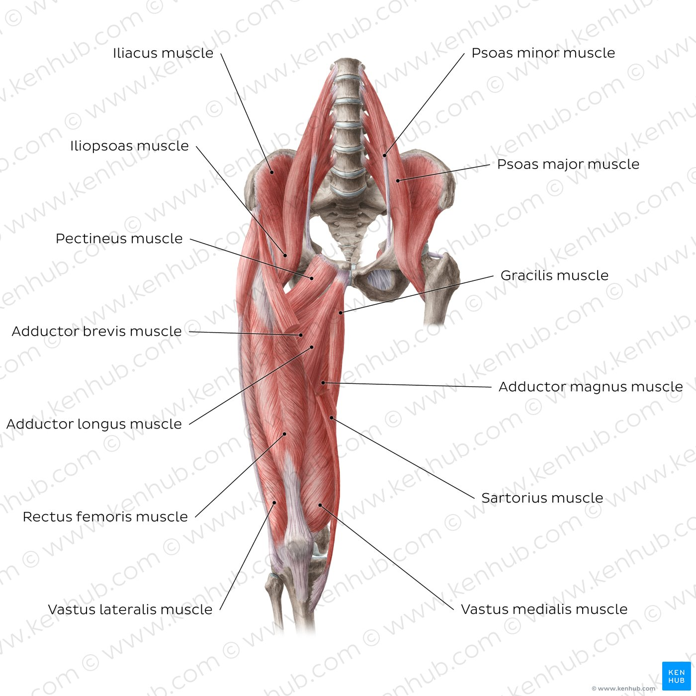
What Medial Hip Muscles? (COVERED)?
Gracialis
Adductor Brevis
Adductor Longus
ADductor Magnus
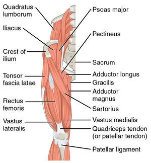
What Posterolateral Hip Muscles (COVERED?)
Glutues Maximus
Gluteus Medius
Gluteus Minimus
Tensor Fascia Lata
Priformis
Gemellus Superior / Inferior
Obturator Femoris
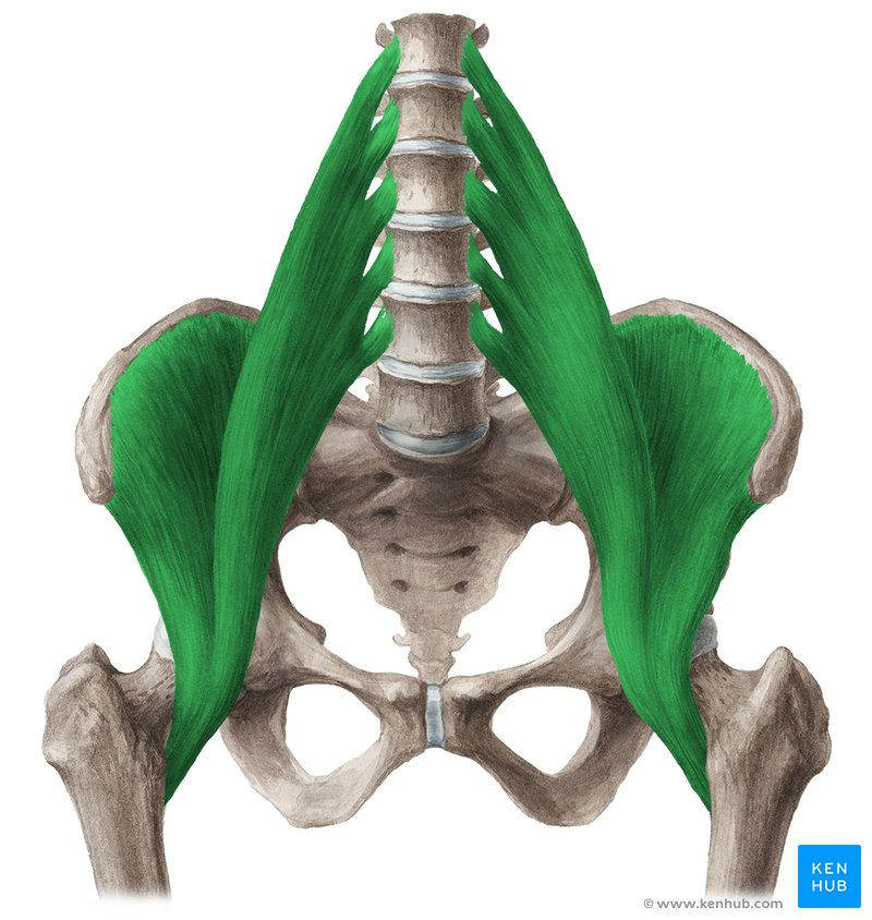
Anterior Hip Muscles
Iliopsoas
O; Psoas major; Bodies; transverse processes / discs of t12-l5
Iliacus; Iiliac fossa
I; lesser trochanter of femur
A; flexion / external rotation of femur (femoroactabular joint)
IN; Femoral Nerve
most diffentiate psoas major / iliacus / iliopsoas
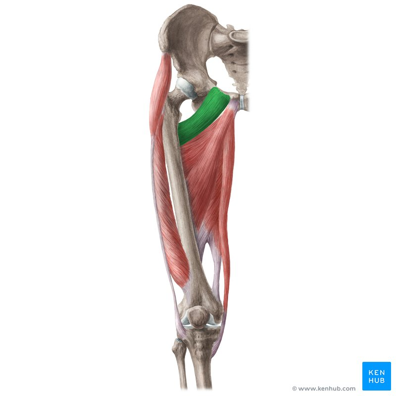
Anterior Hip Muscles
Pectineus
O: Superior Ramus of Pubis
I: Pectineal Line of Femur
A: Flexion / Adduction of Femur (Femoracetubular Joint)
IN; Femoral Nerve
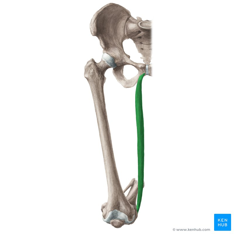
Medial Hip Muscles
Gracilis
O: Pubis
I: Anteromedial Tibial Condyle (pes anserine)
A: Adduction of Femur (femroactebular joint); weak flexion / interal roatation (when flexed) of knee/leg
IN; Obturator Nerve
Cross both the hip / knee joints > knee actions
(Notes) part of Pes anserine (Distal tendons of sartorius / gracilis / semitendinosus coverage @ proximal / medial / anterior tibia/ apears as a goose foot
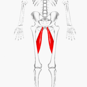
Medial Hip Muscles
Adductor Longus
O: Pubis
I: Linea Aspera (Middle Portion)
A: Adduction / Flexion of Femur (Femoractebular Joint)
In; Obtruator Nerve
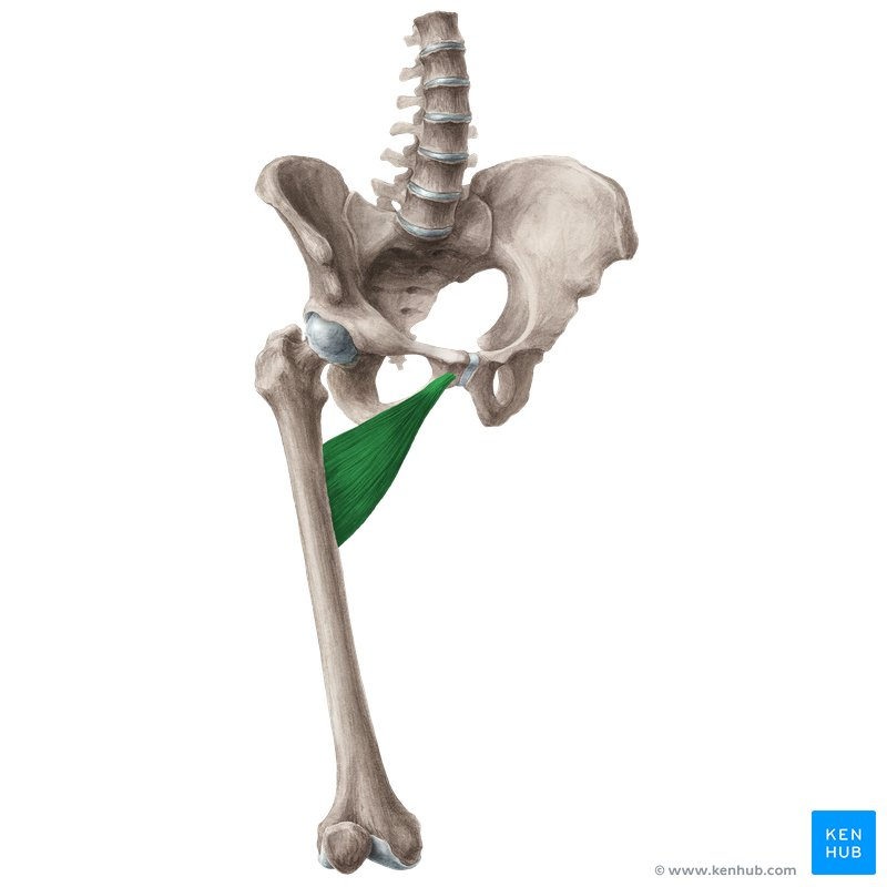
Medial Hip Muscles
Adductor Brevis
O: Pubis
I: Linea Aspera (middle portion)
A: Adduction / Flexion of Femur (femoractebular joint)
In; Obtruator Nerve
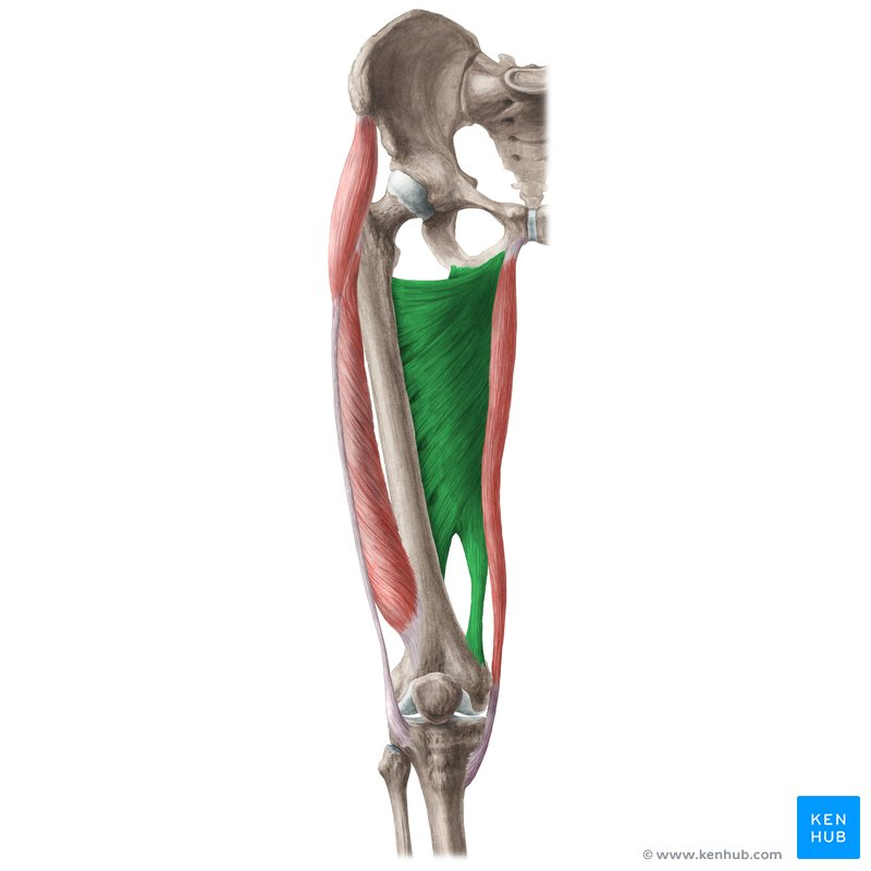
Medial Hip Muscles
Adductor Portion
O: Inferior Pubic Ramus , Ischial Ramus
I; Gluteal Tuberoisty / Entire length of linea aspera / medial supracondylar line
A; Adduction / Flexion of Femur (femoroacetabular joint) - stabilzing hip rotation
In; Obturator Nerve
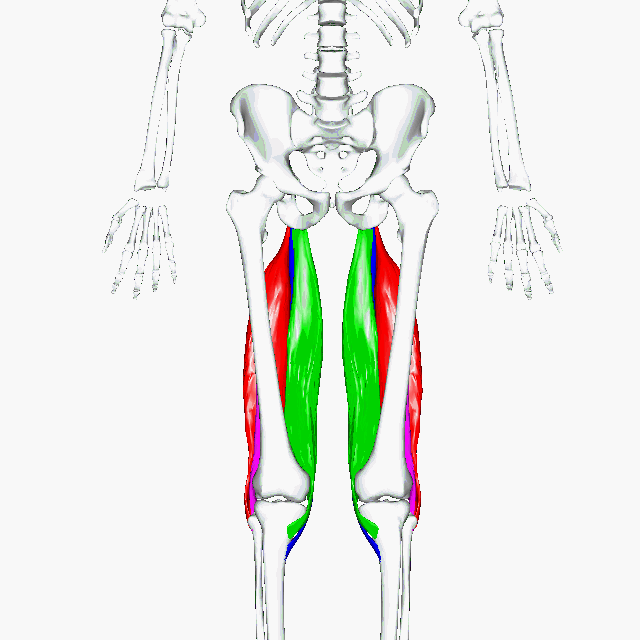
Medial Hip Muscles
Hamstring (ischiocondular) portion
O: Ischial Tuberosity
I: Adductor Tubercle of Femur
A: Adduction / Extension of Femur (femoroactevular joint) ; stabiling hip rotation
IN; Sciatic Nerve
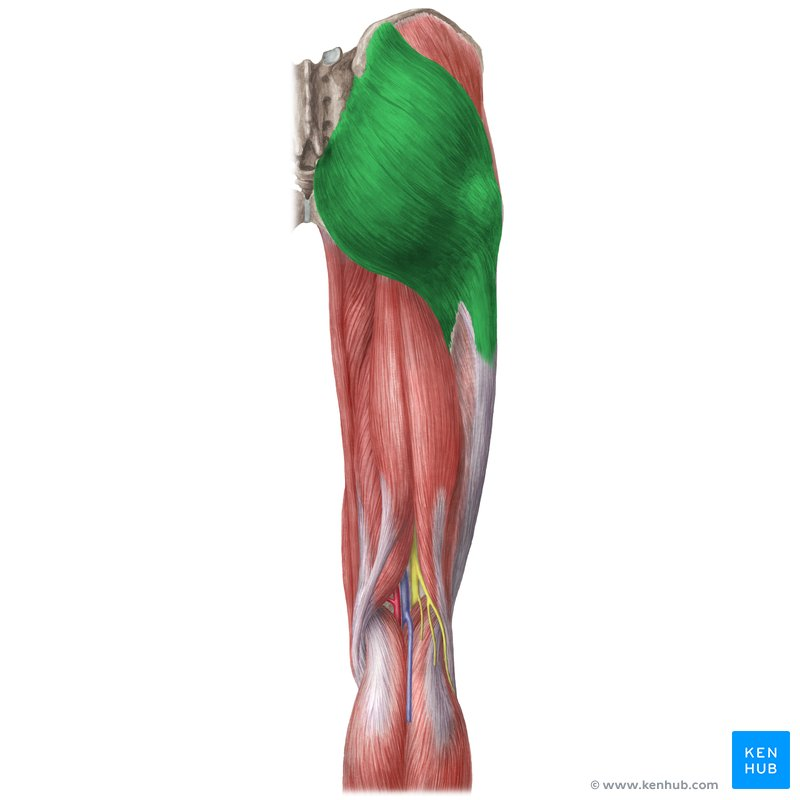
Posterior / Lateral Hip Muscles
Gluteus Maximus
O: Posterior Gluteal Line / Sacrum / Coccyx / Lumbar Fascia
I: Gluteal Tuberosity / Iliotibial band
A: Ext / External rotation of femur (femoroacetabular joint) ; tension iliotibial band
Superior Fibres; Abduction of Femur
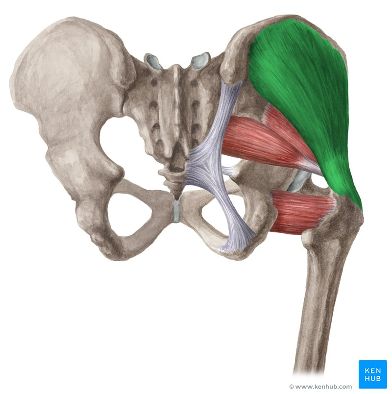
Posterior / Lateral Hip Muscles
Gluteus Medius
O: Ilium between posterior / middle (anterior) gluteal lines
I: greater trochanter (lateral aspect)
A; Abduction / internal rotation of femur (femoracetabular joint) pelvic stabilization
posterior fibres; external rotation of femur
In; Superior Gluteal Nerve
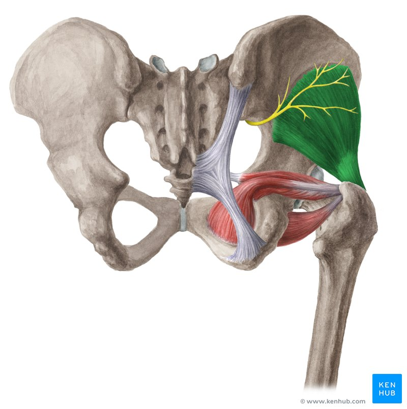
Posterior / Lateral Hip Muscles
Gluteus Minimus
O: Iliium between middle (anterior) / inferior gluteal lines
I: Greater trochanter (anterior aspect)
A: Abduction / Internal Rotation of Femur (femoroacetabular joint) - pelvic stabilization
In; Superior Gluteal Line
Posterior / Lateral Hip Muscles
Tensor Fascia Lata (TFL)
O; Anterior Iliac Crest (immediatiely posterior to ASIS)
I; Iliotibial Band
A; Abduction / Flexion and Internal rotation of Femur (femoroacetabular joint; Tensions Iliotibial band
In; Superior Gluteal Nerve
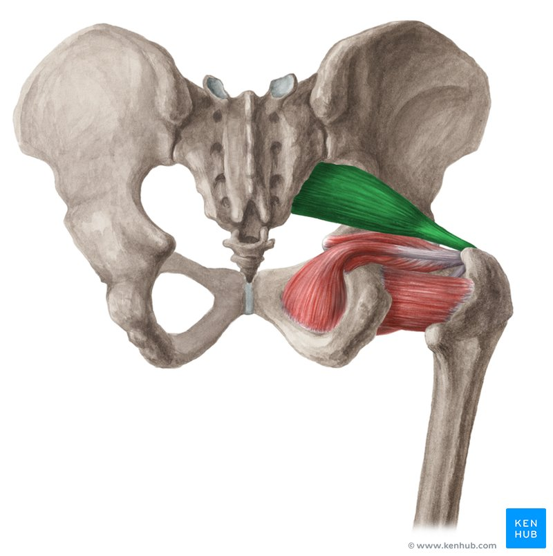
Deep Hip External Rotators
Priformis
O: Anterior Sacrum
I: Greater Trochanter
A: External rotation of Femur when Extended; Abduction of Hip/Thigh when flexed; (femroactebular joint)
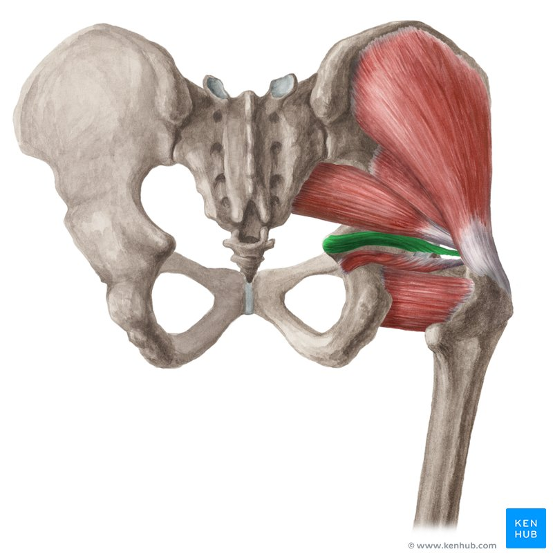
Deep Hip External Rotators
Gemellus Superior
O; Ischial Spine
I; Greater Trochanter
A; External Rotation of Femur when Extended; abduction of hip/thigh/femur when flexed
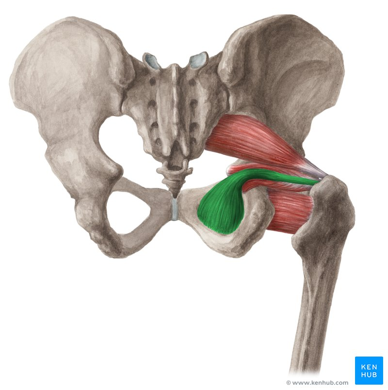
Deep Hip External Rotators
Obturator Internus
O: Inner (posterior) surface of obturator membrane (and boundaries of obturator forearm)
I: Greater Trochanter
A: External Rotation of Femur when extended (abduction of hip/thigh/femur when flexed
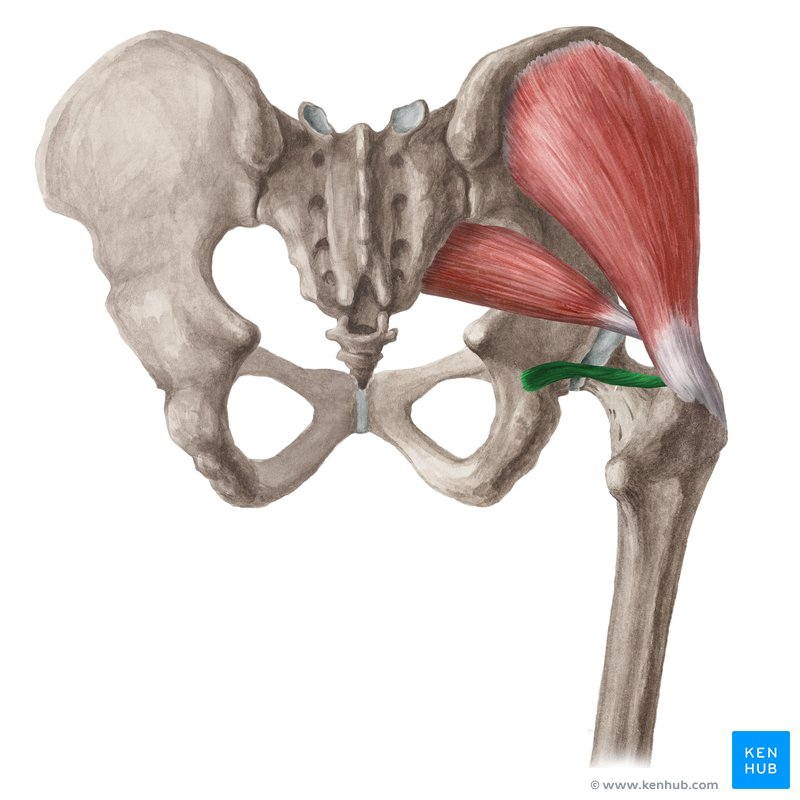
Deep Hip External Rotators
Gamellus Inferior
O: Ischial Tuberosity
I: Greater Trochanter
A: External Rotation of Femur when Extended; Abudction of hip/thigh/femur when flexed
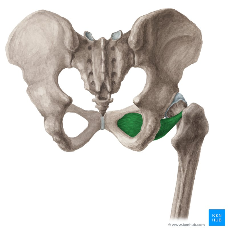
Deep Hip External Rotators
Obturator Externus
O: Outer (anterior) surface of obturator membrane (and boundaries of obturator foramen)
I: Trochanter fossa of Femur
A: External rotation of Femur when extended; Abuction of hip/thigh/femur when flexed
In; Obturator Nerve
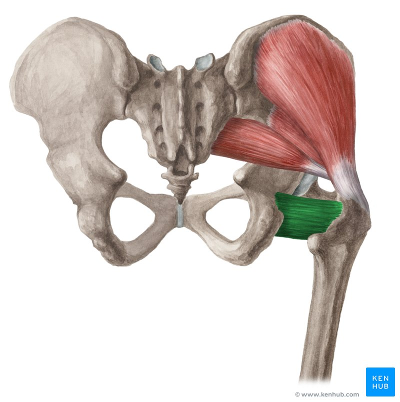
Deep Hip External Rotators
Quadtratus Femoris
O: Ischial Tuberosity
I: Intertrochanteric Crest of Femur
A: External Rotation of Femur
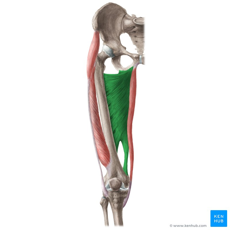
Deep Hip External Rotators
Adductor Magnus
Adductor / Hamstring / Ischiocondylar Portions
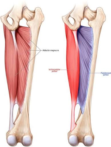
Deep Hip External Rotators
Hamstring (Ischiocondylar) Portion
O: Inferolateral aspect of the ischial tuberosity (the "sit bone").
I: Adductor tubercle on the medial condyle of the femur, with some fibers attaching to the medial supracondylar line
A: Extension of the hip (thigh). It also acts as a powerful adductor of the hip joint.
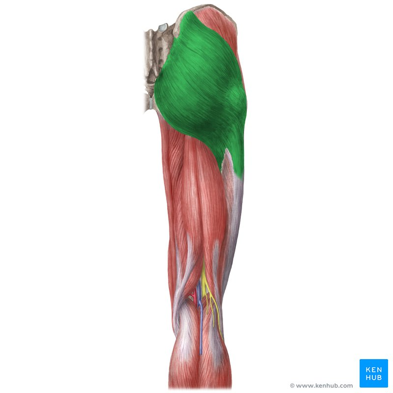
Posterior / Lateral Hip Muscle
Gluteus Maximus
O: Posterior Gluteal Line, Sacrum, Coccyx, Lumbar Fascia
I; Gluteal Tuberosity, Illiotibial Band
A; Ext / external rotation of femur (femoroactabular joint) - tension iliotibial band
Superior Fibres - Abduction of Femur
Inferior Fibres - Adduction of Femur
In; Inferior Gluteal Nervef
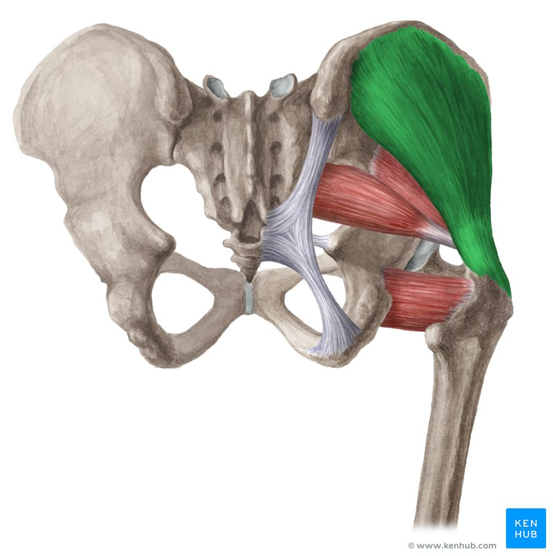
Posterior / Lateral Hip Muscle
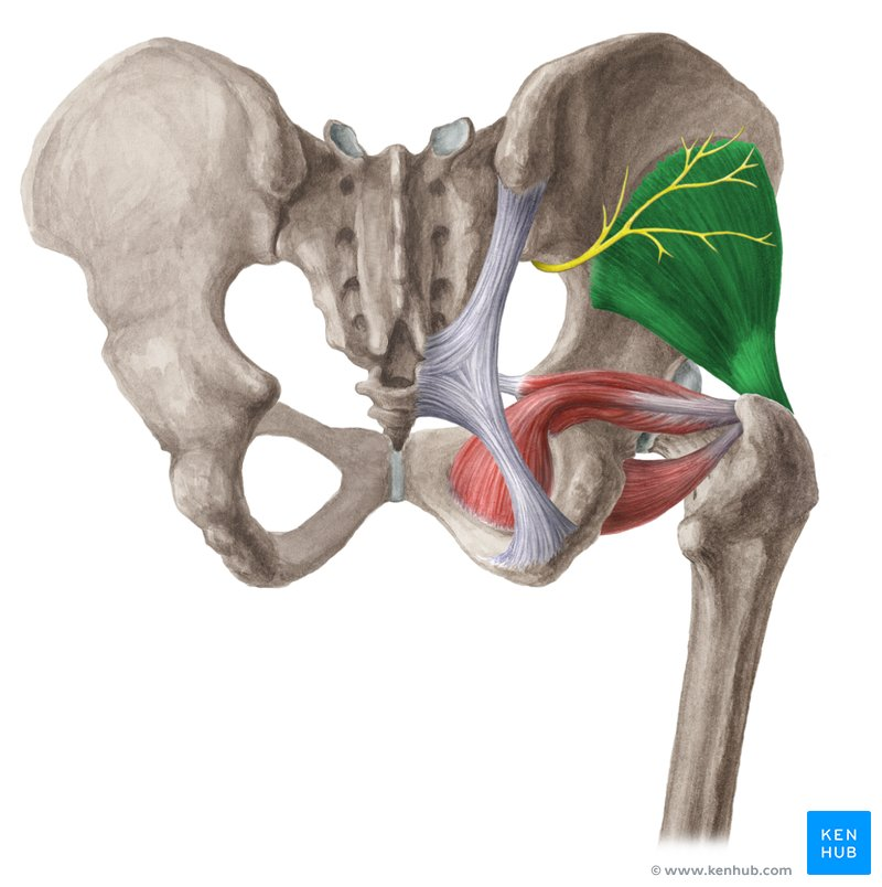
Gluteus Medius
O: The external surface of the ilium (between the iliac crest and the posterior/anterior gluteal lines) and the gluteal aponeurosis. [1, 2]
I: The lateral surface of the greater trochanter of the femur.
A: Abducts the hip (moves the leg away from the midline). Anterior fibers assist with hip flexion and internal rotation; posterior fibers assist with hip extension and external rotation. It is also the primary stabilizer of the pelvis when standing on one leg
Posterior / Lateral Hip Muscle
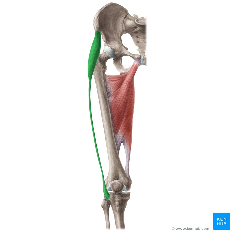
Gluteus Minimus
O: The external surface of the ilium (the large, broad part of your pelvic bone), specifically in the space between the anterior and inferior gluteal lines
I: The anterior facet of the greater trochanter of the femur (the bony bump on the upper/outer part of your thigh bone) and the anterosuperior hip joint capsule. [1]
A: Hip Abduction: Moves the thigh outward, away from the body's midline.
Medial Rotation: Rolls or rotates the thigh inward.
Pelvic Stabilization: Crucial for walking and standing; it keeps the pelvis level and prevents it from sagging to the opposite side when you stand on one leg. [1, 2, 3, 4]
Posterior / Lateral Hip Muscle
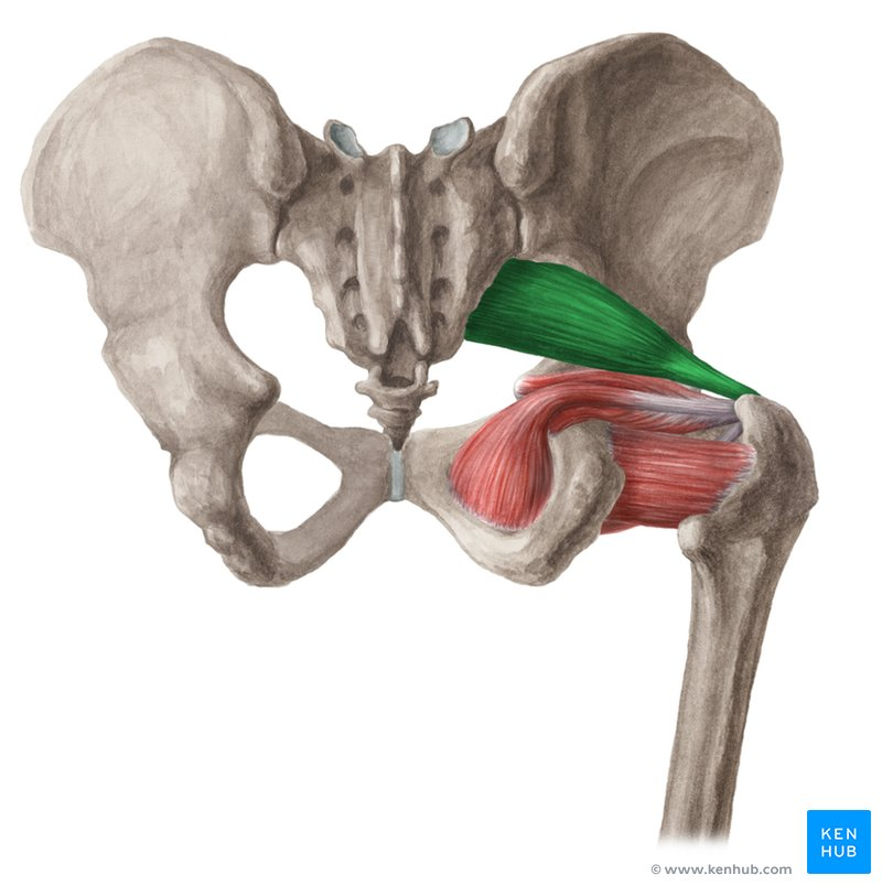
Tensor Fascia Lata (TFL)
O; Anterior Illiac Crest (immeditaelu posterior to asis_
I Iliotibial band
A; Abduction / flexion and internal rotation of fmeur (femoroactebular joint); tension iliotibial band
IN; Superior Gluteal Nerve
Deep Hip External Rotators (Deep Six)
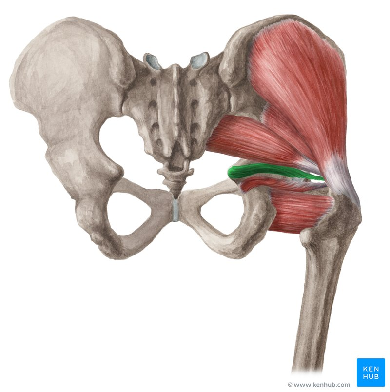
Priformis
O: Anterior (pelvic) surface of the sacrum, the gluteal surface of the ilium near the greater sciatic notch, and the sacrotuberous ligament.
I: Superior border of the greater trochanter of the femur.
A: Laterally rotates the extended thigh and abducts the flexed thigh. It also acts to stabilize the hip joint. [1, 2, 3, 4]
Deep Hip External Rotators (Deep Six)
Gemellus Superior
O: The outer/gluteal surface of the ischial spine of the pelvis.
I: The medial surface of the greater trochanter of the femur, merging into a common tendon with the obturator internus muscle
A: It acts to externally (laterally) rotate the extended thigh and abducts the thigh when the hip is flexed. It also stabilizes the hip joint by keeping the femoral head securely in the socket.
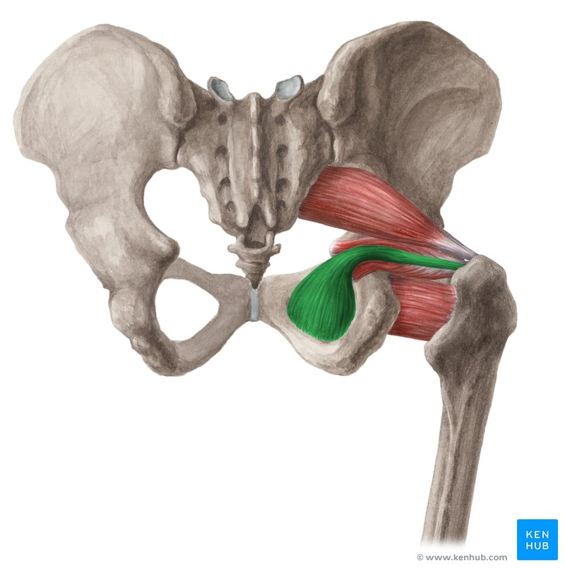
Deep Hip External Rotators (Deep Six)
Obturator Internus
O: Inner (posterior) surface of obturator membrane / boundaries of obturatr foramen
I: greater trochanter
A: External Rotation of Femur when Extended - Abduction of hip/thigh/femur when flexed
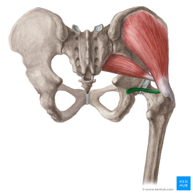
Deep Hip External Rotators (Deep Six)
Gemllus Inferior
O; Ischial Tuberoisity
I; Greater Trochanter
A; External Rotation of Femur when Extended - Abduction of hip/thigh/femur when flexed
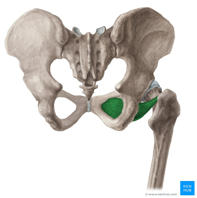
Deep Hip External Rotators (Deep Six)
Obturator Externus
O: Outer surface of the obturator membrane and the bony margins of the obturator foramen (on the pubis and ischium).
I: (Insertion): Trochanteric fossa of the femur.
A: Primarily externally (laterally) rotates the thigh at the hip joint, and also acts as an adductor of the hip (especially when the hip is flexed). [1, 2, 3, 4, 5, 6]
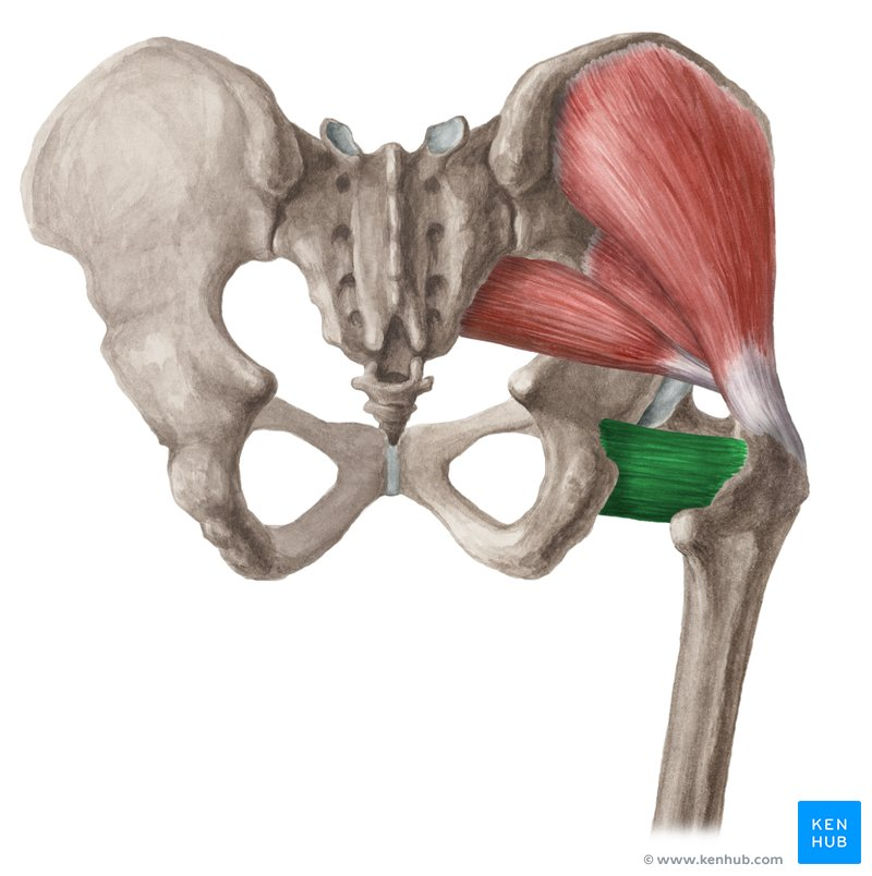
Deep Hip External Rotators (Deep Six)
Quadratus Femoris
O:Ischial Tuberosity
I: Introchanteric Crest of Femur
A: External Rotation of Femur
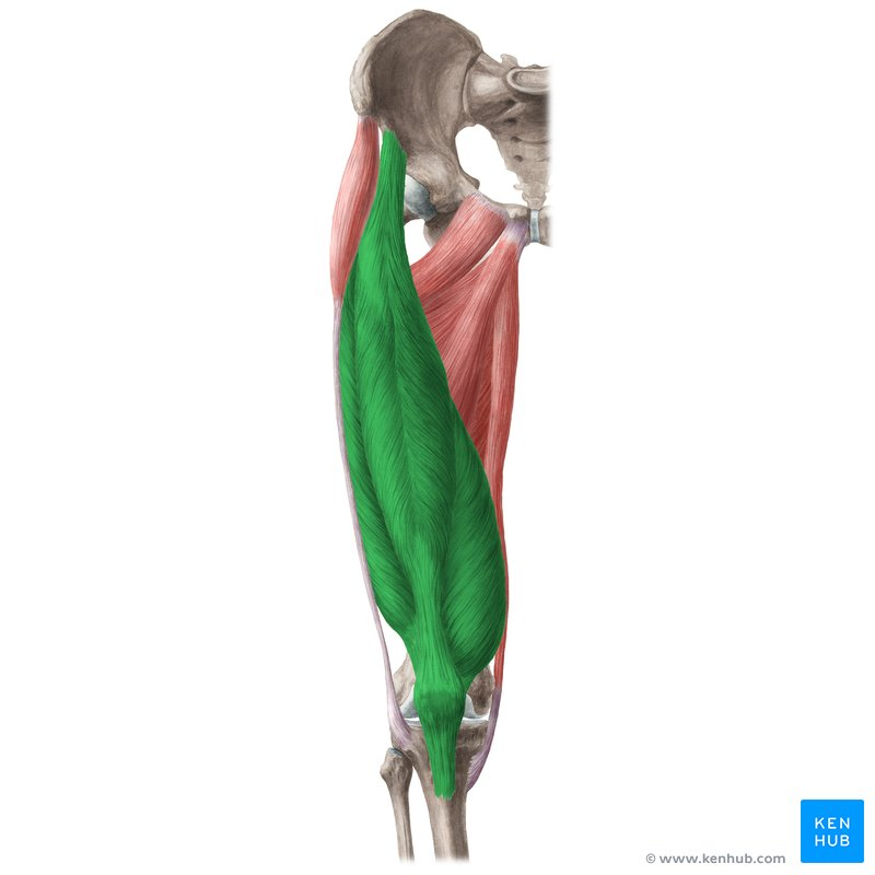
Muscles that Move the Leg?
Quadriceps Femoris
Quadriceps Tendon
Patellar Tendon / Ligament
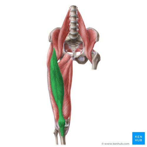
Muscles that Move the Leg?
Rectus Femoris
O: Anterior Inferior Iliac Spine (AIIS)
I: Patella (via quadriceps tendon) / tibial tuberosity; (via pateallar tendon/ligament)
A: Flexion of thigh/hip - extension of leg/knee (tibiofemoral joint)
IN Femoral Nerve
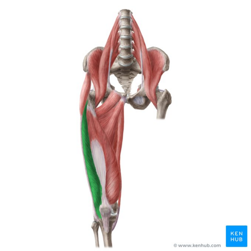
Muscles that Move the Leg?
Vastus Lateralis
O: Greater Trochanter / Lateral lip of Linea aspera
I: Patella (quad tendon) / tibial tuberosity - (patelar tendon / ligament)
IN: Femoral Nerve
A: Extension of leg/knee (tibiofemoral joint)
(Common intramuscular injecion site in infants - vaccinations)
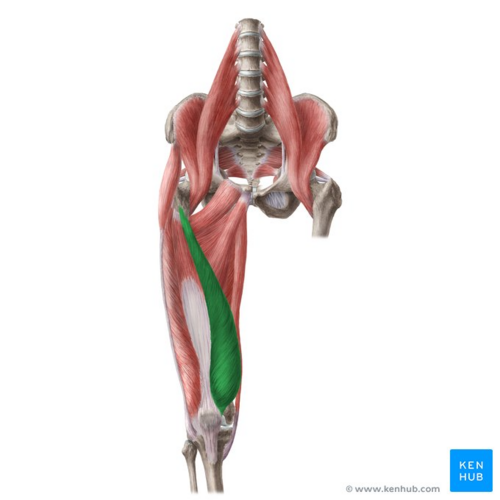
Muscles that Move the Leg?
Vastus Medialis
O: Interochanteric line / medial lip of linea aspera / medial supracondylar ridge
I; Patella (via quadtriceps tendon) / tibial tuberosity (via patellar tendon/ligament)
A: extension of knee/leg (tibiofemoral joint)
In; Femoral nerve
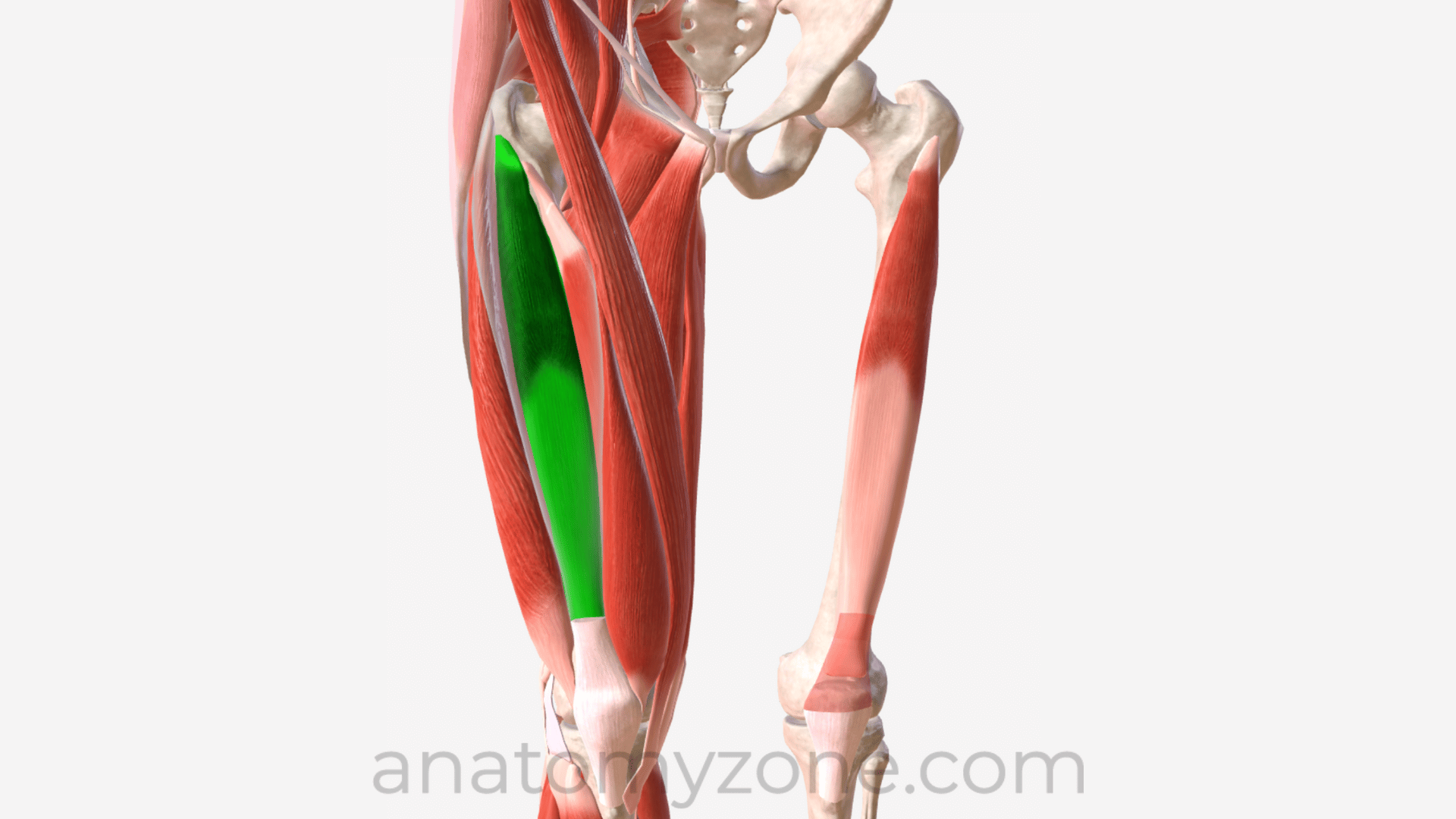
Muscles that Move the Leg?
Vastus Intermedius
O: Anterior Femur
I: Patella (via quadriceps tendon) / tibial tuberosity (via patellar tendon/ligament)
A: Extension of leg/knee - tibiofemoral joint
In; Femoral Nerve
Deep to Rectus Femoris
What muscle? (posterior thigh)
Hamstrings
Biceps Femoris
Semitendinosus
Semimembranosus = cross the hip / knee - contrb to hip extension / knee flexion
Short head of Biceps Femoris > (Monoarticular) @ knee
(Three muscles of posterior thigh that act as prime movers of knee flexion / tg with gluteus maximus - hip extension (short head of biceps femoris is monoarticular / acts only at knee
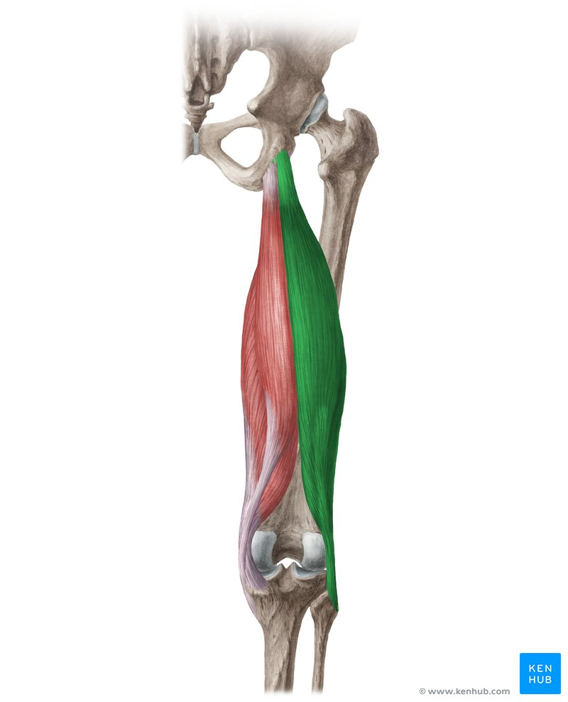
What muscle?
Biceps Femoris (long / short heads)
O: Long Head; Ischial tuberosity / Short Head; Linea asperia (distal) / lateral supracondylar ridge
I: Head of Fibula
A: Flexion of Knee/Leg / External Rotation of Knee / Leg when Flexed
Long head: extension of Hip/Thigh / External rotation of hip/thigh
IN: Sciatic nerve
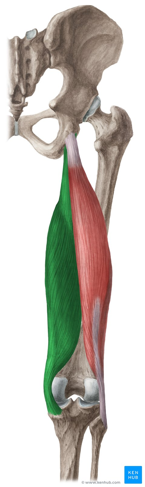
What muscle?
Semimembranosus
O: Ischial tuberosity
I: medial condyle of tibia
A: flexion of knee/leg / interal rotation of knee/leg when flexed / extension of hip/thigh
In; Sciatic Nerve
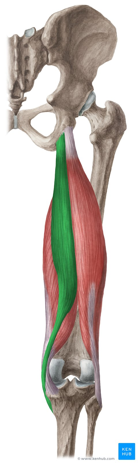
What muscle?
Semitendinosus
O: Ischial Tuberosity
I: Medial Condyle of Tibia
A: Flexion of knee/leg / internal rotation of knee/leg when flexed / extension of hip /thigh ; internal rotation of hip/thigh
IN; Sciatic Nerve
What muscle?
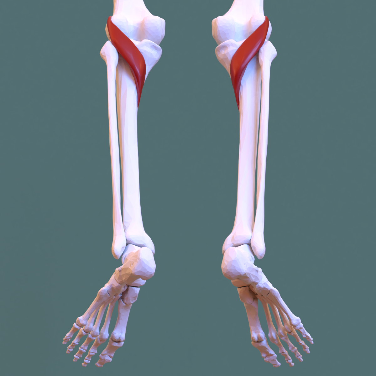
Popliteus
O: Lateral Femoral Condyle / Posterior horn of lateral meniscus
I: Posterior Tibia (superior to soleal line)
A: Flexion / Internal rotation of knee/leg (unlocking the fully extended knee okc)
IN Tibial Nerve
What is Knee Locking / Unlocking
Screw-Home Mechnaism (SHM)
Largely passive locking mechanisms that occur during the final -20 degrees - of stability whie reducing muscular effort during standing
OCCURS as the quads extend the knee into terminal extension - tibia/femur rotate relative to one another - prod a locked pos
Popliteus activiely unlocks the knee to intitate flexion
mechanis is esp important during actitivies such as walking, where the knee alternatives between stability (locked) / mobility (unlocked)
Knee Locking / Unlocking
Non-weight Bearing (OKC)
Tibia externally (Laterally) rotates on fixed femur
Knee Locking / Unlocking
Weight Bearing (CKC)
Tibia is fixed / Femur internally (medially) rotates on tibia
What is occuring
Unlocking The Knee (popliteus muscle)
Knee Locking / Unlocking (poplitues muscle)
Weight-bearing (CKC)
Tibia is fixed - so poplitues externally rotates the femur on fibia
What is the Anterior Comp
Tibialis Anterior
Extensor Digitorum Longus
Extensor JHallucis Longus
What is Lateral Comp
Fibularis
Peroneals
What Post Comp
Superficial - Gastrcenemius / Soleus / Plantaris
What post comp
Deep - Tibialis Posterior / Flexor Digitorum Longus / Flexor Hallucis Longus
What Anterior Comp Muscle
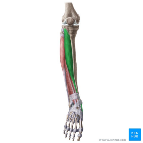
Tibialis Anterior (Anterior Comp)
O: Anterolateral Tibia
I: Plantaar aspect of 1st metatarsal and medial cuneiform
A: Dorsiflexion (Talocrual Joint) and inversion / subtalar and intertarsal joints - of anke/foot
In; Deep peroneal nerve
What Anterior Comp Muscle
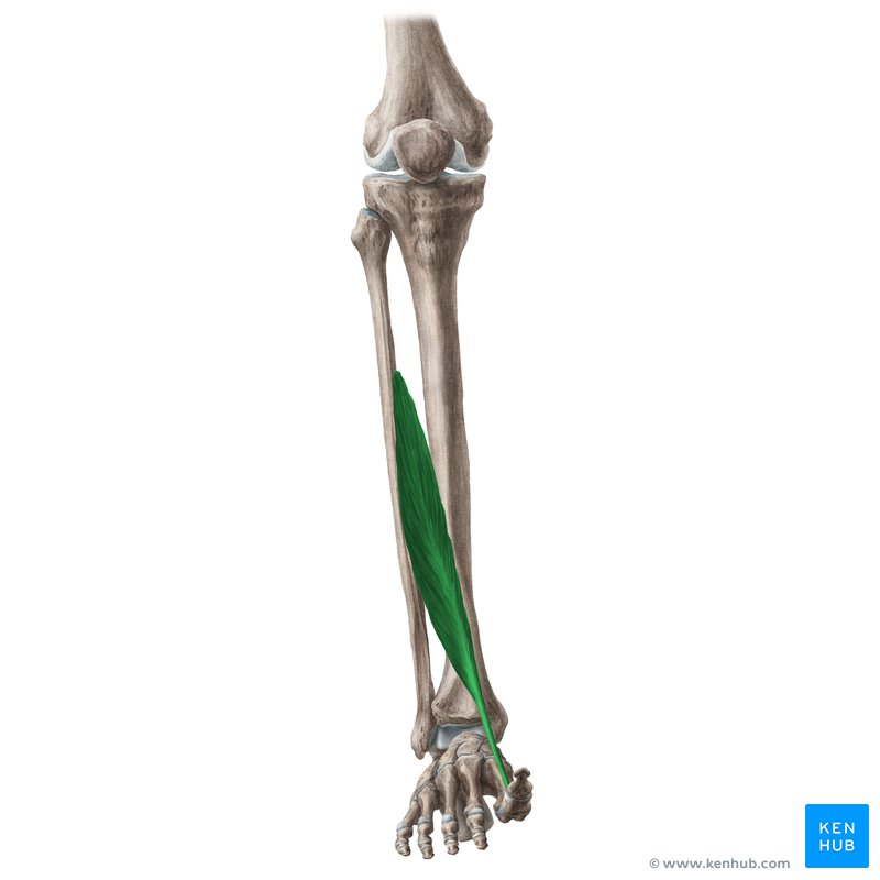
Extensor Hallucis Longus
O: Middle / Anterior Fibula
I: Dorsal Aspect of Distal Phalanx of great (1st) toe / hallux
A; Extension of Great toe / hallux (mtp / ip joints) dorsiflexion of ankle / talocural joint
IN; Deep peroneal nerve
What Anterior Comp Muscle
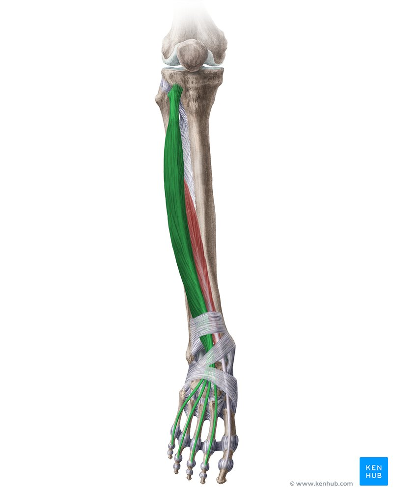
Extensor Digitorum Longus
O: Proximal ½ of anterior fibula / lateral tibial condyle / proximal interoseous membrame
I: Dorsal Aspect of middle / distal phalanges - 2nd/5th toes (4 seperate tendons)
A: Extension / toes/digits 2/5 (mtp / ip joints); dorsiflexion of ankle (talorcrual joint)
In; Deep peroneal nerve
Small muscle called Peroneus Tertilis exists in the digital anterior compartment (not req to know)
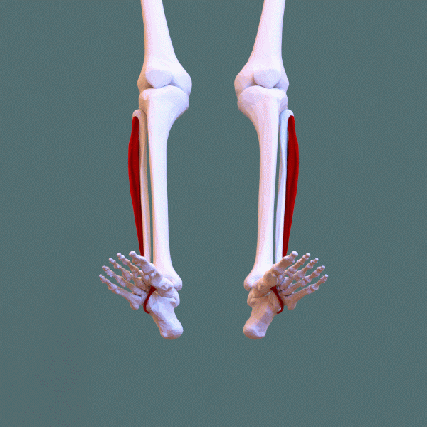
What Lateral Comp Muscle
Peroneus Longus (Fibularis Longus)
O; Proximal 2/3 of lateral fibula
I; Plantar aspect of 1st metatarsal / medial cuneiform
A; Eversion of ankle/foot (subtalar / intertarsal joints) ; plantarflexion of ankle (talocrual joint)
In; superfiical peronael nerve
(important muscle for supporting arches of foot)
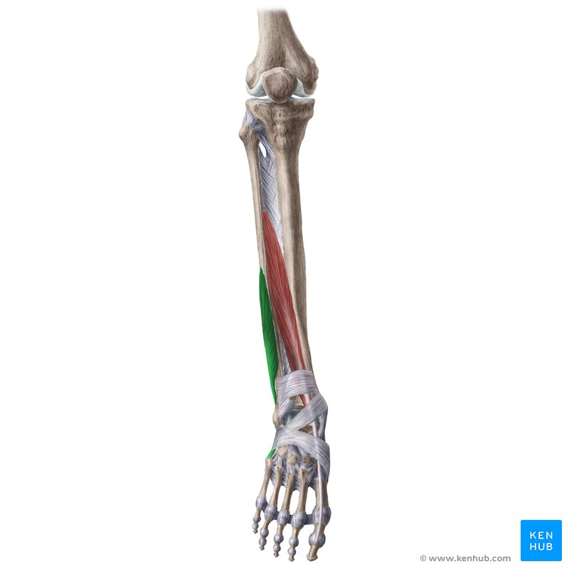
What Lateral Comp Muscle
Peroneus Brevis (Fibularis Brevis)
O: Distal 2/3 of Laterral Fibula
I Base of 5th metatarsal
A; Eversion of ankle/foot (subtalar / intertarsal joints) / plantarflexon of ankle (talocrual joint)
IN: Superfical peroneal nerve
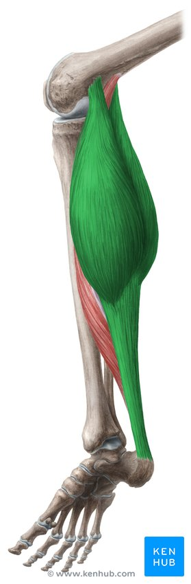
What posterior comp muscle
Gastrocenemius (Superficial)
O; Posterior aspectof medial / lateral femoral condyles
I; Calcaneus (via Achilies Tendon)
A; Plantarflexion of Ankle/foot (talocrural joint); Flexion of leg/knee (tibiofemoral joint)
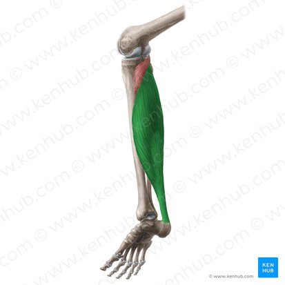
What posterior comp muscle
Superfifical
Soleus
O - Proximal aspect of medial / lateral femoral condyles
I - Calcaneus (achiles tendon)
A - Plantarflexion of ankle/foot (talocrual joint)
In; Tibial Nerve
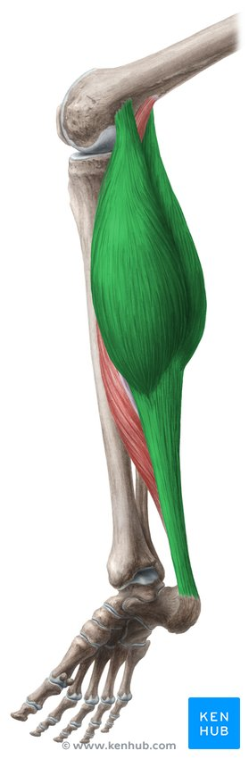
What posterior comp muscle
Superfiical
Gastrocenemius
O; Posterior aspect of medial / lateral femoral condykle
I; Calcaneus (achillies tenon)
A; Plantarflexion of ankle/foot (talocrual joint)
in; tibial nerve
What Tendon
Achileies Tendon
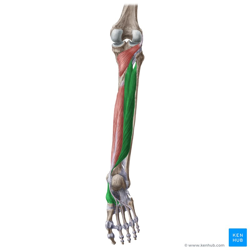
What is Deep Posterior Muscle
Tibialis Posterior
O; proximal posterior tibia and fibula
I; Calcaneus / navicular tuberoisty / cuboid / all cunniforms and metatarsals 2/4
A; Plantarflexion (talocrual joint) / inversion (subtalar / intertarsal joints) of ankle/foot
In; Tibial neve
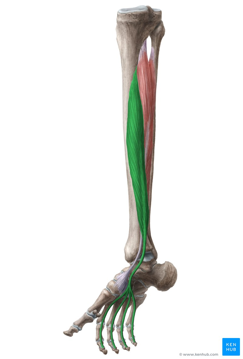
What is Deep Posterior Muscle
Flexor Digitorum Longus
O; Posterior Tibia (Inferior-to soleal line)
I; Plantar aspect of distal phalanges pf 2nd/5th toes/digits - 4 sep tendons
A; Flexion of 2nd/5th toes (mtp/ip joints); Plantar flexion of ankle/foot (talocrual joint)
In; Tibial Nerve
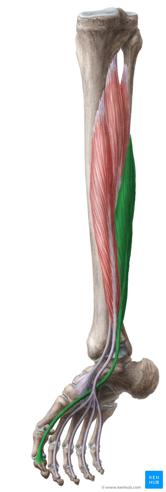
What is Deep Posterior Muscle
Flexor Hallucis Longus
O; Distal 2/3 posterior fibula
I; Disltal phlanx of great 1st toe/hallux
A; Flexion of great toe/hallux (mtp / ip joints); plantarflexion of ankle/ foot (talocrural joint)
IN; Tibial nerve
IMPORTANT FOR SUPPORTING ARCH FOR FOOT
How many Intrinsic Muscles of Foot
19 Muscles are located entirely within the foot
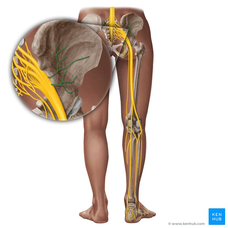
What nerve / Innervation
Superior Gluteal nerve - supplies lateral hip muscles - important for pelvic stability during gait - exits the pelvis - superior - prirformis
Glutues medius, minimus, tensor fascia lata
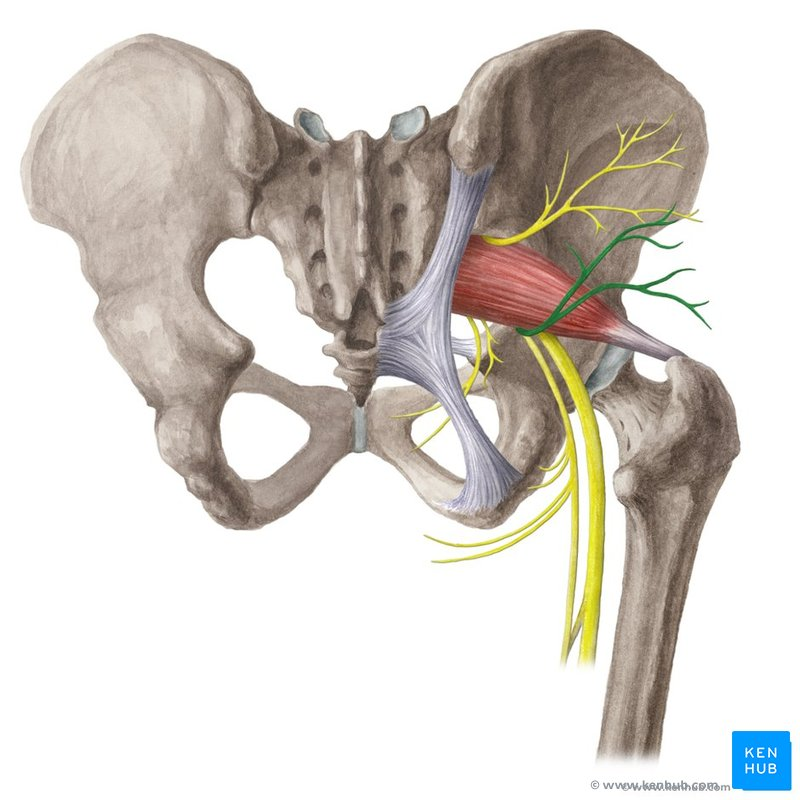
What nerve / Innervation
Inferior Gluteal Nerve - Supplies primary gluteal hip extensor ; exists the pelvis inferior - priroformis
Gluteus Maximus
What nerve / Innervation
Femoral Nerve
Iliopsoas (via branches)
Sartorius
Pectineieus (partailly
quadriceps femoris
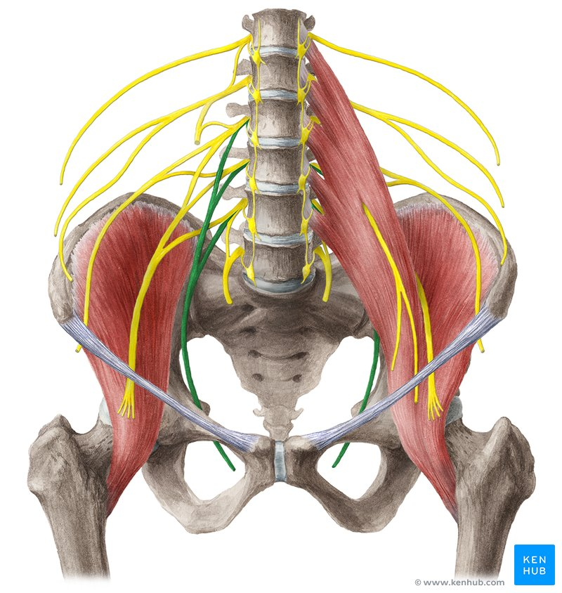
What nerve / Innervation
Obturator Nerve
adductor longus
adductor brevis
adductor magnus
gracilis
obturator externus
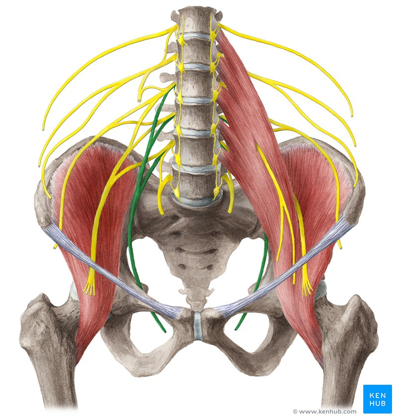
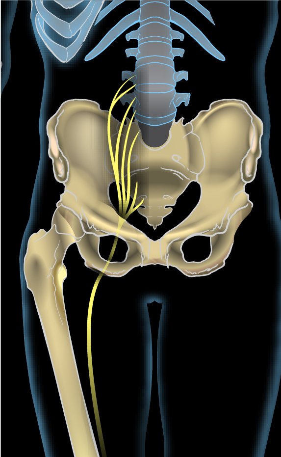
What nerve / Innervation
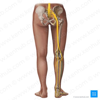
Sciatic Nerve
Hamstrings (bipceps femoris - long head, semitendinosis, semimembranosus)
hamstring portion - adductor magnus
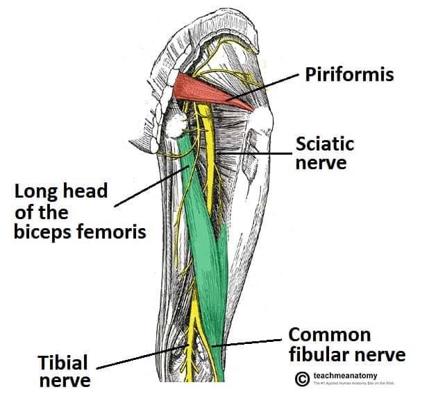
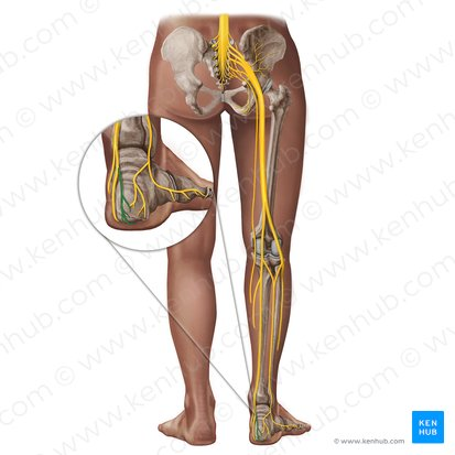
What nerve / Innervation
Tibial Nerve
Posterior Leg (superfifical) ; Gastrocenmous, soleus, plantaris
Posterior leg (deep); Tibialis posterior, flexor digitorium longus, flexor hallucis longus
plantar foot - (medial / lateral plantar nerves) - most intrinisic foot muscles
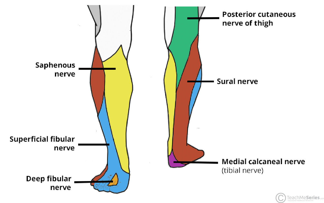
What nerve / Innervation
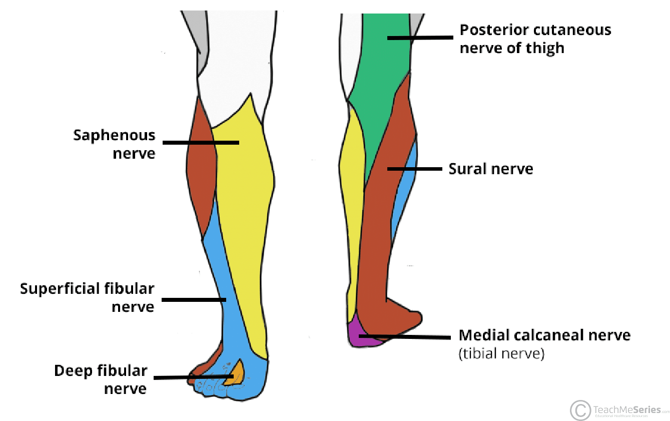
Common Fibular (Peroneal) Nerve
no innervatio
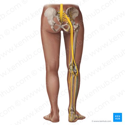
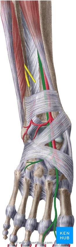
What nerve / Innervation
Deep Fibular (peroneal) nerve
Tibialis Anterior
Extensor Digitorum Longus
Extensor Hallucis Longus (anterior leg)
Dorsal Foot
Dorsal foot intrimsic muscles - (extensor digitorum brevis, extensor halllucis brevis)
What nerve / Innervation
Superficial Fibular (Peroneal) Nerve
Fibularlis (peroneus) longus
Fibularis (peroneus) brevis - (lateral leg)
Pathophysiology
Hamstring Strain
Pathophysiology
Patellar Tendinopathy (Jumpers knee)
Pathophysiology
Achilles Tendinopathy
How many intrinsic muscles of the foot?
19 Muscles located entirely within the foot; (Origin / inserition within the foot)
What muscle (superfical postrior comp)
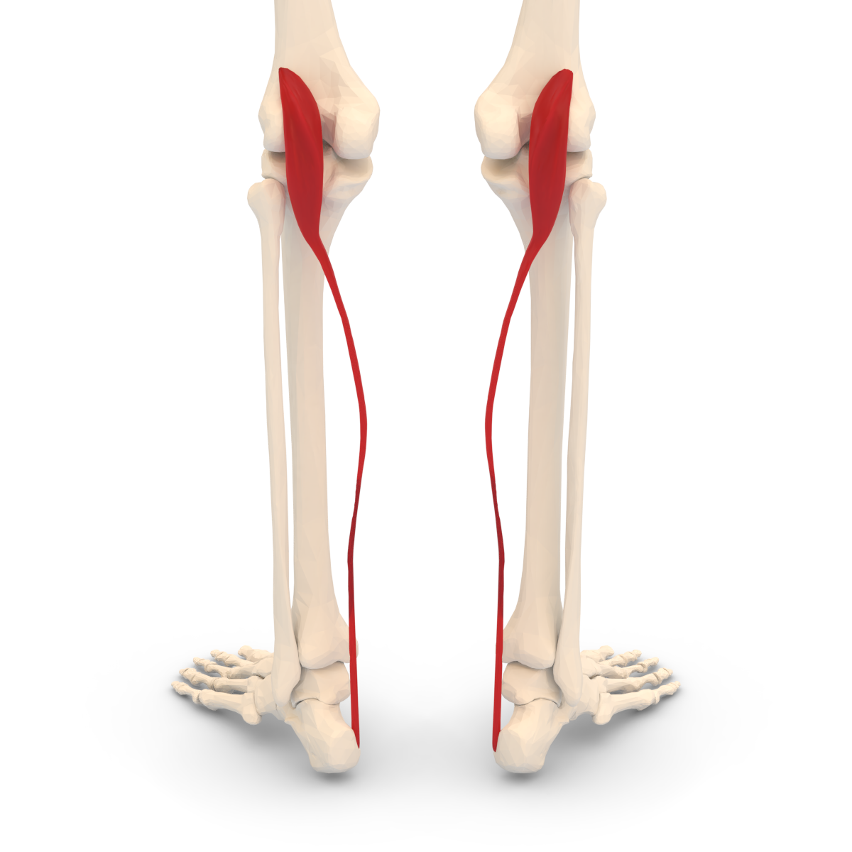
Plantaris
O - Lateral Supracondylar ridge of femur
I Posteromedial calcaneus of may joint achillies tendon
Plantarflexion of ankle/foot (talocrural joimt) weak flexion of knee leg
In tibial nerbe
(NOT EVERYONE HAS A PLANTARIS MUSCLE)
What os the Flexion of Proximal Phalanx MCP Joint ; with Simutaneoues Extension of Dstal / Middle Phalange? (MUSCLE)
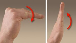
Lumbricals
Lumbricals ½ - Median Nerve
Lumbricals ¾ - Ulnar Nerve
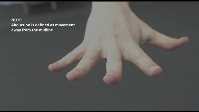
Abduction of D2/D5 - (MCP Joints) WHAT MUSCLES
Dorsal Interosseal - Ulnar Nerve
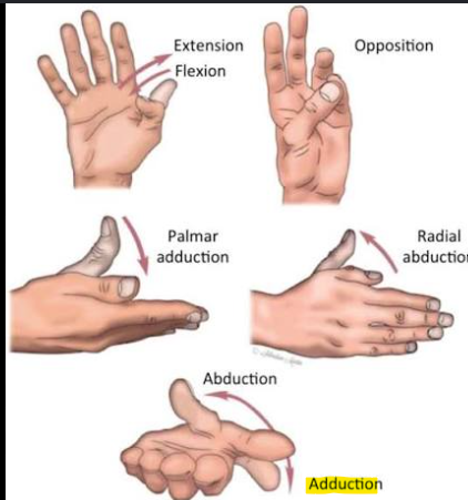
Adduction of D2/D5 - (MCP Joints) WHAT MUSCLES
Palmar Interossei - Ulnar Nerve
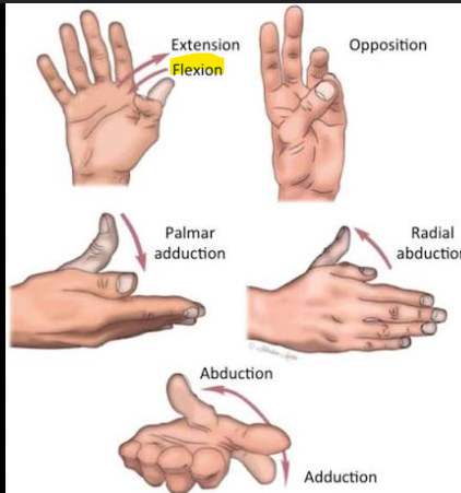
Flexor of Pollex (Cmc / MCP / IP joints)
Flexor Pollcis Longus - Median Nerve
Flexor Pollcis Brevis - Median Nerve
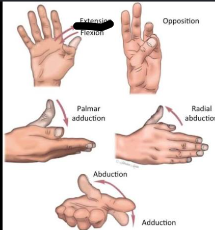
Extension of pollex (CMC / MCP / IP JOINTS)
Extensor Pollcis Longus
Extensor Pollcis Brevis
Abductor Pollcis Longus = Radial Nerve
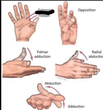
Ext of Pollex
CMC / MCP / IP JOINTS
Adductor Pollicis Longus - Radial Nerve
Adductor Pollicis Brevis - Median nerve
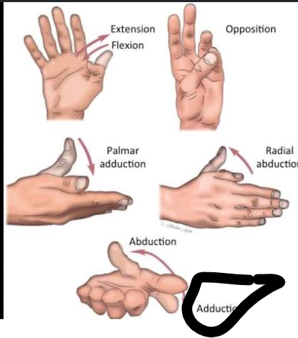
Adduction of Pollex (CMC JOINT)
Adductor Pollcis - Ulnar Nerve
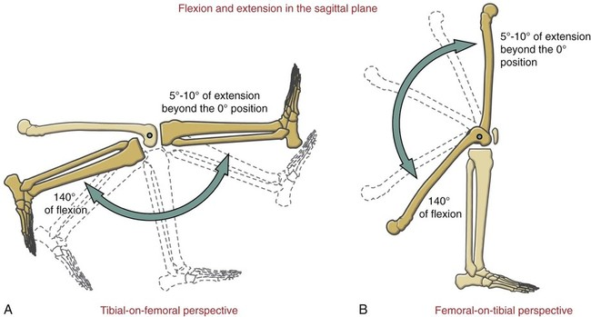
Flexion of Leg / Tibia at Knee / Tibiofemoral Joint
Biceps Femoris - Sciatic Nerve
Semtindonosus - Sciatic Nerve
Semimebranosis - Sciatic Nerve
Gastrocenmeius - Tibial Nerve
(Sartorius / Gracialis / Plantaris - not req)
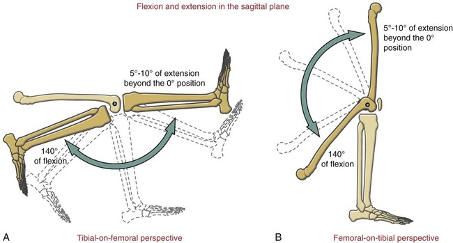
Extension of Leg / Tibia at Knee / Tibiofemoral Joint
Rectus Femoris
Vastus Medialis
Vasus Lateralis
Vastus Intermedius
= Femoral Nerve (ALL)
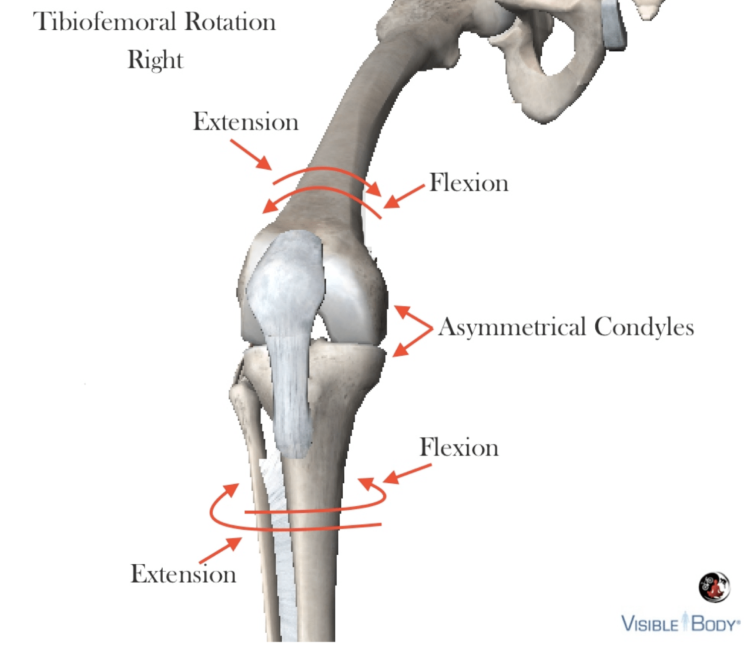
Internal Medial of Leg / Tibia at Knee / Tibiofemoral Joint
Poplitues - Tibial Nerve
Semitiendinosus - Sciatic Nerve
Semimebranosus - Sciatic Nerve
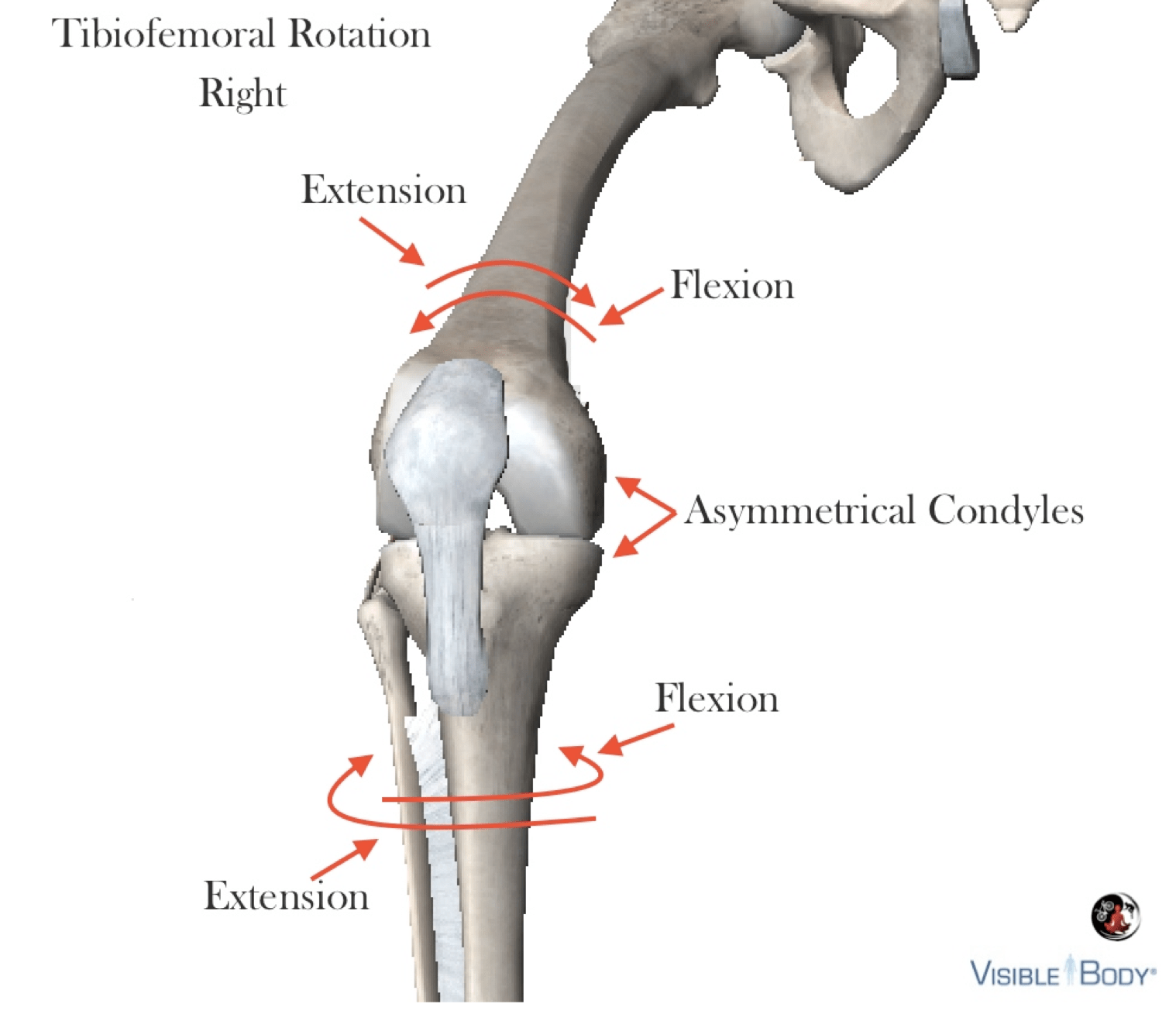
External lateral rotation of Leg / Tibia at Knee / Tibiofemoral Joint
Biceps Femoris
Sciatic Nerve
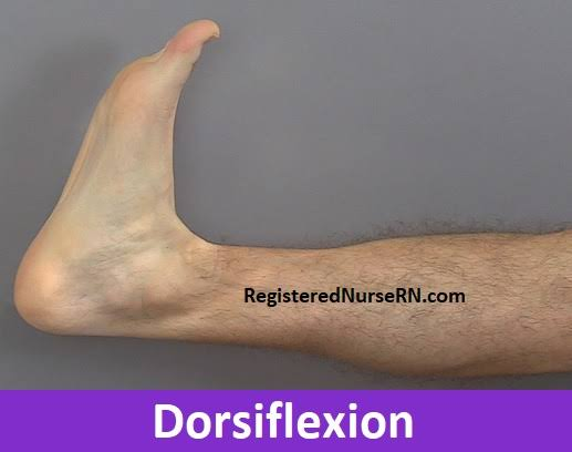
Movement of Ankle / Foot
(Talocurual Joint): Dorsiflexion
Tibialis Anteiror
Syntergists (WEAKER CONTRIBUTION):
Tibialis Posterior, Flexofr Digitorum Longus, Flexor Hallcius Longus
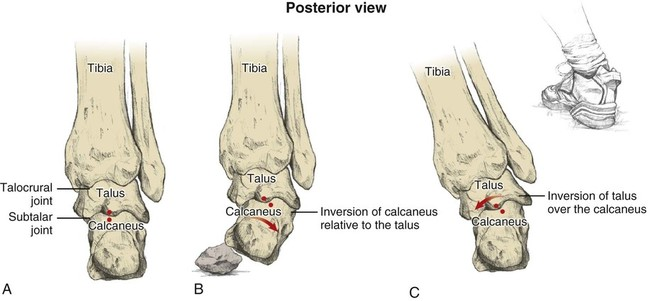
Inversion
(Subtaar Joint)
WHAT MUSCLES?
Tibialis Anterior (When ankle is in dorsiflexion) - Deep peroneal Nere
Tibialis Posterior (Ankle in Plantarflexion) - Tibial Nerve
Eversion Subtalar Joint
What muscles
Peroneus Longus
Peroneous Brevis
Superficial Peroneal Nerve
How many intrincsic muscles?
19 muscles located entirely within the foot
17 arranged into 4 plantar layers - superficial to deep
FUNCTIONS:
Flexion, ext, abd, add of toes - (digits of foot)
Maintain foot aignment, biomechaniachs proprioception / bal
stabilze / maintain tendons of extrinsic muscles thro direct atchment / bleding w them
support arches of foot - working with extrinsic muscles such as tinailis posterior fibularis, flexor hallucis longus