Anatomy of Speech & Hearing
 Call Kai
 Learn
Learn Practice Test
 Spaced Repetition
 Match
Match Flashcards
 Knowt Play
1/70

Earn XP
Description and Tags
Anatomy of Speech & Hearing Exam 1
Name | Mastery | Learn | Test | Matching | Spaced | Call with Kai | Chat |
|---|
No analytics yet
Send a link to your students to track their progress
71 Terms
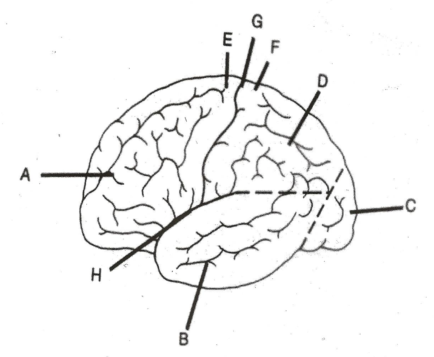
label A
frontal lobe

label B
temporal lobe

label C
occipital lobe

label D
parietal lobe

label E
pre-central gyrus

label F
post-central gyrus
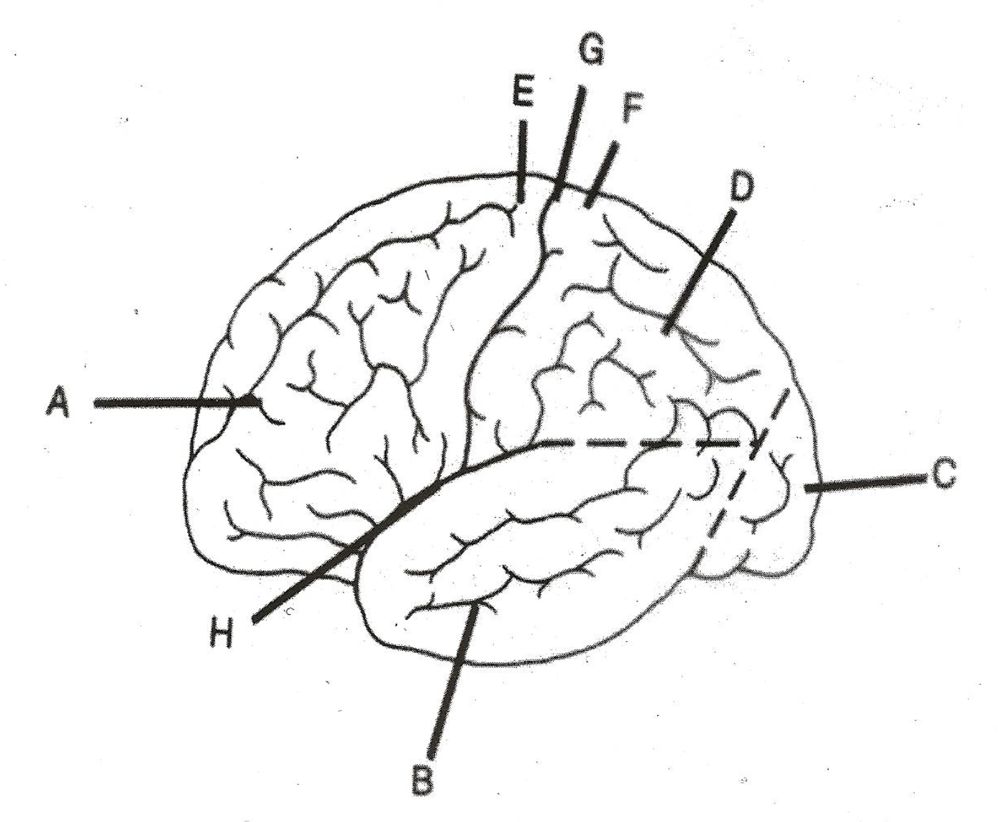
label G
central sulcus/central sulcus of Rolando/Rolandic fissure/fissure of Rolando

label H
deep sulcus/sulcus of Sylvian/lateral sulcus/Sylvian fissure
Brodmann’s area 44 & 45
Broca’s area
Brodmann’s area 22
Wernicke’s area
Brodmann’s area 6
pre-motor cortex
Brodmann’s area 40
supramarginal gyri
Brodmann’s area 39
angular gyrus
Brodmann’s area 41 & 42
primary auditory context
Brodmann’s area 4
pre-central gyrus
Brodmann’s area 1, 2, & 3
post-central gyrus
projection pathways function
communication between lower & higher regions in cerebrum
associational pathways function
communication within the same hemisphere
commissural pathways function
communication between hemispheres
dura mater
superficial layer of the meninges - tough mother
arachnoid mater
middle layer of the meninges - spider-like web
pia mater
deepest layer of the meninges - soft mother
diancephalon
contains the thalamus, epithalamus, hypothalamus, subthalamus
thalamus function
sensory processing relay station - every sensation passes through (excluding olfactory) before going to the somatosensory cortex
hypothalamus function
body temperature, hunger, thrist, mood, sex drive, blood pressure, sleep
epithalamus
pineal body, gonad development, olfactory reflex
control circuits (basal ganglia & cerebellum) function
coordinate, integrate, & help control the diverse activities of the many sturcture pathways involved in motor performance (integrate activities of direct and indirect pathways)
basal ganglia
contains caudate nucleus & lenitform nucleus (made of globus pallidus & putamen)
basal ganglia function
helps maintain a stable musculoskeletal environment & modulates excessive commands from cortex
damage to basal ganglia
results in hypokinesia or hyperkinesia dysarthria- greater impact on speech than lesions of the cortex
hypokinesia
reduced mobility/rigidity as a result of a loss of dopamine, i.e., Parkinsons
hyperkinesia
involintary movements as a result of excessive dopamine, i.e., tourrettes
hindbrain
contains cerebellum, pons & medulla oblongata
cerebellar control circuit
responsible for coodination of trunk zone, modulates equilibrium - dentate nuclei (in cerebellar hemispheres) are important for speech control, active in initiating movement, executing pre-planned motor tasks, and regulating posture
damage to cerebellum
ataxia (various forms of incoordination, i.e., gait disturbances, intention tremor, hypotonia), dysarthria (area impacted when intoxicated, think of someone insanely drunk)
intention tremor
occurs when trying to complete a movement
terminal tremor
tremor’s amplitude increases as the movement gets closer to completion
rest tremor
tremor/involuntary movement occurs at rest
pedoncles
how the pons connects to cerebrum (inferior, middle, superior)
sensory info travels on the ___ side
dorsal
motor commands travel on the ___ side
ventral
fasciculus gunneatis
relays sensory info from the waist up - contralateral processing
fasciculus gracilis
relays sensory info from below the waist - contralateral processing
decussation of pyramids
majority of motor commands cross over to the opposite side of the body to the target muscles to complete the movement
direct activation pathway/pyramidal system/motor system
contains corticobulbar tracts & corticospinal tracts - fiber tracts originate in motor cortex and terminate in cranial nerve motor nuclei in brainstem (corticobulbar) or originate in motor cortex and terminate in spinal nerve nuclei in spinal cord (corticospinal)
indirect activation pathway/extrapyramidal system/indirect motor system
“milk run,” originates in motor cortex and synapse with other structures (basal ganglia) before terminating in C.R. N and SP. N. motor nuclei in brainstem and spinal cord
upper motor neuron system
contains direct motor system and indirect motor system
final common pathway/lower motor neuron pathway
cranial nerves and spinal nerves that exit the brainstem and spinal cord- the peripheral mechanism through which all motor activity is mediated; the final link to speech muscles - carries out upper motor neuron commands for voluntary movements
damage to lower motor neuron pathway
prevents normal activation of muscle fibers - denervation (partial=paresis, complete=paralysis); voluntary & reflexive motor activities equally; can be bilateral or unilateral - atrophy resulting in hypotonia - spontaneous motor activity on the surface of the skin (fasciculations)
flaccid dysarthria
injury or malfunction of one or more of the cranial or spinal nerves - lesion is always between the brainstem or spinal cord and the muscle - results in hypotonia, diminished reflexes, atrophy, weakness (paralysis or paresis), fasciculations
damage to direct activation pathway
a loss or reduction in its ability to facilitate fine, discrete movements - reduced tone and weakness - positive Babinski sign - pathological oral reflexes (suck, snout)
damage to indirect activation pathway
usually occurs with damage to DAP - loss of inhibitory role in motor control, positive signs (overactivity) - increased muscle tone & spasticity - hyperactive reflexes
spastic dysarthia
bilateral damage to direct and indirect activation pathways - speech characteristics reflect a combo of weakness and spasticity (slow movement and reduced range and force)
cranial nerves that recieve bilateral input
C.R. N. V, VII, IX, X, XI
cranial nerves that recieve unilateral input
C.R. N. VII, XII
cranial nerve V - trigeminal
superficial facial sensory - for pressure, pain, temperature in oral cavity, not taste - dentist infuses numbing medication into this nerve
cranial nerve VII - facial
muscles for facial expression - taste for anterior 2/3 of tongue - controls salivary glands
cranial nerve IX - glossopharyngeal
sensation & taste for posterior 1/3 of tongue & entryway to pharynx - mediates gag reflex (sensory) - senses food/liquid & helps to trigger the pharyngeal swallow response - stylopharhyngeal muscle, elevates larynx during swallowing
cranial nerve X - vagus
closes velum during speech to prevent unwanted nasal sounds - pitch elevation - add/abduction of vocal folds (weak side is immobile) - sensory below larynx - prevents choking
cranial nerve XI - accessory
laryngeal & pharyngeal muscles w/ vagus nerve, innervates in stenocleidomastoid muscle & trapezius muscle
cranial nerve XII - hypoglossal
all intrinsic and extrinsic muscles of the tongue - not responsible for sensory
damage to cranial nerve VII - facial
facial drooping on the weak side - when retracting (smile), the movement will deviate to the strong side - when protruding (pucker), the movement will deviate to the weak side
damage to cranial nerve IX - glossopharyngeal
trouble swallowing
damage to cranial nerve X - vagus
damage can occur on any of the three branches or where it runs on the brainstem, superior damage impacts the inferior branches functions
pharyngeal branch of vagus nerve
soft palate & velum - responsible for closure during speech to prevent unwanted nasal sounds
superior laryngeal branch of vagus nerve
divides into external motor nerve and internal sensory nerve
recurrent laryngeal branch of vagus nerve
add/abduction of vocal folds - sensory below the larynx - prevents aspiration
external motor branch of vagus nerve
motor innervation to the cricothyroid muscle of the larynx (stretches and lengthens the vocal cords to control high-pitched vocalization)
internal sensory branch of vagus nerve
provides sensation to the larynx above the vocal folds - prevents choking
damage to cranial nerve XI - accessory
weakens head rotation (opposite side of weakness), reduce ability to shrug shoulders on side of lesion
damage to cranial nerve XII - hypoglossal
extraoralputrusion (sticking out your tongue) will deviate to the weak side - movements should be symmetrical