L2: Neurophysiology I
 Call Kai
 Learn
Learn Practice Test
 Spaced Repetition
 Match
Match Flashcards
 Knowt Play
1/45

There's no tags or description
Looks like no tags are added yet.
Name | Mastery | Learn | Test | Matching | Spaced | Call with Kai | Chat |
|---|
No analytics yet
Send a link to your students to track their progress
46 Terms
Nervous System Organization Overview

Nervous tissue is composed of two types of cells:
Neurons
Glial Cell
Neurons
they are “the” nerve cells able to transmit information
Neuron composition
Axon = info moves away from cell body
Dendrite = info moves towards cell body
Cell body = integrates in- and outgoing information
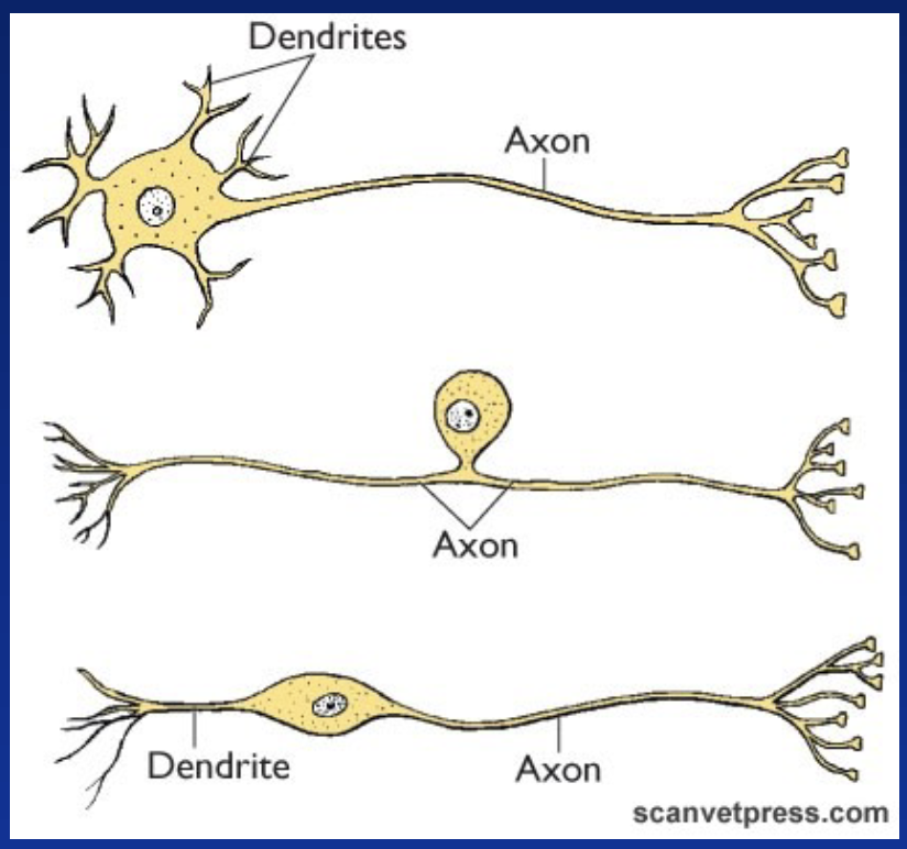
How can neurons be categorized?
Number of processes:
Multipolar, mainly in CNS (top)
Pseudounipolar, mainly in PNS (middle)
Bipolar, mainly sensory organs (bottom)
Function:
Sensory (afferent): from PNS to CNS
Motor (efferent): from CNS to muscles and glands
Interneurons (association): relay info between neurons within the CNS
Specialized “receptors”: transducers = convert stimuli to signal

Glial cells
non-neuronal cells; 10X more abundant than neurons (oligodendrocytes, astrocytes, ependymal cells, microglia)
Glial cells functions:
Provide structural support to nervous tissue
Participate in myelin formation (oligodendrocytes)
Secrete glutamate: can modulate excitatory level of neurons (astrocytes)
Some possess phagocytic activity (microglia)
Contact both blood vessels and neurons = transport of nutrients → neurons do not store glucose or Oxygen
White/Grey Matter
Grey matter corresponds to cell bodies; integration centres
White matter corresponds to bundles of neuron processes with the white appearance due to myelin sheaths; predominantly axons
Nerves
bundles of axons; run from or to the CNS
Cell bodies location
Cell bodies of sensory neurons are located in clusters named ganglia (outside of the CNS)
Cell bodies of motor nerves are located in well-defined area of the CNS (brain and spinal cord)
Myelin characteristics
white lipid (sphingomyelin) around nerve fibres, specifically axons
only in white matter (not all fibres)
electrical insulations
transmission of AP faster in myelinated fibres
How are myelin formed?
Glial cells wraps around an axon, and much of their cytoplasm is lost, leaving layers of lipid membrane around the axon
What are the interruptions between myelin sheaths
Nodes of ranvier
every 1-2mm
denuded axon points will allow depolarisation = transmission of action potential (AP)

How does Resting Membrane Potential (RMP) occur
RMP results from a difference in charge across the cell membrane (between cytosol and extracellular fluid)
Inside of membrane is negative RELATIVE to outside
every cell of the body possesses a membrane potential
What does absolute value of RMP depend on
differs between cell type and depends on the amount of charges, ion channels and the thickness of the membrane
Average RMP in a nerve cell
–70 to –90 mV
Charges of intra- and extracellular compartments
Are electroneutral
In the cytosol, negative charges carried by large organic molecules are attracted to the membrane by positive charges on the outside.
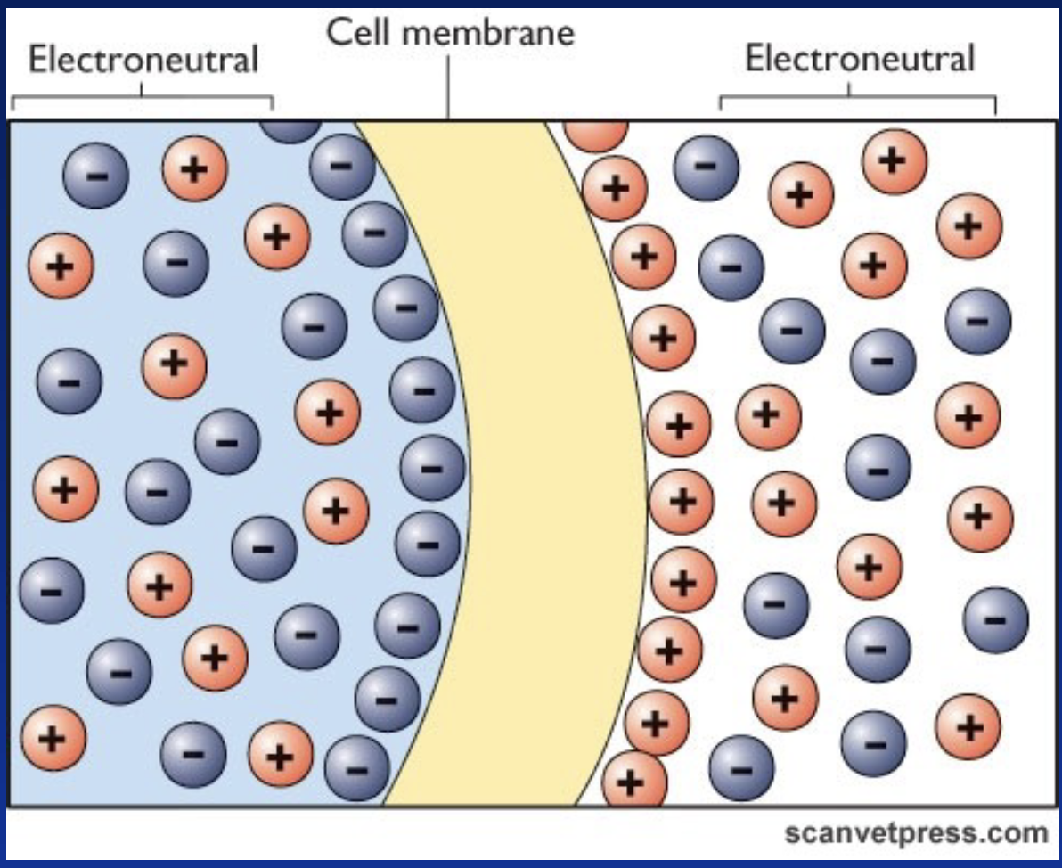
What maintains the RMP?
Combination of:
Selective permeability (passive based on diffusion)
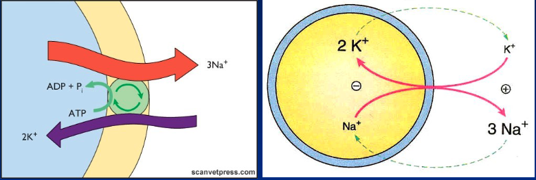
Na+/K+ pump (3 Na+ out 2 K+ in)
Large anions trapped on the inner surface of membrane
Selective permeability (diffusion) and examples
Passive leakage of ions through channels (concentration gradient)
Resting membrane permeable to K+, barely permeable to Na+, Ca2+ and Cl- → positive charges accumulate outside

Ion pumps
Concentration of ions remains relatively constant inside the cell → needs to compensate for diffusion leakage
Na+/K+ pump: pumps 3 Na+ out and brings 2 K+ in
Goes against concentration gradients and for Na+, against the membrane polarity (outside already positive relative to inside)
Requires a lot of energy up to 40% of ATP availability
Excitable cells
Cells that can generate electrical impulses (Action Potentials)
How are excitable cells stimulated
Chemical, electrical or physical stimulations induce a change in membrane potential to reach a THRESHOLD provoking the opening of voltage gated ion channels
How does depolarization occur
If Na+ channels open (or Ca2+ in certain nerve endings and smooth and cardiac muscle cells), Na+ rushes inside the cell (gradient concentration) → potential less negative, then inverted (positive)
How does repolarization occur
Subsequent opening of K +channels results in an outflow of K+ returning the potential to RMP
Generation of Action Potentials in Neurons steps
Depolarization= an initial depolarization (stimulation) needs to reach threshold to provoke the opening of Na+ voltage gated channels
After about 0.5 ms, opened Na+ channels close rapidly
Repolarization= K+ voltage gated channels then open (delayed compared to Na+ channels) → outflow of K+
Hyperpolarization= K+ voltage gated channels then progressively close, outflow of K+ continues after reaching the RMP
Once all gated channels are closed, ions rejoin their respective compartments by diffusion and Na+/K+ pumps
Refractory period= Neurons cannot be re-stimulated until RMP is restored

Types of gated channels
Voltage-gated channels
Ligand-gated channels: binding sites for neurotransmitters
Each channel is composed of several subunits, and has various degrees of specificity

All-or-none rule
Nerve cells follow the all-or-none rule
When threshold is met, an AP is generated
The amplitude of the AP is fixed for that cell
Intensity is based on frequency of APs, not the amplitude

Conduction of action potential in unmyelinated axons
Depolarization and repolarization processes (AP) propagate along the cell membrane
Need for the change in potential to reach threshold on the nearby microdomain to trigger opening of gated channels

Conduction of action potential in myelinated axons
AP occurs the same way but only at the nodes of Ranvier
Myelin prevents ion leakage, current jumps from one node to the other = SALTATORY conduction
Velocity increased as less membrane affected = less energy required to transport ions

Nerve velocity depends on:
Dissipation of current which is dependent on:
thickness of myelin
diameter of the fire (thicker=faster)
range from 100 to 0.5 m/sec and from 2500 to 250 impulses/sec
Synpatic transmission
Continuity of signal between a neuron and other neurons or between a neuron and target cells such as skeletal muscles (neuromuscular synapse)
Signal sent by neurotransmitter, and neuron is triggered to reach action potential
Electric insulator
cell membrane made of phospholipids
Synaptic gap/cleft and its characteristics
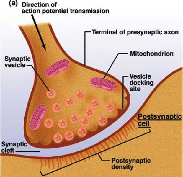
a gap that exists between pre- and post- synpatic cell membranes
rarely, direct continuity in electric impulse = gap junction (cardia and some smooth muscles)
in vertebrates, neuronal synapses = predominantly CHEMICAL synapses
Neurotransmitters
Molecules able to transmit information from a neuron and convert the electrical signal (AP) into a chemical signal
How does a neurotransmitter work
Released by pre-synaptic neuron into the gap
Bind to specific receptors on post-synaptic membrane
Elicit a response
How are neurotransmitters classified and types
classified based on molecular size and composition
Small molecules:
synthesized in the nerve terminals by specific enzymes
amino acids derivatives: biogenic amines
Neuropeptides (3-40 AA):
synthesized in the cell body
packaged in secretory vesicles
transported to the site of release
Steps of action in neuromuscular synapse
Action potential (AP)
At the end of the neuron, AP opens voltage gated Ca2+ channels = in-flux of Ca2+
Ca2+ triggers exocytosis
Diffusion in the cleft
Binding (ex. ACh) to specific receptors and Na+ gated channel opens
Depolarization = Ion channels open on post-synaptic membrane and reaches threshold
Neurotransmitter inactivated termination of signal

Postsynaptic folding purpose
is common in neuromuscular synapse (not in interneurons) which increases surface
Acetylcholine (ACh)
neuromuscular synapse transmitter
Termination of transmission for small molecules
Picked back up by presynaptic neuron via endocytosis and recycled for next time
Deactivated in the cleft by enzymes released by post- synaptic cell (ie Acetycholine Esterase)
Termination of transmission for neuropeptides
After binding to its receptor, can be internalized by post-synaptic cell via endocytosis and be degraded by cellular enzymes
Broken down by extracellular peptidase in the gap
*Receptor can be desensitized
Integration of Multiple Synapses Between Neurons: neuro-muscular synpase
1 neuron = AP = muscle cell depolarization
Integration of Multiple Synapses Between Neurons: neuron-neuron synpase
1 neuron can receive impulse from multiple other neurons
Synapses can be either excitatory or inhibitory
1 impulse does not always lead to a response (need to reach threshold
Excitatory synapse =
depolarization = entry of Na+
Inhibitory synapse =
hyperpolarization = entry of Cl- and/or outflow of K+
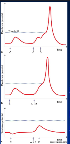
Explain these 3 graphs
A & B = excitatory neurons; C = inhibitory neurons
a) Only A activated: Excitatory postsynaptic potential (EPS) not sufficient to reach threshold as 3 impulses needed
b) A and B activated: A and B for EPS reaches threshold as meets AP
c) A,B, and C activated: C able to decrease EPS and block excitatory action of A+B
