Week 1-rads (abdominal and thoracic)
 Call Kai
 Learn
Learn Practice Test
 Spaced Repetition
 Match
Match Flashcards
 Knowt Play
1/183

There's no tags or description
Looks like no tags are added yet.
Name | Mastery | Learn | Test | Matching | Spaced | Call with Kai | Chat |
|---|
No analytics yet
Send a link to your students to track their progress
184 Terms
Pylorus is on the right side of the abdomen, so the dog should be placed in left lateral view
What side of the dog abdomen can the pylorus be found and what lateral position should the dog patient be placed?
The fundus is located on the left side of the abdomen, so the patient should be placed in right lateral view.
What side of the abdomen is the fundus and what position should the dog patient be placed to accurately view this structure?
Pancreas, adrenals, lymph nodes, ureters, and ovaries/uterus
What organs of the abdominal cavity are not normally seen on a radiograph?
Number, size, shape, position, and opacity
What are the Roentgen signs in radiology?
Release tensions in legs
How can one get rid of skin folds on a patient during abdominal radiographs?
Spondylosis deformans
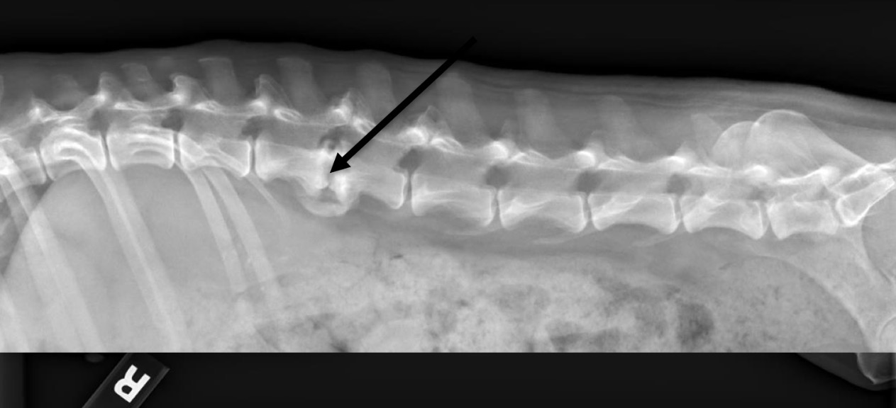
What is this structure?
Diaphragmatic hernia
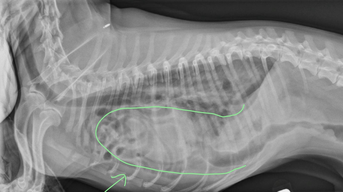
What is occurring in this patient?
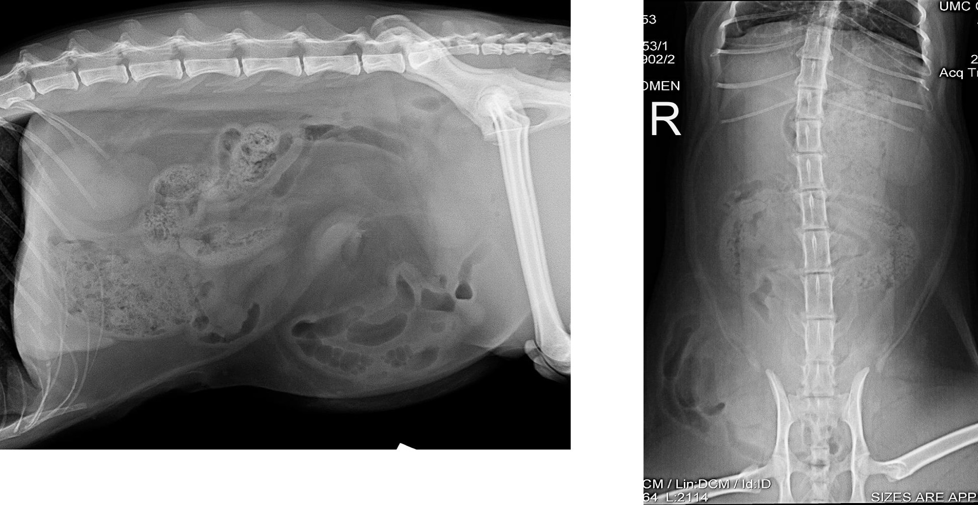
Right Inguinal hernia
What is occurring in this patient?
Serosal definition
What term refers to the ability to discern abdominal organs due to contrast with surrounding peritoneal fat?
If the patient is emaciated (loss of fat) or neonate (Brown fat)
Why would there be reduced/lost serosal detail in an x-ray in relation to body condition?
Free fluid, peritonitis (steatitis), carcinomas, mass effect, post op changes
What are some pathological reasons as to why an abdominal radiograph would have reduced serosal detail?
Up to 6 months
How long will neonates and juveniles have brown fat?
Peritoneal effusion
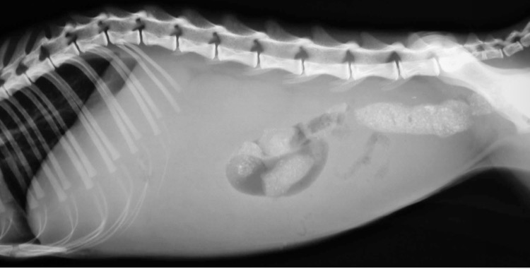
What is occurring in this patients abdomen?
Steatitis
What condition is common in cats due to vit E deficiency, chronic pancreatitis, or high fish diet and is defined as inflammation of fat tissue?
FIP
What disease in cats can often lead to peritoneal effusion?
Pneumoperitoneum
What term refers to increased contrast of serosal definition in abdominal radiographs?
Secondary to surgery, penetrating trauma, rupture of hollow viscera, and necrotic neoplasia/abscesses
What are some causes to pneumoperitoneum?
Highlighted serosal surfaces, geometric-shaped gas opacities
On a radiograph of a patient with pneumoperitoneum, what are some common findings one can see?
Horizontal beam lateral and VD
What x-ray position makes viewing pneumoperitoneum easier?
Pneumoperitoneum
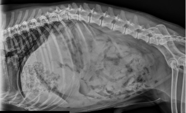
What is occurring in this patient?
Dystrophic mineralization/tissue death, metabolic disorders (Cushing), inflammation, neoplasia, mesenteric cyst/Bates body
What are some causes of intrabdominal mineralization?
Mesenteric cysts/Bates body
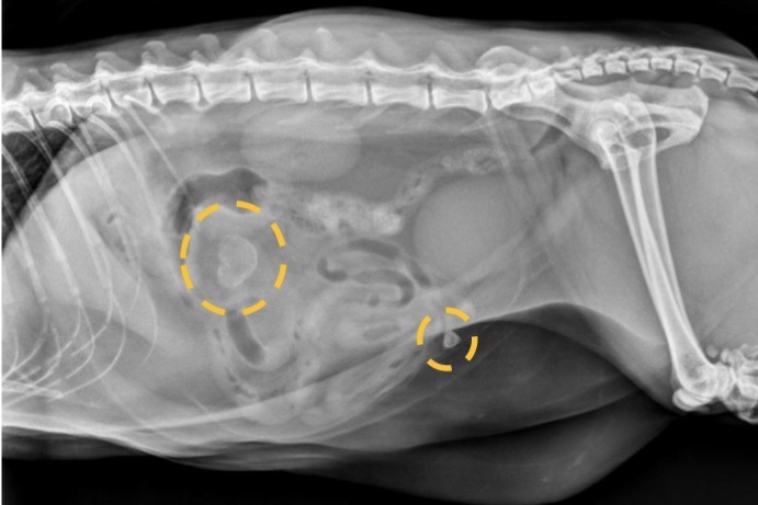
What are these incidental findings often observed in cats?
The retroperitoneum
What is an extraperitoneal space situated ventral to the vertebrae and paraspinal muscles that extends along the length of the entire abdomen from the diaphragm to the anus?
Kidneys, ureters, adrenals, aorta, caudal vena cava, celiac and cranial mesenteric arteries, renal and deep circumflex vessels
What structures are found within the retroperitoneal space?
Retroperitoneal effusion
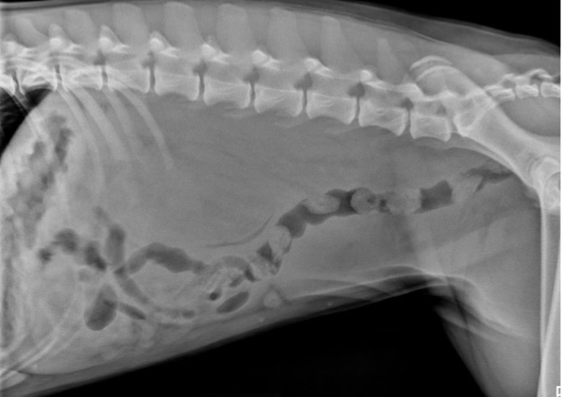
What is occurring in this patient?
Bone tumor at L4
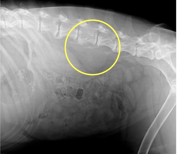
What is happening at this location that is causing the observed retroperitoneal effusion?
Pneumoretroperitoneum
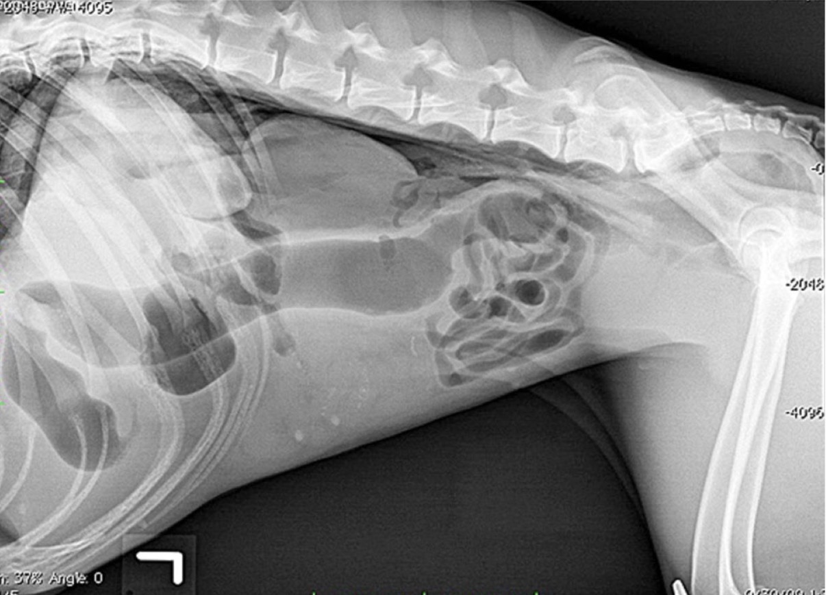
What is occurring in this patient that is causing the enhanced vision of the kidneys?
Falciform fat
In obese cats, what can displace the liver?
Rounding or blunting of the caudoventral liver margin, extension beyond the costal arch, and caudal displacement of the gastric axis
On a lateral view of a patient with generalized hepatomegaly, what are some common findings?
Perpendicular to spine
In a normal view of the stomach and the liver in a lateral view of the abdomen, how should the gastric axis be situated?
Generalized hepatomegaly
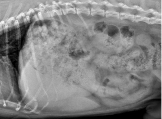
What is occurring in this patient?
Right sided heart failure
What is a cardiovascular cause to hepatomegaly?
Hyperadrenocorticism, hypothyroidism, and diabetes mellitus
What are some endocrine related causes to hepatomegaly?
Possible micro hepatica
What can be said if the gastric axis is displaced cranially?
Normal variant, porto-systemic shunt, microvascular dysplasia, and cirrhosis (older patients)
What are some causes to micro hepatica?
Gallbladder mineralization
What is a common finding amongst cats in their cranial ventral aspect of the liver?
Right cranial
Where is the pancreas found in the abdomen?
Pancreatitis
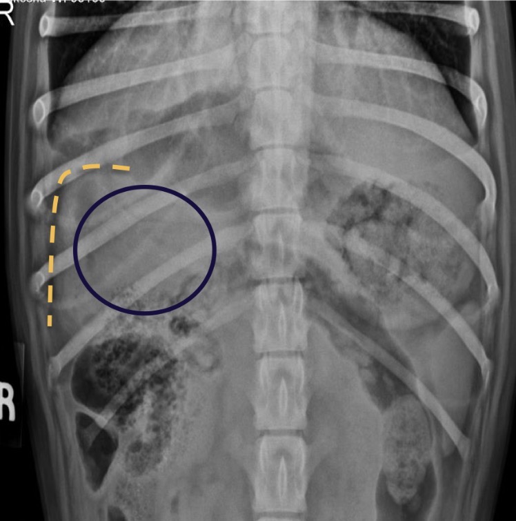
What is occurring in this patient in the highlighted area?
Tail of spleen on the right side
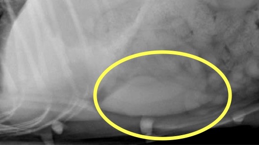
What specific part of this organ is circled and what side of the body is it found in canines?
No (if seen usually splenomegaly)
Is the spleen usually seen in cat abdominal radiographs?
Often associated with GDV in large breed dogs due to stricture
Splenomegaly is often associated with what? and because of why?
C-shaped soft tissue opacity in central abdomen
The spleen of a dog with splenomegaly will often take what shape?
Splenomegaly
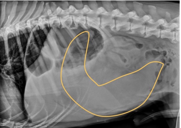
What is occurring in this patient?
Head of spleen- rightward
small int and desc colon- caudally
In the case of splenomegaly, where will the head of the spleen, small intestine and descending colon be displaced to?
Splenic mass
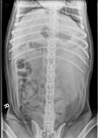
What is occurring in this patient?
Clockwise
Which way is the torsion often oriented in GDV?
dorsal and cranial
Which way does the pylorus move in GDV?
Ventral and right
Where does the fundus and greater curvature of the stomach shift in GDV?
Pylorus
In a right lateral view of a patient with GDV, which compartment of the stomach will fill with air?
Gastric dilation without volvulus
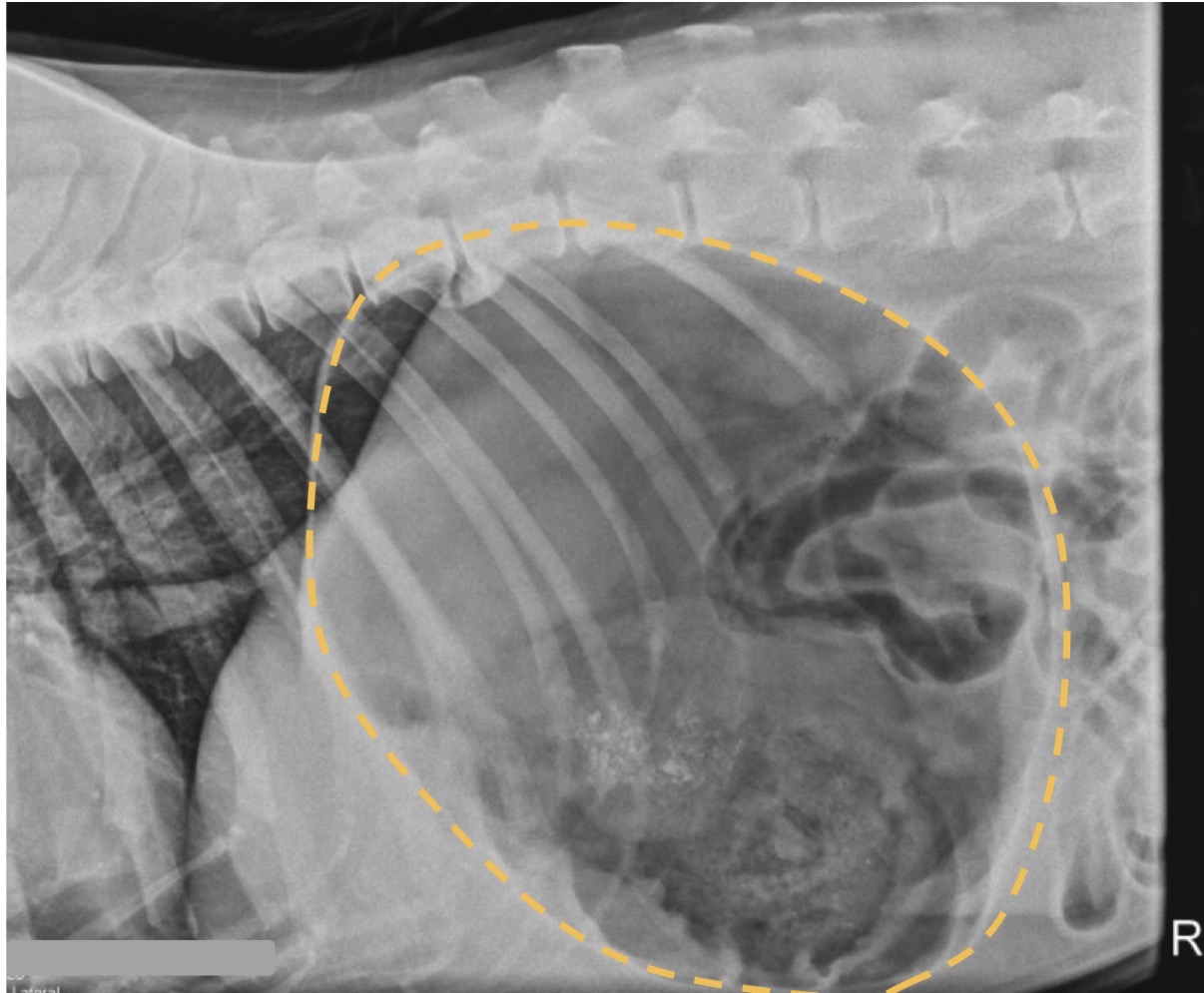
What is this patient experiencing?
GDV
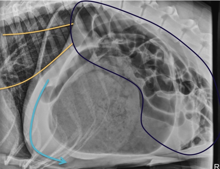
What is the patient experiencing in this x-ray?
Pyloric outflow obstruction
What is a common cause to chronic vomiting in dogs?
Pylorospasm
What is a functional cause to pyloric outflow obstruction?
Mass, fibrosis, inflammation, hypertrophic pyloric stenosis, chronic hypertrophic pyloric gastropathy
What are mechanical causes to pyloric outflow obstruction?
Gastric dilation and “gravel”signs
What are some radiographic findings for pyloric outflow obstruction?
Pyloric outflow obstruction
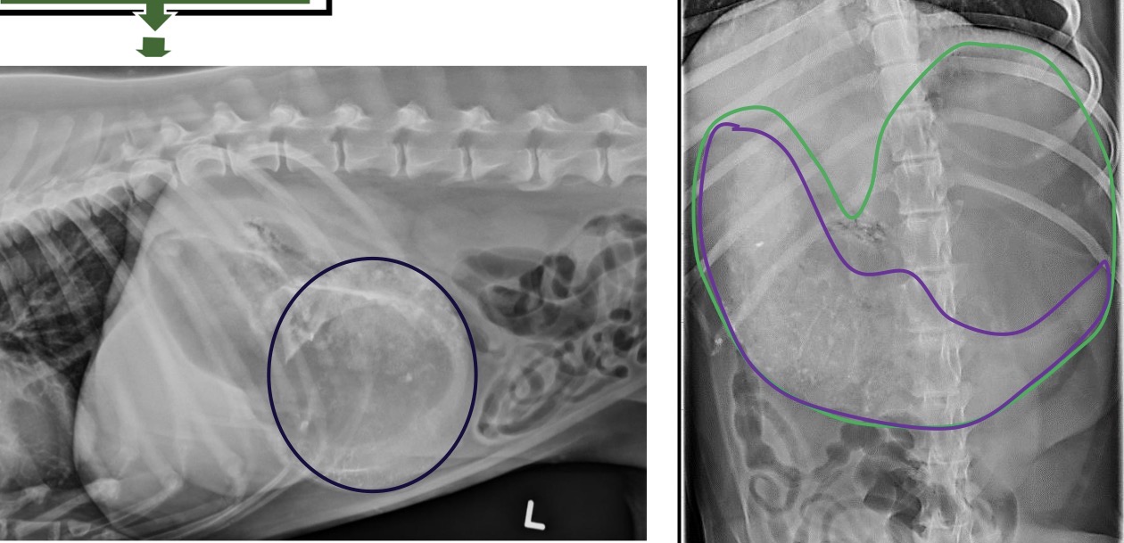
What is occurring in this patient that is evident in these radiographs?
Contrast or U/S
What can one do when suspecting a FB, but can’t see it on radiographs?
Gastric wall thickening, thickening or inc number of rural folds, and may see mineralization with uremic gastritis
What are some radiographic findings that suggest gastritis?
Gastritis
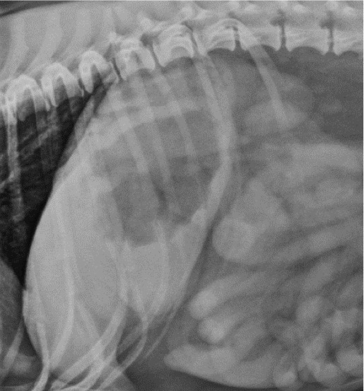
What is evident in the stomach of this dog?
Gastric thickening most likely due to neoplasia
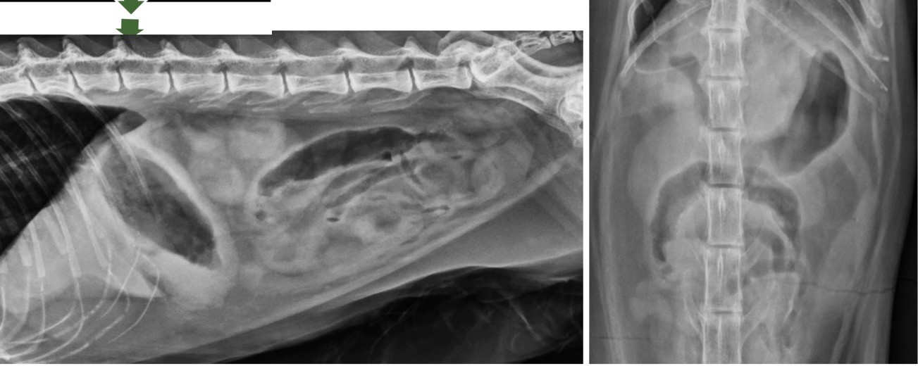
What is evident in these x-rays?
PPDH (peritonealpericardial diaphragmatic hernia)
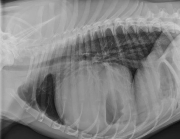
What is evident in this radiograph?
Incidental in cats and serious in dogs
In which species can PPDH be a incidental finding and in which species is it more serious?
Hernia and rupture of the diaphragm
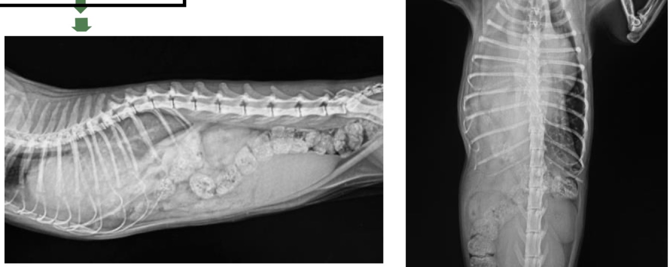
What is occurring in this patient that is evident in these radiographs?
Perianal hernia
X-rays were taken of a 13 year old mix breed dog. Right off the bat shit looks fu*kd. Most of the small intestines are coming out the but hole?? and oh what is that?!?! the bladder. Now you tell me what is most likely occurring in this patient?
Diarrhea
What is the most common clinical sign of intestinal ds without ileus/enteritis?
Corrugation and bunching of small int, possible gravel signs
What is a radiographic finding of the intestines that infers severe enteritis?
Bacterial overgrowth will produce endotoxins which will generate gas
What is a toxic cause to ileus?
Hypokalemia
What metabolic imbalance can cause ileus?
Dysautonomia causes dynamic ileus
What is a neurological functional cause to ileus?
Generalized ileus
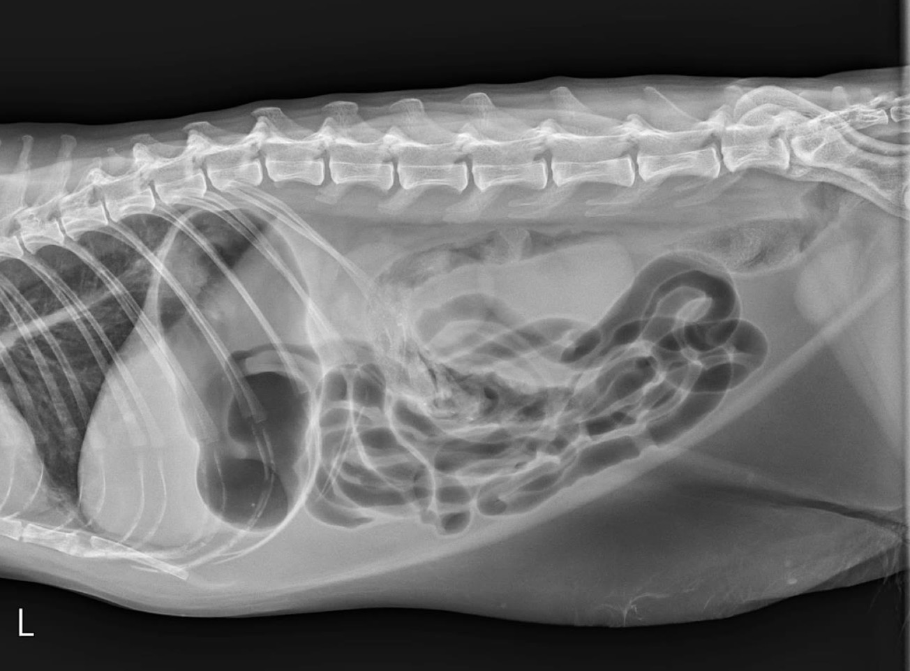
What is occurring in this patient?
Vomiting and severe fluid and electrolyte loss
What clinical signs are often associated with complete proximal small intestinal obstruction?
anorexia, and intermittent vomiting
What clinical signs are associated with a distal small intestinal obstruction?
Early stages the intestinal dilation will be gaseous and eventually will fill with fluid
How will the radiographic findings change in mechanical ileus over time?
Stacking SI loops giving a hairpin appearance, loss of serosal definition, and gravel sign
What three radiographic findings suggest mechanical ileus is occurring in a patient?
Cranially- dilation of SI in more cranial areas. Normal dilation in SI caudal to FB
Caudally- Dilation of entire SI
What dilation patterns can be seen in SI cranial foreign body and a caudal FB?
Most likely a FB causing two populations bowel. Some SI is gaseous, while the more ventral SI is normal
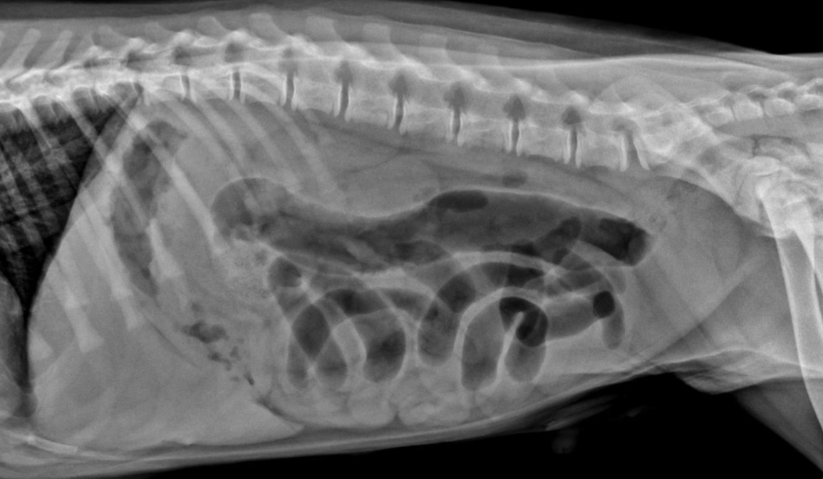
What can this radiograph suggest is occurring in this patient?
Linear foreign body
What important surgical condition is most common in cats and causes intestinal ds without ileus?
In the pylorus
Where will a linear foreign body usually lodge itself in canine patients?
Bunched and plicated
How will the SI appear in a feline patient with a linear foreign body?
Teardrop, crescent shape, triangle or irregular
What shape of gas bubbles are common in patients with intestinal ds without ileus (linear foreign body)?
Linear foreign body
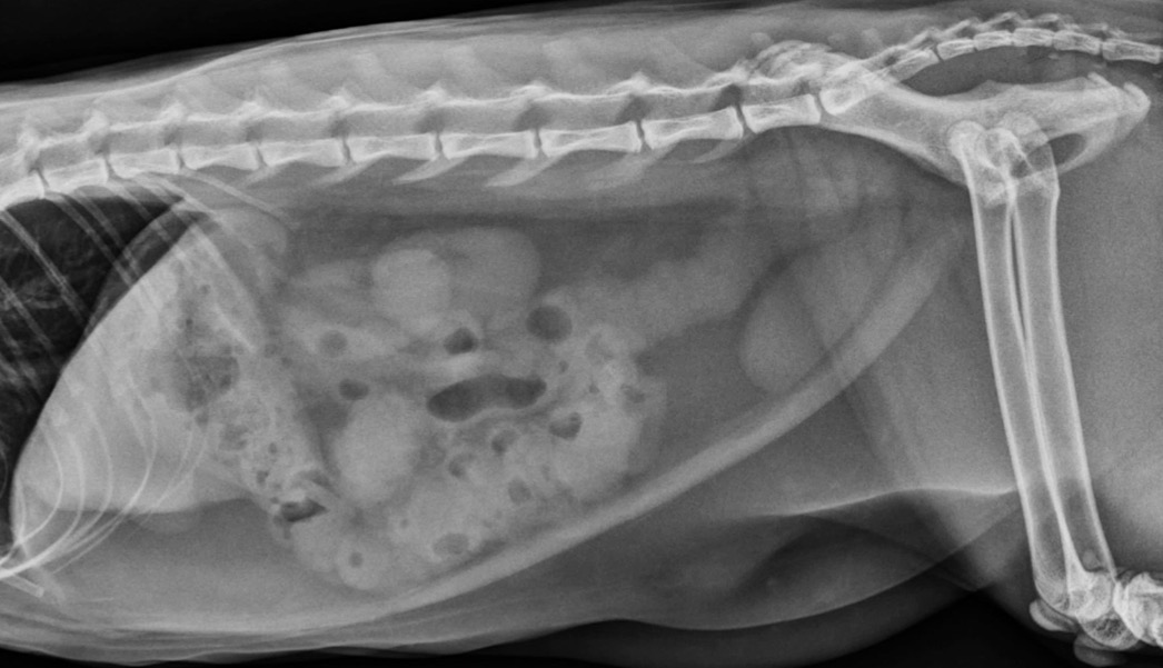
What is occurring in this radiograph?
Mesenteric torsion
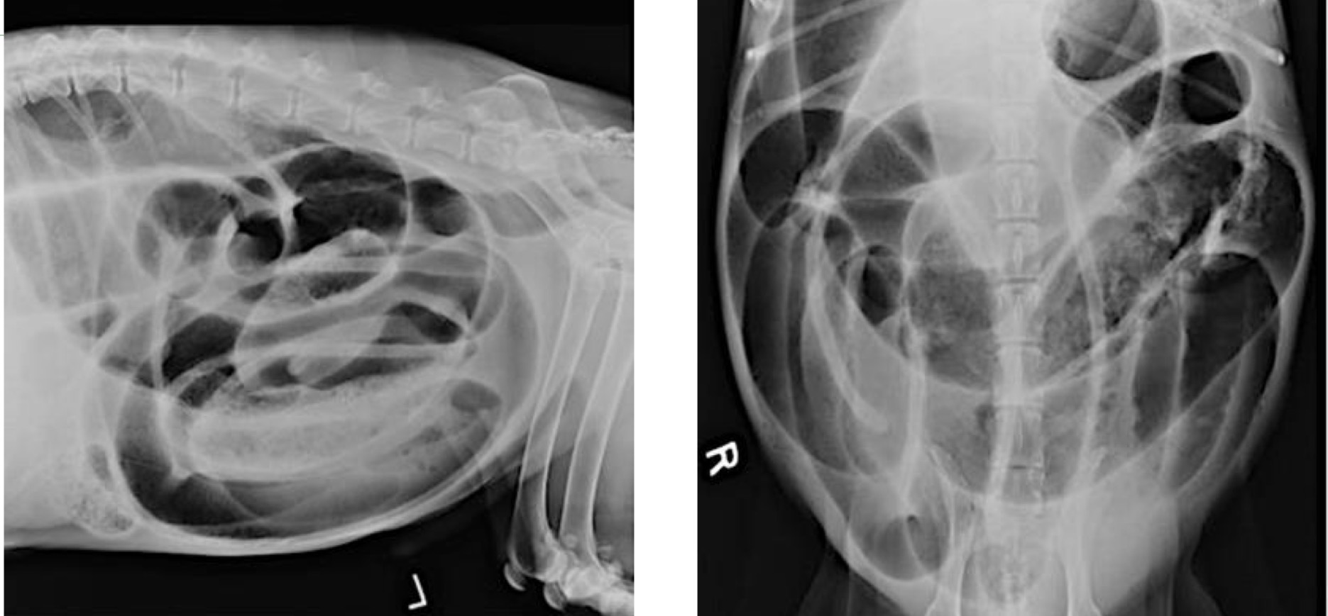
What is occurring in this patient?
Intussusception
What term refers to telescoping or inversion of the bowl that can cause mechanical obstruction?
Peristalsis pathology, intestinal parasites, focal neoplastic lesions, linear foreign bodies and systemic disease
What are common primary or secondary conditions that can cause intussusception?
Intussusception
What is common in the ileocolic junction and is thought to be secondary to hook worm parasitism?
Constipation
Difficult, incomplete or infrequent evacuation of dry hardened feces (often in cats) is called what?
Obstipation
The inability to evacuate a mass of hard dry feces is termed what?
Megacolon
Hypomobility and dilation of the colon which results in constipation and obstipation is termed what?
cats
Which species experiences idiopathic megacolon?
Megacolon
Chronic renal disease and persistent subclinical dehydration can cause what in the colon?
Diameter of the colon to L5 vertebrae body (1.28-1.48 constipation, greater than 1.48 is mega)
How can one differentiate constipation from megacolon using radiographs?
Ileus of the colon
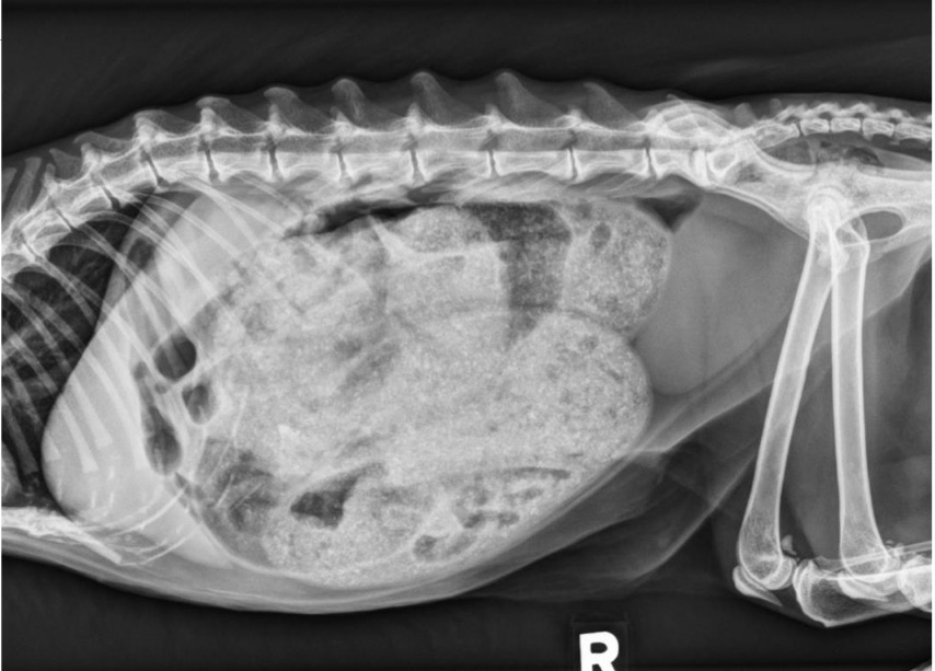
What is occurring in this patient?
Ileocolic junction
Where is the most common place of intussception in the colon?
Intussusception and cecal inversion
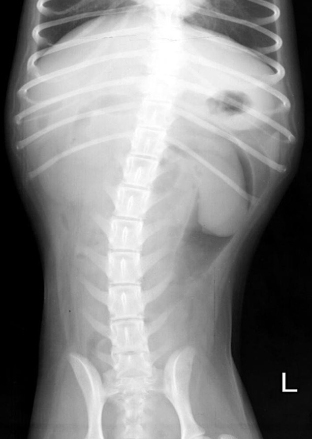
What is occurring in this patient?
Lymphosarcome, hydronephrosis, perinephric pseudocyst, and acute nephritis
What are some causes to renomegaly with smooth contour?
Bilateral splenomegaly due to lymphoma
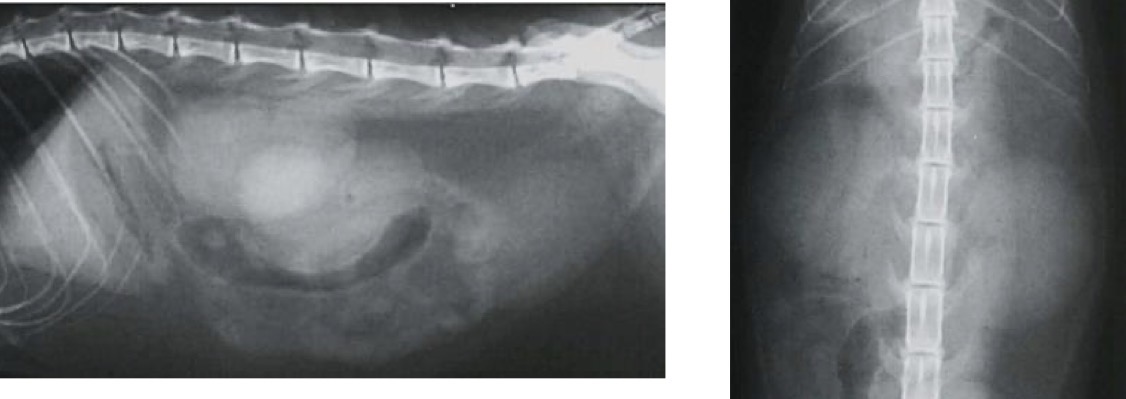
What is occurring in this patient?
Left renomegaly due to hydronephrosis
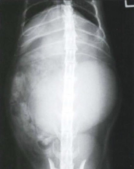
What is occurring in this patient?
Neoplasia, focal renal cyst, polycystic kidney dz, FIP, abscess/hematoma, renal cyst adenoma
What can cause renomegaly with irregular contour ?
Right renomegaly
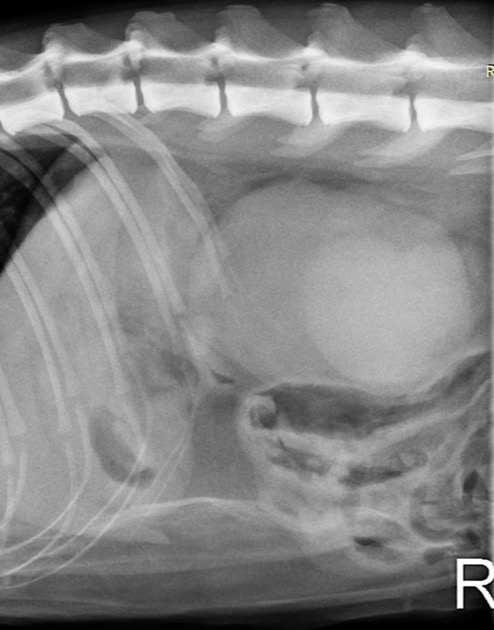
What is occurring in this patient?
Congenital renal disease, end-stage renal disease
What can cause a decrease in size of kidneys?