TopHat Objectives Anatomy Exam 2
 Call Kai
 Learn
 Practice Test
 Spaced Repetition
 Match
 Flashcards
 Knowt Play
1/46

There's no tags or description
Looks like no tags are added yet.
47 Terms

What side of the dog is this distal femur & tibia/fibula
Left leg

On the dorsal view of the tarsus, the arrow A is on the ______ bone, arrow B is on the _______ bone, and arrow 3 is on the _______ bone.
talus, calcaneus, 4th tarsal

The bone indicated by the X is the _______ tarsal bone
central
What is the common pelvic (proximal) attachment for the biceps femoris, semitendinosus, and semimembranosus
isciatic tuberosity
What is the prominent fusiform-shaped muscle used as a caudal landmark for the femoral triangle?
pectineus
The flexor surface of the tarsus is _____
dorsal
The ____, ____, and ______ muscles act as antagonists to the middle gluteal muscle action of extension at the hip joint
tensor fascia latae, iliopsoas, sartorius

The ___ is labeled X, and it articulates with a bony depression, the _____.
femoral head, acetabulum
The flexor surface of the tarsus is the _____, while the flexor surface of the digits of the hindlimb is the _____ surface
dorsal, plantar

The red arrow is labeling the bony structure called the _____ tuberosity
ischiatic
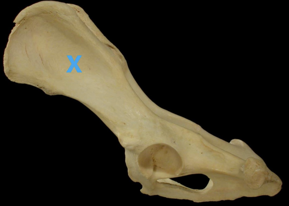
The label X is on what bone?
ilium
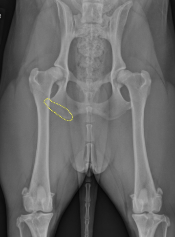
The outlined area is a portion of which bone?
ischium
What is the primary function of the cranial and caudal cruciate ligaments?
They prevent excessive cranial or caudal movement of the tibia
A luxation of the femoral head from the acetabulum is most often in the ______ direction. The pull of the _____ muscles aid in this displacement
craniodorsal, gluteal muscles

What is the bone that the yellow arrow is pointing to and where is it found
popliteal sesamoid bone, found within tendon of a muscle

This image is being viewed from the cranial surface. The yellow arrow indicates the _____ ligament. It is from the _____ side of the dog
cranial cruciate, left

On the caudal view of this equine pelvis, the structure indicated by the green arrow is the ______
tuber sacrale

The bone being viewed from the cranial surface is the…
right equine femur

On this equine tarsus, what is the bone indicated by the green arrow
fourth tarsal

Osteoarthritis commonly affects the _____ joint, which is indicated by the green arrow
distal intertarsal joint

Muscles that attach to the bony surface labeled X have what action at the coxofemoral joint?
extension

Identify bone X
Lateral splint bone, Metatarsal 4

On the bovine, identify the bone indicated by the pink arrow
fused central and fourth tarsal

On the osseous bovine crus, the _____ is indicated by the green arrow
lateral malleolus

The ____ ridge of the femur is an extremely prominent distal featured labeled by the green arrow. It provides a surface for the ____ to rest on during standing
medial trochlear, patella

Muscles attaching to this labeled area have what action at the hock
extension

What is the labeled structure
greater trochanter

The muscle labeled is a major propulsive muscle of the pelvic limb. What action is muscle X responsible for on the pelvic limb
extension of the hip
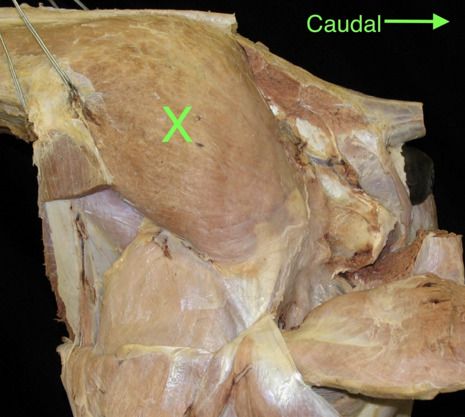
The muscle labeled attaches to what bony structure distally
greater trochanter
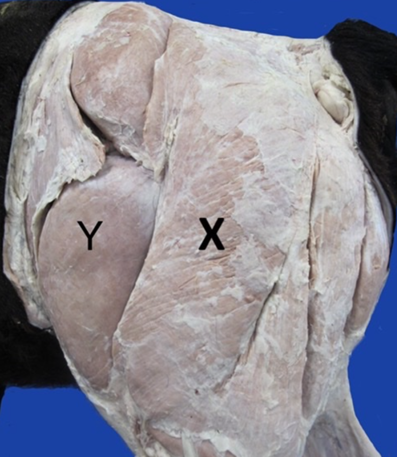
In the calf, the ______ muscle is indicated by X. The muscle Y acts primarily to ______ the stifle
gluteobiceps, extend

Identify the connective tissue structure deep to the pink arrows
sacroischiatic ligament

On this image of an equine thigh, which nerve is indicated by the arrow labeled A?
tibial

The ____ muscle is indicated by A. The fluid filled sac, the _____ has been opened and is labeled B
deep gluteal, trochanteric bursa

The label X is indicating the _____ muscle that acts to ____ the hip, stifle, and hock
biceps femoris, extend

in the bovine, the muscle that attaches to the bony surface labeled by the pink arrow has which actions
extend the tarsus, flex the digits
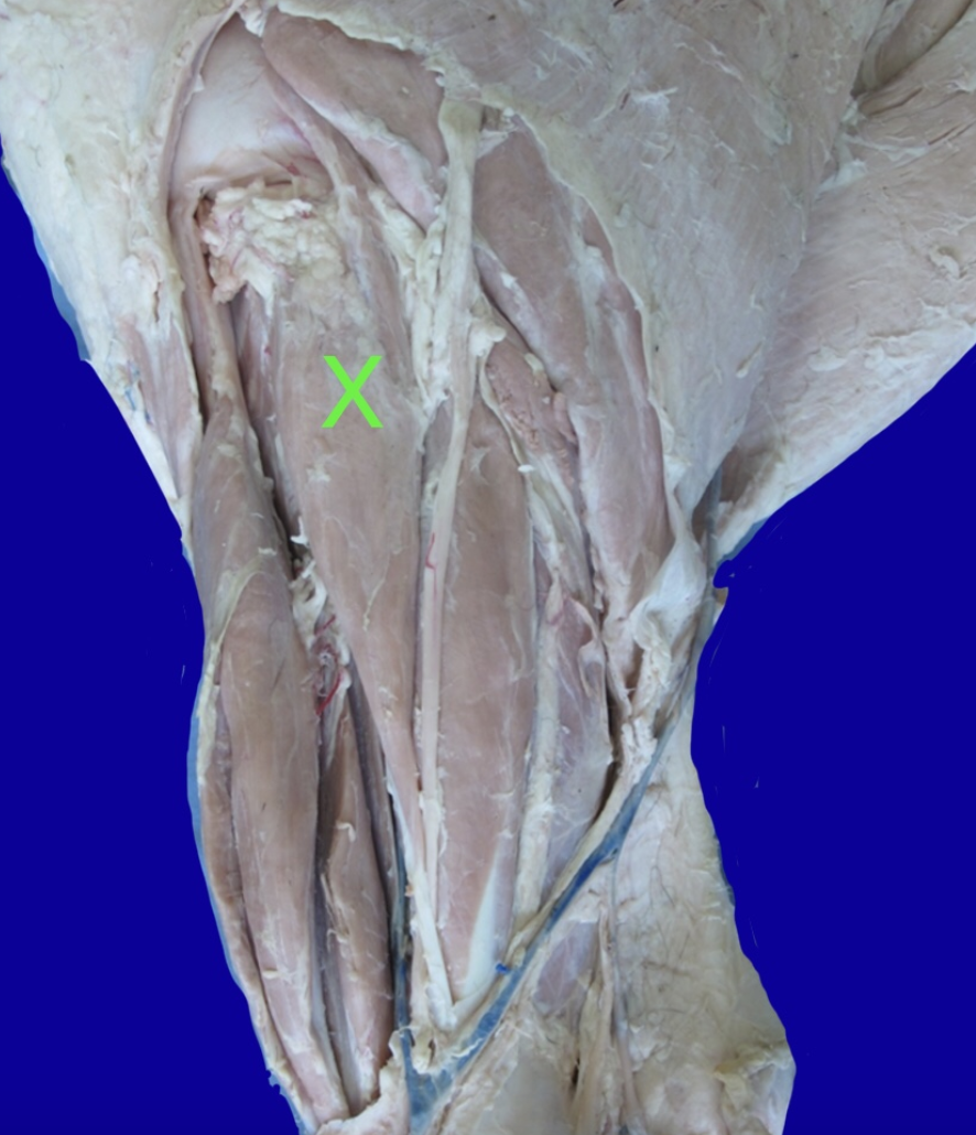
Identify the muscle labeled X from a canine dissection
fibularis longus

The medial tendon of which muscle in the equine is indicated by the red arrow
cranial tibial

In this bovine crus dissection, identify the muscle labeled X
peroneus tertius

Muscles crossing the surface labeled by the yellow arrow have what action at the tarsus
flexion

Muscles X and Y both attach to the tuber calcanei via the _____ tendon and can therefore act to ____ the hock
common calcaneal, extend
What is the significance of a positive cranial drawer sign test
it indicates a likely tear of the cranial cruciate ligament

identify the structure highlighted in green, which serves as an attachment for biceps femoris and superficial gluteal muscle
sacrotuberous ligament

the ____ muscle is labled X and it’s major function at the hip joint is to ____
biceps femoris, extend the hip
What muscles are components of the common calcaneal tendon in small animals
biceps femoris, SDF, gracilis, gastrocnemius, semitendinosus
which of these muscles extend the digits and flex the hock
long digital extensor

Identfiy the highlighted CT structure between the femur and the head of the fibula (lateral view left stifle)
lateral collateral ligament
the right and left sides of the ischium and pubis are joined on the median plane at the ______
pelvic symphysis