Unit 2 Exam
 Call Kai
 Learn
Learn Practice Test
 Spaced Repetition
 Match
Match Flashcards
 Knowt Play
1/151

There's no tags or description
Looks like no tags are added yet.
Name | Mastery | Learn | Test | Matching | Spaced | Call with Kai | Chat |
|---|
No analytics yet
Send a link to your students to track their progress
152 Terms
Tissue
Group of similar cells that perform a common function
Types of Tissues:
Epithelial, Connective, Nervous, Muscular
Characteristics of Epithelial Tissue
Closely fit by tight junctions
Apical (free) Surface
Basement membrane
Avascular
Highly sensitive
Types of Epithelial Tissue
Simple squamous, simple cuboidal, simple columnar, stratified squamous, stratified cuboidal, pseudostratified columnar
Simple VS. Stratified
Simple: single layer
Stratified: multiple layers
Simple Squamous
• Single layer of flat cells.
• Function: Rapid diffusion of substances .Secretes serous fluid
• Location: Alveoli in lungs, Capillaries, Serous membranes

Simple Cuboidal
• Single layer of cube shaped cells
• Absorption and secretion
• Thyroid gland

Simple Columnar
• Single layer of columnar cells: May have cilia and goblet cells
• Absorption and secretion: Goblet cells secrete mucus
• Lines the gastrointestinal tract (ex. Intestines)
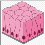
Pseudostratified Columnar
• Single layer of columnar cells: Cells of different heights with nuclei at
different levels makes it appear stratified. May have cilia and goblet cells
• Cilia propel mucus, goblet cells secrete mucus
• Lines the respiratory tract (Trachea and bronchi)

Stratified Squamous Keratinized
• Multiple cell layers with dead cells that are flat and scaly at surface
• Resists mechanical injury (abrasion), prevents water loss through skin; resists
penetration by pathogenic organisms
• Epidermis of skin
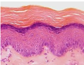
Stratified Squamous Non-Keratinized
• Same as keratinized epithelium without the surface layer of dead cells
• Resists mechanical injury (not as much as keratinized though) and penetration
of pathogenic organisms
• Tongue, oral mucosa, esophagus

Transitional
• Multiple cell layers, surface cells change from round to flat when stretched
• Allows an organ to distend for filling
• Ureters and urinary bladder

Connective Tissue Characteristics
Highly vascular
Composed of cells and extracellular matrix
Extra Cellular Matrix
Made of ground substance and fibers
• Ground substance: very small, gelatinous
• Fibers: Three types of fibers
Collagen: Strength
Reticular: support
Elastic: allow stretch and recoil
Types of Connective Tissue
• Connective Tissue Proper (Fibrous): Loose & Dense
• Bone
• Cartilage
• Blood
Types of Loose Fibrous CT
Areolar, Adipose, Reticular
Areolar CT
Fibroblasts, collagen, elastic, and reticular fibers in matrix
Connects tissues and organs
Papillary layer in dermis of skin
Allows space for edema
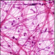
Adipose CT
• Empty-looking cells with nucleus pressed against cell membrane
• Adipocytes are fat cells filled with triglycerides
• Energy storage, cushions organs
• Hypodermis (subcutaneous layer of skin: what we typically call
our fat) and around the heart and kidneys
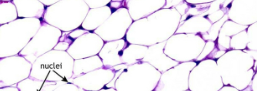
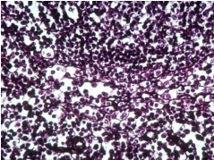
Reticular CT
• Mesh of short intertwining fibers and fibroblasts
• Supportive framework for lymphatic organs
• Lymph nodes and spleen
Types of Dense CT
Regular & Irregular
Dense Regular CT
• Densely packed, parallel collagen fibers
• Provides tensile strength in one plane of movement
• Tendons attach muscles to bones, ligaments hold bones together

Dense Irregular CT
• Densely packed, randomly arranged collagen fibers and few visible cells
• Withstandlas stresses in different directions
• Reticular dermis of skin

Cartilage
• Supportive connective tissue with flexible, rubbery matrix
• Main cell is chondrocytes
• Fibers differ with type of cartilage
Types of Cartilage
Hyaline, Elastic, Fibrocartilage
Hyaline Cartialge
• Chondrocytes surrounded by clear, glassy matrix
• Creates smooth surface for joint movement
Keeps airways open
• Articular cartilage at end of long bones
Trachea

Elastic Cartilage
• Chondrocytes surrounded by elastic fibers
• Provides flexibility and stretch
• External ear
Tip of nose

Fibrocartilage Cartilage
• Chondrocytes surrounded by thick bundles of collagen fibers
• Shock absorption
• Pubic symphysis, intervertebral discs

Blood CT
• Fluid connective tissue:
Plasma is the matrix.
Contains RBCs, leukocytes,
and platelets
• Transports oxygen, nutrients, hormones, and removes wastes
• Inside heart and blood vessels
Nervous Tissue
• Nervous tissue is found in brain, spinal cord and nerves.
• Main function is to receive stimuli from body and send back responses.
• Cells in nervous tissue are neurons and neuroglia
Neurons
• Cell body with many extensions
• Conduct nerve impulses
• Brain, spinal cord, nerves
Neuroglia
• Many different types
• Protect and assist neuron
• Brain, spinal cord, nerves
Types of Muscular Tissue
Skeletal, Cardiac, Smooth
Skeletal Tissue
• Long cylindrical cells (muscle
fibers)
Multiple nuclei per cell
Striations: alternating dark and
light bands
Voluntary: conscious control
• Attach to bone
Responsible for
movement
• Skeletal muscles
Muscles of the face

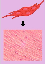
Smooth Muscle Tissue
• Short fusiform cells (thick in middle, tapered at ends)
One centrally located nucleus
Unstriated and involuntary
• Propels contents through an organ
Changes diameter of blood vessels
• Wall of digestive and urinary tract
Walls of blood vessels
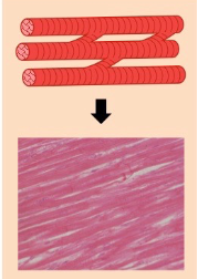
Cardiac Muscle
• Short and branched
One centrally located nucleus
Intercalated discs join cardiac muscle cells
Striated and involuntary
• Contraction of cardiac muscle will pump blood to body organs
• Located only in the heart
Glandular Epithelial
ET that secretes substances
Endocrine Glandular ET
• Secrete into bloodstream. They are ductless glands.
• Endocrine glands secrete hormones. They are few in number.
• Example: Thyroid and adrenal glands
Exocrine Glandular ET
• Secrete out to the surface by a duct.
• Very numerous
• Example: Sweat glands
Types of Cell Junctions
Tight Junctions: seal cells together to prevent passage of water or ions: Ex. Blood brain barrier and epithelium lining urinary bladder
Gap Junctions: membrane proteins form tunnels that allow ions to pass from one cell to the other. Allows rapid spread of electrical signals. Ex. Cardiac muscle, skeletal muscle
Adhesion (desmosome) junctions: anchor adjacent cells that are subjected to mechanical stress. Ex. Cardiac muscle
Layers of Skin
Epidermis, Dermis, Hypodermis
Epidermis
• Keratinized stratified squamous epithelium
• Thickness of the epidermis determines whether skin is “thick” or “thin”
Dermis
Two layers
• Papillary layer
Areolar connective tissue
• Reticular layer
Dense irregular connective tissue
Hypodermis
• Not a part of the skin, but usually studied with the skin
• Adipose connective tissue (subcutaneous fat)
Thick Skin
on palms and soles, fingers and toes
• Has sweat glands, but no hair follicles or sebaceous (oil) glands
• Epidermis is thicker
Thin Skin
covers rest of the body
• Possesses hair follicles, sebaceous glands, and sweat glands
5 layers of epidermis (bottom to top)
Stratum basale
Stratum Spinosum
Stratum granulosum
Stratum lucidum
Stratum corneum
Stratum basale
Single layer of stem cells. Scattered melanocytes and Merkel cells
Stratum spinosum
Keratinocyte attachments appear spiny. Few dendritic cells
Stratum granulosum
Granular keratinocytes
Stratum lucidum
only in thick skin. No nucleus or organelles
Stratum corneum
dead keratinocytes flaking away
Cells in epidermis
stem, melanocytes, merkel, keratinocytes, dendritic/langerhans
Stem cells
mitotic dividing cells that produce keratinocytes
Melanocytes
synthesize the pigment melanin. Shields DNA of cells from UV radiation
merkel cells
touch receptors
Keratinocytes
squamous “skin cells” - synthesize keratin
Dendritic/langerhans cells
macrophage - perform phagocytosis of harmful pathogens or toxins
Layers of dermis
Papillary, Reticular
Papillary Layer
• Superficial layer composed of areolar tissue
• Contains dermal papillae
Dermal papillae
upward fingerlike extensions of the dermis. They fit into epidermal ridges on fingertips that leave fingerprints.
Reticular Layer
Deeper and thicker layer composed of dense irregular CT
Structures in Dermis
Meissner’s corpuscles, free nerve endings, sweat glands, sebaceous glands, Pacinian corpuscles, hair bulbs
Meissners corpuscles
touch receptors
Pacinian corpuscles
pressure receptors
Hair bulbs
located in dermis but they originate from epidermis
3 layers of hair from root to out
Medulla, Cortex, Cuticle
Arrector pili muscle
muscle attached to hair bulb; Bundles of smooth muscle cells - contract to cause goose bumps
Skin glands
Sudoriferous, sebaceous
Sudoriferous gland
Sweat gland; • Eccrine (merocrine) sweat glands - duct to skin surface
• Apocrine sweat glands - duct to hair follicle
• Sweat is mostly water and minerals. It is slightly acidic which
inhibits overgrowth of bacteria.
Sebaceous gland
oil
Hemoglobin
Red pigment
Carotene
yellow pigment
Pallor
decreased blood flow to skin makes more white
Emotional stress, low blood pressure, circulatory shock, cold, anemia
Cyanosis
bluish discoloration from deficiency of oxygen.
Airway obstruction (drowning or choking) and lung diseases (emphysema)
Jaundice
yellowish discoloration of skin and sclera due to excess of bilirubin in blood
Cancer, hepatitis, compromised liver function
Basal cell carcinoma
- Most common type
- Least dangerous because it seldom metastasizes
- Forms from cells in stratum basale
Squamos cell carcinoma
- Arise from keratinocytes in stratum spinosum
- Lesions appear on scalp, ears, lower lip, or back of the hand
- Have raised, reddened, scaly appearance later forming a concave
ulcer
- Chance of recovery good with early detection and surgical removal
- Tends to metastasize to lymph nodes and may become lethal
Malignant melanoma
- Skin cancer from melanocytes; often in a preexisting mole
- Less than 5% of skin cancers, but most deadly form
- Treated surgically if caught early
- Metastasizes rapidly; unresponsive to chemotherapy; usually fatal
- High incidence in men, redheads, people who experience severe
sunburn in childhood
Hair bulb
swelling at base. Contains
Matrix: mitotically active cells
Papilla: blood vessels for nutrition
Hair receptors
• Nerve fibers that entwine each
follicle
• Respond to hair movement
Piloerector muscle (arrector pili)
• Bundles of smooth muscle cells
• Goose bumps
Types of tissue repair
Regeneration, Fibrosis
Regeneration tissue repair
replacement of dead or damaged cells by the same type of cell as before
• Restores normal function
• Skin injuries and liver regenerate
Fibrosis
replacement of damaged cells with scar tissue
• Holds organs together
• Does not restore normal function
• Severe cuts and burns, healing of muscle injuries
Dep dermal tissue repair
Hemostasis: Severed blood vessels will make a clot closing the gap. The surface will harden and form a scab to seal
Organization: beneath the scab, macrophages will gradually digest the blood clot
Granulation tissue: Fibroblasts lay down collagen fibers and new blood vessels restore circulation.
Maturation and Regeneration : The fibrosed area will mature and contract. Surface epithelium will divide and regenerate.
Function of nervous system
• Sensory input: Gathering information. Monitoring changes in and outside
body. Sense organs detect stimuli and send signals to the brain and spinal cord.
• Integration: Process and interpret sensory input to determine appropriate
response
• Motor output: Response to stimuli. Send commands to activate muscles or
glands in response to the input
Central Nervous System
Brain + Spinal Cord
Peripheral nervous system
Cranial nerves + spinal nerves
Neurons
Actual nerve cells that transmit signals
Neuroglia
Support system for the neurons
Soma (cell body)
Contains organelles except centrioles (no cell division). Metabolic functions
Dendrites
Receptive: receives stimuli from other neurons
They convey stimuli to the soma by graded potentials.
Axon
Axon hillock: Nerve impulse (action potential) is generated in the trigger zone.
Impulse travels down the axon
Axon: conducting area. Will conduct the action potential to the terminal
Axon terminal
Secretory area. Terminal branches have knobs containing synaptic vesicles of
neurotransmitters.
Organization of Peripheral nervous system
Somatic, visceral
Somatic
Signals from skin, muscles, joints. Response to
skeletal muscles
Visceral
signals from viscera (internal organs). Response to
cardiac muscle, smooth muscle and glands
Myelination
• Myelin sheath is an insulating layer of material that is mostly lipid.
• Myelinating glia wrap several layers of myelin around the cell membrane of
a segment of the axon.
• Schwann cells insulate peripheral nerves while oligodendrocytes insulate
the CNS.
• Areas not covered with myelin are called nodes of Ranvier
Synapse
The synapse is a connection
between two neurons. It transmits
signals between neurons
Presynaptic neuron
axon bulges to form synaptic knob that contains synaptic vesicles which store neurotransmitter (Acetylcholine)