Auditory Physiology
 Call Kai
 Learn
 Practice Test
 Spaced Repetition
 Match
 Flashcards
 Knowt Play
1/47

There's no tags or description
Looks like no tags are added yet.
48 Terms
Sound intensity
How much energy is present in the sound wave, perceived as loudness
Auditory nerve
Innervates the bony labyrinth
Modiolus
Central, bony axis which houses fibres of cochlear nerve that run to the auditory nerve
Base
Part of the cochlea sensitive to high frequency sounds
Apex
Part of the cochlea sensitive to low frequency sounds
Stria vascularis
Produces endolymph; supplies oxygen, other nutrients to cochlea
Inner hair cells
Arranged in one row, 90-95% afferent neurons
Scala vestibuli
Between cochlear wall and Reissner’s membrane
Scala media
Between Reissner’s membrane and basilar membrane
Scala tympani
Between basilar membrane and cochlear wall
Outer hair cells
Arranged in 3 rows, 90-95% efferent neurons
Stereocilia
Hair cells that respond to fluid motion and are responsible for mechano-electrical transduction
Alternating current
In phase with frequency (at lower Hz sounds), up to about 1000 – 3000 Hz is when you lose it
Direct current
Overall increase at beginning of sound, decrease at end
Spiral Ganglion cells
Cells that sit in the modiolus of the cochlea
Place coding
Tonotopic organization of frequencies in the cochlea
Rate coding
Phase locking of afferent auditory nerve fibres to the period / frequency of the sound itself
Cochlear nucleus
Part of the auditory pathway that begins to process frequency (pitch) and intensity (loudness)

Superior Olivary Complex (SOC)
Part of the auditory pathway that compares intensity and time differences between the two ears

Lateral lemniscus
Part of the auditory pathway which is a major tract of axons carrying information to brainstem nuclei. It is the integrative relay station, reduces spectral splatter, suppresses sound from reverberation

Inferior colliculus
Part of the auditory pathway that is involved in multi-modal sensory perception and integration, assists with temporal patterns and auditory space

Medial Geniculate
Part of the auditory pathway that distributes sensory information, important role in attention to sound

Cocktail party effect
Having to orient your attention when there is a bunch of competing sounds involved, the medial geniculate is involved
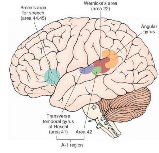
Heschl’s gyrus
Part of the brain associated with discrimination of frequency & temporal cues, association of sound with past experience

Secondary auditory cortex
Part of the brain associated with harmonic, melodic, rhythmic patterns
Binaural hearing
Excitatory and inhibitory neurons happening in both sides of the brain, but different innervation pathways

Congenital inner ear issues
Rubella, syphilis, toxoplasmosis, cytomegalovirus, cochlear dysplasia/aplasia
Acquired inner ear issues
Noise damage, sudden idiopathic, ototoxic drugs, skull fractures, presbycusis, radiation treatment
Sudden idiopathic
Sudden onset of illness that affect your inner ear
GJB-2
Most common genetically-linked hearing loss
Presbycusis
Age-related hearing loss, gradual loss of hearing
“Hidden” hearing loss
Neural impairments but little change to cochlea
Psychoacoustics
Study of the experience of sounds, holds that sound is an auditory experience
Subjective experience
The experience of sound depends on the individual listener, dictated from an individual to individual level
Auditory threshold
The lowest hearing level that a person can detect
Frequency
Number of oscillations/second
dB SPL
Raw physical sound pressure
dB HL
Severity of hearing loss relative to a "normal" baseline
dB SL
Sound intensity above a single individual's threshold
Air conduction
Most auditory perception is through this pathway
Pure tone testing
Test used to determine hearing sensitivity (threshold) across frequencies important for understanding speech
Audiogram
Plots auditory thresholds for a range of frequencies important for speech
20
Reference sound pressure for dB SPL is __ 𝜇Pa
Cerebellopontine angle
Where the cerebellum, medulla oblongata, & pons join together
Dorsal
Fibres from base of cochlea (high freq) end in the ____ part of the dorsal cochlear nucleus
Ventral
Fibres from apex (low Hz) end in the ____ part of the dorsal cochlear nucleus
Ventral cochlear nucleus
From here, 2/3 of fibres go to contralateral superior olivary complex (SOC), remaining to ipsilateral SOC and lateral lemniscus
Dorsal cochlear nucleus
From here, fibres cross the midline to end on cells of the lateral lemniscus