L12: Microscopy II
 Call Kai
 Learn
 Practice Test
 Spaced Repetition
 Match
 Flashcards
 Knowt Play
1/47

There's no tags or description
Looks like no tags are added yet.
48 Terms
What is darkfield microscopy useful for?
low contrast, absorption, and high diffusion
What type of light is collected in DF microscopy?
scattered light
What is the difference in DF and BF ability to produce an image that accurately represents the morphological information of an object?
BF mogs
What is DF best for?
revealing outlines, edges, and boundaries
In DF which regions appear bright?
scattering regions
In DF, what regions appear dark?
those that don’t scatter
How can DF microscopy be performed?
both in reflection and in transmission
How is the objective andmicroscope set up so light illuminating the sample isnt collected by the objective?
there is a light stop that blocks the central rays
only oblique illumination reaches the sample
What makes sodium chloride optically isotropic?
its cubic crystalline lattice
What makes calcite optically anisotropic?
its anisotropic crystalline lattice
What characterizes an amorphous polymer?
no periodic crystalline structure or repeating unit
What is birefringence?
double refraction of light
What causes birefringence?
orientation dependent differences in a compound’s refractive index
What type of material exhibits birefringence?
transparent, molecularly ordered material
Describe the geometry of the axis of isotropic crystal (salt)?
all axes are the same length and perpindicular to one another
salt has a single refractive index
Describe the geometry of an anisotryopic crystal’s (quartz) axis.
3 equal horizontal axes at 220° to each other, and 1 vertical axis, perpendicular to the others
How many refractive indices does Quartz have?
two
Describe the orientation of refractive indexes in an optically isotropic material?
constant throughout
How does light travel through optically isotropic materials?
one single velocity
What determines how an anisotropic material interacts with light?
it depends on the orientation of the crystalline lattice w respect to the incident light
What happens after light enters a non-equivalent axis in an optically anisotropic material?
it is refracted into two rays and each is polarized w their vibration directions oriented perpendic to each other and traveling at different velocities
Describe the steps of polarized micrsocopy.
a linear polarizer placed before the objective polarizes light parallel to its axis
polarized light enters anistrropic crystal
light is refracted and divided to 2 components vibrating parallel to the crystallographic axes & perpendic to eachother
ligh reach a 2nd polarizer that is oriented perpendic to the first polarizer
retardation of one ray w respect to the other causes interference to occur between the waves as they pass through analyzer
Birefringent samples acquire a spectrum of color when observed through crossed polarizers.
What is the main result of polarized microscopy?
enabling the quantifying birefringence
Why does collagen exhibit birefringence?
its ordered helical structure
fiber has dif refractive indexes along it length vs across it
What is the assembly protocol for polarized microscopy targeting extrinsic birefringence?
aq silk fibroin is mixed w crosslinkers
mechanical tension is introduced
during drying, silk fibroin undergoes βsheet transformation and the formation of a nanofibrillar network
What is birefringence useful for w/ respect to mechanical properties?
quantifying stress-induced alignment of silk fibrils in aerogels
What else is observed in correspondence with minor nanofibril alignment?
low birefringence, in correlation to low-stress regions
What is the main mechanism fluorescence microscopy uses to increase contrast of an object being imaged?
absorption of light, quantum yield of fluorophore
What are some biomedical applications of fluorescence?
detecting individual fluorescent molecules
studying macromolecules distribution and dynamics inside tissues
study of rxn mechanisms by fluorescence energy transfer
dynamics of single tagged molecules
determ of ionintracell concentration
study of cell viability and cell functions
What are different ways a specimen can fluoresce?
intrinsically fluorescent
conjugation between tagged macromolecule and fluorochrome
What are the different parts of a widefield fluorescence microscope?
eyepieces, camera, fluorescent filter set, objectives, specimen, xyz stage, epi-illumination source
What does an excitation (Ex) filter do in a widefield fluorescence microscope?
selectively transmits a band of short wavelengths for exciting a specific fluorophore in the specimen
What does a dichroic mirror do in a widefield fluorescent mircroscope?
reflects the Ex light towards the objective and specimen, while transmitting returning Em fluorescent light towards the detector
What does an emission (Em) filter do in a widefield fluorescence microscope?
transmits Em light and it removes unwanted background light
What type of objectives are preferred?
color corrections (apochromatic) and high NA
What is the problem that confocal microscopy addresses?
thick fluorescent specimens, bright fluorescent signals from objects lying outside focal plane increase backgroun and yield low-contrast images
How does confocal microscopy address the low contrast issue for thick fluorescent specimens?
rejects signals from sources above and below the focal plane
illuminates specimen w a focused scanning laser beam
places a pinhole aperture in the image plane in front of an electronic photon detector
How does confocal microscopy work?
uses a stepper motor that changes the microscope focus in small steps along the z-axis
Are confocal images generated in a microscope?
no, built up electronically
What does the photomultiplier transducer do in confocal microscopy?
transduces the intensity of the photons at the pinhole in a voltage
signal is digitizeed and converted into pixels that form an image
How many pinholes can be used in some confocal configurations?
2! additional pinhole allows transmission only through a small area
What role does raster movement of the laser play in confocal microscopy?
one mirror scans along x-axis
the other on y-axis
both are coordinated to generate a pattern of a raster on the specimen
What role does electronic magnification play in confocal microscopy?
magnification can be adjusted electronically by varying the area scanned by the laser (zoom factor) w/o having to change the objective
How does decreasing the size of the pinhole affect thickness of the focal plane along the z-axis and contrast?
reduces thickness of focal plane along z-axis
improves contrast by excluding out of focal plane sources of light
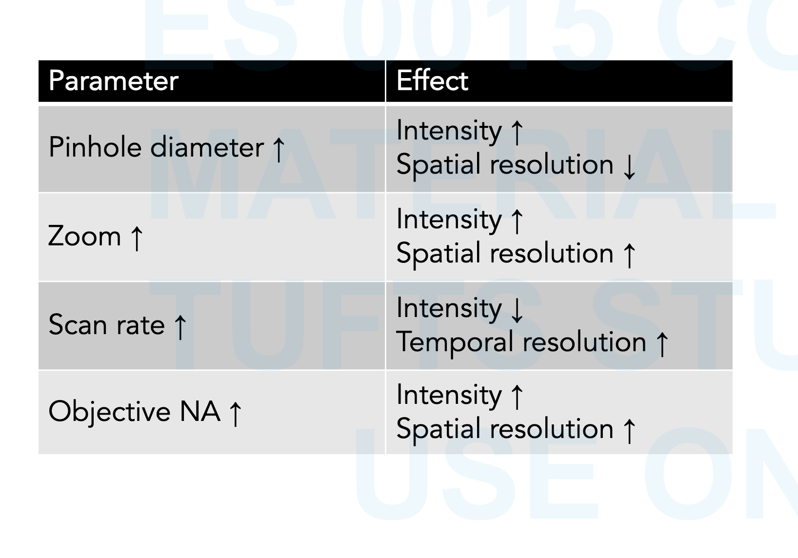
What is another factor that can affect spatial resolution?
scan rate
effects of different parameters
What are the advantages of confocal over widefield?
sharp image
dont have to physically slice the sample
can investigate the spatial arrangement
composite or projection view
3D views
5D views (x,y,z, timed, multiple colors)
List a biomedical application of confocal microscopy.
disease development monitoring