Respiratory and Renal Systems
 Call Kai
 Learn
 Practice Test
 Spaced Repetition
 Match
 Flashcards
 Knowt Play
1/364

There's no tags or description
Looks like no tags are added yet.
365 Terms
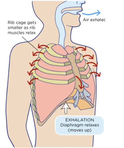
diaphragm contracts
during inhalation, this occurs where this moves down. The rib cage expands as the rib muscles contract.

diaphragm relaxes
during exhalation, this occurs where this moves up. The rib cage gets smaller as the rib muscles relax.
external respiration
the movement of gases between the environment and the body’s cells.
the exchange of oxygen and carbon dioxide between the air in the lung's alveoli and the blood in the pulmonary capillaries. This passive process moves oxygen into the bloodstream and carbon dioxide out of the blood through simple diffusion.
functions of the respiratory system
Exchange of gases between the atmosphere and the blood - this system and the circulatory system coordinate the transfer of oxygen and carbon dioxide between the atmosphere and the cells
Homeostatic regulation of body pH
Protection from inhaled pathogens and irritating substances
Vocalization

respiratory system bulk flow
Flow takes place from regions of higher to lower pressure. A muscular pump creates pressure gradients. Resistance to air flow is influenced primarily by the diameter of the tubes through which air is flowing
alveolocapillary membrane
external respiration occurs here, the ultra-thin blood-gas barrier in the lungs that separates inhaled air from pulmonary blood flow, allowing oxygen to enter the blood and carbon dioxide to leave.

Four processes in external respiration
Exchange of air between atmosphere and lungs (Ventilation) - Inspiration and Expiration
Exchange of O2 and CO2 between lungs and blood
Transport of O2 and CO2 by blood
Exchange of gases between blood and cells
respiratory tract
the site for the mechanics of breathing. The path that air travels through your body during breathing. It splits into the upper respiratory tract (nose, mouth, sinuses, pharynx, and upper larynx) and the lower respiratory tract (lower larynx, trachea, bronchi, bronchioles, and lungs), working together to deliver oxygen and remove carbon dioxide.
Conducting system, or airways
Upper respiratory tract: Mouth, nasal cavity, pharynx, larynx
Lower respiratory tract: Trachea, 2 primary bronchi, their branches, lungs

alveoli
Singular alveolus: site of gas exchange
Microscopic, cup-shaped air sacs at the end of the bronchioles in your lungs. They are the primary units where the respiratory system performs gas exchange, passing inhaled oxygen into your bloodstream while releasing carbon dioxide to be exhaled
thoracic cage
bones and muscle of thorax and abdomen
Bones and muscles of the thorax surround the lungs
Spine and rib cage
Diaphragm, intercostal muscles, sternocleidomastoids, scalenes
Pleural sacs each surround a lung
pleural sacs
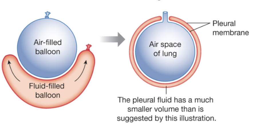
a double-layered serous membrane that surrounds each lung, consisting of the inner visceral pleura (attached to the lung) and the outer parietal pleura (attached to the chest wall and diaphragm). A thin layer of fluid fills the space between them to reduce friction during breathing
Enclose the lung

airways
connect the lungs to the external environment
Pharynx → larynx → trachea (windpipe)
Larynx contains vocal cords
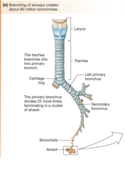
The system of tubes that carry air into and out of your lungs. They start at your nose and mouth, lead down through your windpipe (trachea) and branching bronchial tubes, and end in tiny air sacs (alveoli) where oxygen and carbon dioxide swap places in your blood
bronchioles
Primary bronchi. . They branch off the larger bronchi and deliver oxygen-rich air to the tiny air sacs called alveoli. Unlike larger airways, bronchioles lack cartilage and are surrounded by smooth muscle that controls airflow.
functions of airways
warm, humidify, and filter inspired air
Warming air to body temperature
Adding water vapor
Filtering out foreign material through mucus and cilia

honey comb pattern
increases the surface area for the exchange
Together, they create a vast surface area—about the size of a tennis court—to maximize gas exchange.
smooth muscle in the lungs
surrounds the bronchial tree from the trachea down to the terminal bronchioles, controlling how wide or narrow the air passages are.
Bronchiole dilation or constriction. Airways - sympathetic/parasympathetic
alveoli
the site of gas exchange. Microscopic, cup-shaped air sacs at the end of the bronchioles in your lungs. They are the primary units where the respiratory system performs gas exchange, passing inhaled oxygen into your bloodstream while releasing carbon dioxide to be exhaled.
Type I alveolar cells
gas exchange.
Large, flat, squamous epithelial cells that cover about 90% to 95% of the alveolar surface area in the lungs. These thin cells form the primary air-blood barrier and allow for the rapid diffusion of oxygen and carbon dioxide
Type II alveolar cells
produce surfactant. Cuboidal cells in the lung alveoli. They produce pulmonary surfactant to lower surface tension and prevent lung collapse, act as progenitor stem cells to repair damaged alveolar tissue, and help regulate fluid balance and immune defense
connective tissue
elastin and collagen. One of the four main tissue types in the body. It supports, protects, binds, and separates organs and other tissues. It is made of living cells, protein fibers (such as collagen and elastin), and a gel-like or fluid ground substance.
elastin
a primary structural protein in the lungs that provides the elastic recoil necessary for passive breathing and exhalation
collagen
the most abundant structural protein in lung connective tissue, providing tensile strength, mechanical support, and structural integrity to the alveolar-capillary network.
capillaries in the lungs
Alveolar cells have close association with these.
Tiny blood vessels measuring 5 to 8 µm in diameter that wrap around the alveoli. They form a ultra-thin blood-gas barrier (~0.2–0.3 µm) where deoxygenated blood releases carbon dioxide into the air sacs and absorbs fresh oxygen

pulmonary circulation
high flow, low pressure.
Right ventricle → pulmonary trunk → pulmonary arteries → lungs → pulmonary veins → left atrium
Blood flow through lungs = blood flow through rest of body
Low resistance due to shorter total blood vessel length

surfactant
(short for surface-active agent) is a chemical compound that lowers the surface tension between two liquids, a gas and a liquid, or a liquid and a solid. Pulmonary _________ is a fluid-lipid-protein mix made by Type II alveolar cells that lines the lungs. It cuts surface tension in the alveoli to stop them from collapsing during exhalation and assists with immune defense.
pleura
Double walled- lines inside of thorax and covers surface of lung
Visceral
pleural fluid
Lowers friction between membranes. Holds lungs tight against the thoracic wall. Holds lungs stretched in partially inflated state.
Reduces abrasion or friction between membranes. Fixes the lung to the wall → wants to naturally recoil and collaspe.
pleural sac
forms a double membrane surrounding the lung, similar to the fluid-filled balloon surrounding an air-filled balloon.
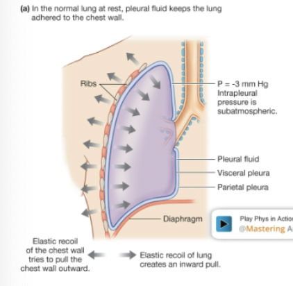
There is negative pressure in the thoracic cavity based on lung stretched and stuck to thoracic wall (-3 to -4 mmHg)
airway branching
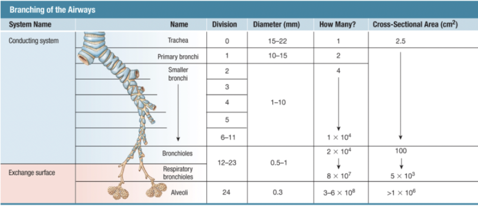
to deliver air from the external environment to the exchange surface. Increased cross-sectional area because of more branching through divisions
The human tracheobronchial tree splits repeatedly into roughly 20 to 25 generations of branches, starting from the single trachea down to the gas-exchanging alveoli. This structure divides the right and left main bronchi into lobar, segmental, and terminal airways before reaching the respiratory zone.
trachea
maintains shape with cartilage
a 10 to 12 cm long cartilaginous tube that connects the larynx to the bronchi. As a vital part of the respiratory system, it channels air into and out of the lungs while warming, humidifying, and filtering it via mucus and tiny cilia.
Primary bronchi
bifurcates into right and left bronchi to each lung, branch until bronchioles- cartilage and smooth muscle
Bronchioles
no cartilage just smooth muscle. Branches until respiratory bronchioles. Diameter decreases but total cross-sectional area increases
epithelial cells in the lung
lining the airways and submuscosal glands secrete saline and mucus. Airways lined with ciliated epithelium bathed in saline layer.
line the entire respiratory tract from the airways to the alveoli, forming a protective barrier and managing gas exchange. Major types include ciliated cells, mucus-secreting goblet cells, and basal cells in the airways, alongside gas-exchanging Type I and surfactant-producing Type II pneumocytes in the alveoli.
goblet cells
Debris trapped by mucus secreted by __________, move upward by mucociliary escalator
specialized, cup-shaped epithelial cells that line the respiratory, gastrointestinal, and reproductive tracts. Their primary function is to synthesize and secrete gel-forming mucins that create a protective mucus layer to trap pathogens, lubricate tissues, and defend against mechanical or chemical damage.
Mucus contains immunoglobulins that disable pathogens
Immune cells are stationed in tissues to destroy pathogens
mucociliary escalator
Cilia brush debris up from branching all the way up
a self-clearing biological defense system in the respiratory tract. It traps inhaled dust, pollen, and pathogens in sticky mucus and sweeps them upward using microscopic, hair-like cilia, moving debris out of the lungs to be swallowed or coughed away.

air
a mixture of gases at atmospheric pressure. Gases move down pressure gradients. invisible, odorless, and tasteless mixture of gases—primarily 78% nitrogen and 21% oxygen—that surrounds Earth and forms its atmosphere.
Dalton’s law
Total pressure equals sum of all partial pressures (Pgas)
(or the Law of Partial Pressures) states that the total pressure of a non-reacting gas mixture equals the sum of the individual partial pressures of each gas.
Ptot = P1 + P2 + … + PN

Boyle’s Law
describes pressure-volume relationships
P1V1 = P2V2
states that the pressure and volume of a gas have an inverse relationship when the temperature and amount of gas stay the same. If the volume goes up, the pressure goes down. If the volume goes down, the pressure goes up

The ideal gas equation
gases move down pressure gradients. PV=nRT
Where P is pressure, V is volume, n is the moles of gas, T is absolute temperature, and R is the universal gas constant, 8.3145 J/(mol·K)
This relationship says that if the volume of gas increases, the pressure decreases, and vice versa.

higher to lower
Gases, singly or in a mixture, move from areas of _______ pressure to areas of _______ pressure.
If the volume of a container of gas changes, the pressure of the gas will change in an inverse manner
Example: Plunger pulled out- barrel volume increases. Meds flow from high to low pressure into needle
respiratory cycle
1 inspiration followed by 1 expiration.
the process of moving air in and out of the lungs to bring in oxygen and remove carbon dioxide. It consists of two main phases: inspiration and expiration.

inspiration
(Inhalation)
• Active process: The diaphragm and external intercostal muscles contract.
• Volume increase: The diaphragm moves downward and flattens, expanding the thoracic (chest) cavity.
• Pressure decrease: Increased volume lowers the pressure inside the lungs below atmospheric pressure.
• Airflow: Air rushes into the lungs from the outside to balance the pressure.
expiration
(Exhalation)
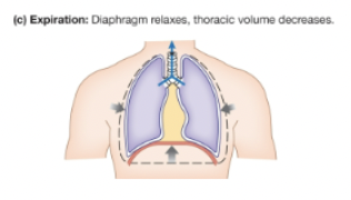
• Quiet/Resting (Passive): The diaphragm and external intercostals relax. The chest cavity shrinks due to the natural elastic recoil of the lungs. This raises lung pressure above atmospheric pressure, pushing air out.
• Forced/Active: During exercise or deep breathing, internal intercostal and abdominal muscles contract to push air out faster.
lung volumes
change during ventilation, pulmonary function tests use a spirometer.
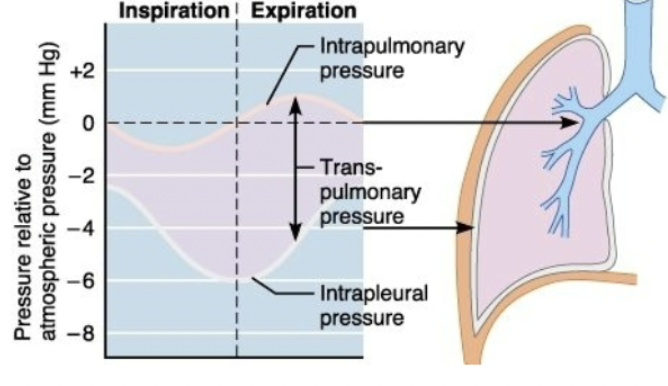
tidal volume
volume that moves during a respiratory cycle
the amount of air that moves in or out of the lungs during a single normal respiratory cycle. In a healthy resting adult, it averages about 500 mL (or 5 to 7 mL/kg of body weight), typically running around 500 mL for males and 400 mL for females
Inspiratory reserve volume (IRV)
additional volume above tidal volume
the extra volume of air that you can forcibly inhale with maximum effort after reaching the end of a normal, quiet tidal inhalation
Expiratory reserve volume (ERV)
forcefully exhaled after the end of a normal expiration
the extra volume of air that you can forcefully exhale past a normal, quiet tidal expiration
residual volume (RV)
volume of air in the respiratory system after maximal exhalation
In healthy adult humans, it averages about 1.0 to 1.2 liters (roughly 20% of total lung capacity) and cannot be measured directly using a standard spirometer.
lung capacity
Vital capacity: (VC) = IRV + ERV + VT
Total: (TLC) = IRV + ERV + VT + RV
total volume of air the lungs can hold
spirometry
measures lung volumes per unit time. a common, painless lung function test that measures how much air you can inhale and exhale, and how fast you can blow it out. Administered using a device called a spirometer, it helps diagnose conditions like asthma and chronic obstructive pulmonary disease (COPD)
FEV1 measures the volume forcefully expired in the first second

factors influencing lung volumes
Lung volumes are based on body surface area and vary with age and condition of the respiratory tract
examples of lung volumes
Tidal volume is normal quiet breathing
Additional air breathed in (inspiration) past tidal volume is inspiratory reserve volume (IRV)
Amount of air forcefully exhaled at the end of normal expiration is expiratory reserve volume (ERV)
capacity
Sum of two or more lung volumes
Vital capacity- maximum air that can be moved in one breath
Total lung capacity = vital capacity + residual volume
pressure gradients
during ventilation, air flows because of these. Flow is proportional to the change of pressure over the resistance.
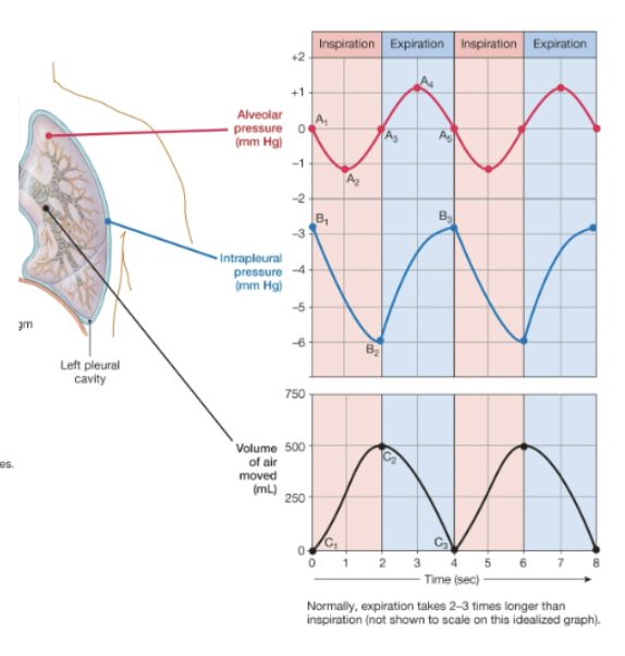
alveolar pressure decreases
Inspiration occurs when ________________
• Time 0: When pressures are equal there is no air flow
• Time 0-2 sec: inspiration
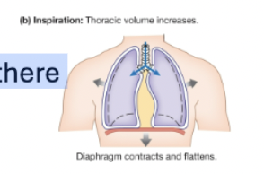
alveolar pressure increases
Expiration occurs when …
Time 2-4 sec: expiration
Time 4 sec: Passive vs. active expiration
intrapleural pressure
changes during ventilation. Subatmospheric: normally negative pressure (-4 mmHg)
During the respiratory cycle
• Inspiration: pressure drops
• Expiration: pressure returns to normal value

elastic recoil
In the normal lung at rest, pleural fluid keeps the lung adhered to the chest wall. This of the chest wall tries to pull the chest wall outward, but the lung creates an inward pull.
lung compliance
the measure of the lungs' ability to stretch and expand, calculated as the change in volume divided by the change in pressure
ability to stretch
lung elastance
ability to return to resting volume when stretching force is released
the tendency of the lungs to recoil to their original shape after being stretched. Mathematically, it is the reciprocal of lung compliance (elastance = Δ P / Δ V), meaning a stiffer lung has high elastance and low compliance.
disease states
Lung Compliance and Elastance May Change in…

high compliance
Stretches easily-low elastance. Emphysema.
the lungs are overly stretchy, pliable, and lose their natural elastic recoil, calculated as a large change in volume per unit change in pressure (Δ V / Δ P). While the lungs expand very easily during inhalation, they struggle to push air back out - gas trapping
emphysema
a chronic lung disease that damages the tiny air sacs (alveoli) in your lungs. This damage makes it hard to breathe, causes shortness of breath, and leads to a long-term cough. Smoking is the main cause of this condition.
low compliance
Requires more force. Restrictive lung diseases
Fibrotic lung diseases (fibrosis). Inadequate surfactant production (NRDS) - problem with air diffusion
the lungs are stiff and hard to expand, requiring higher pressure to achieve a normal change in volume. This condition increases the work of breathing and is commonly associated with restrictive lung diseases and fluid accumulation
fibrosis
excessive accumulation of extracellular matrix proteins, mainly collagen, resulting in scar tissue formation and stiffening within organs. It is a pathological extension of the normal wound-healing response triggered by chronic inflammation, repeated injury, or autoimmune disorders, and it can impair or destroy normal organ function
transpulmonary pressure
the difference between alveolar pressure and intrapleural pressure (Ptp = Palv - Pip). It acts as the primary distending force that keeps the lungs open and drives ventilation. At rest, normal transpulmonary pressure is roughly +4 mmHg to +5 cmH₂O
holds the lung open, and it exists only while intrapleural pressure stays below atmospheric pressure
Law of LaPlace
P=2T/r
The two bubbles shown have the same surface tension (T). According to this, pressure is greater in the smaller bubble

surfactants
surface active agents
Disrupt cohesive force of water
Mixture containing proteins and phospholipids
More concentrated in smaller alveoli
In the lungs, smaller alveoli have more, which equalizes the pressure between large and small alveoli.

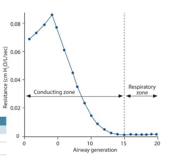
airway diameter
Determines Airway Resistance. Wider airways have less resistance. R is proportional to length*viscosity/radius4
bronchoconstriction
increases resistance - parasympathetic.
the tightening of the smooth muscles that surround the bronchial tubes in the lungs. This narrowing of the air passages restricts airflow, causing symptoms like wheezing, coughing, shortness of breath, and chest tightness. It is commonly linked to asthma, COPD, and physical exertion.
bronchodilation
decreases resistance - sympathetic: β2 receptors on smooth muscles relax in response to epinephrine
the widening of the bronchial air passages in the lungs by relaxing the surrounding smooth muscles, which decreases airway resistance and makes breathing easier. It is primarily achieved using medications called bronchodilators to treat conditions like asthma and chronic obstructive pulmonary disease (COPD).
factors that affect airway resistance
Length of the system → constant, not a factor
viscosity of air → usually constant; humidity and altitude may alter slightly
diameter of airways:
upper airways: affected by physical obstruction mediated by mucus and other factors
bronchioles:
Bronchoconstriction: parasympathetic neurons (muscarinic receptors), histamine, leukotrienes
Bronchodilation: carbon dioxide, epinephrine
Rate and Depth
of Breathing Determine the Efficiency of Breathing.
together determine minute ventilation (the total amount of air moved into and out of the lungs each minute).
total pulmonary ventilation
Volume of air moved in and out of lungs per minute
Ventilation rate x Tidal volume
the total volume of air moved into and out of the lungs per minute. It is calculated by multiplying the respiratory rate (breaths per minute) by the tidal volume (air per breath), averaging about 7,500 mL/min in a resting adult.
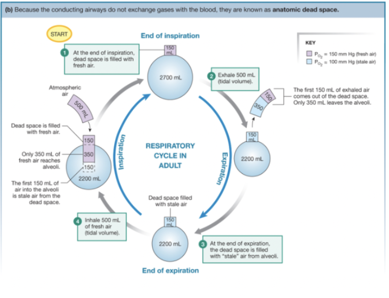
anatomic dead space
fills with air. Exhale mix of fresh and stale air.
The volume of air in the conducting airways (such as the nose, pharynx, trachea, and bronchi) that does not take part in gas exchange. In a healthy adult, this volume is about 150 mL, which makes up roughly one-third of a normal tidal volume. Conducting airways do not exchange gases with the blood.
alveolar ventilation
A better indication of how much fresh air reaches the alveoli. Fresh air remaining in the dead space does not get to the alveoli. ventilation rate x (tidal volume - dead space).
the volume of fresh air that reaches the alveoli per minute to participate in gas exchange.
For example: if dead space is 150 mL: 12 breaths/min x (500-150 mL) = 4200 mL/min
total pulmonary ventilation
the total volume of air moved into and out of the lungs per minute. greater than alveolar ventilation because of dead space
= ventilation rate x tidal volume
For example: 12 breaths/min x 500 mL/breath = 6000 mL/min

Alveolar Gas Composition
varies little during normal breathing. O2 entering alveoli ~ O2 entering the blood. Fresh air into lungs ~ 10% total lung volume at the end of inspiration
at sea level consists primarily of oxygen (PAO2 ~= 100 mm Hg) carbon dioxide (PACO2 ~= 40 mm Hg), water vapor (PH2O ~= 47 mm Hg), and nitrogen (PN2 ~= 573 mm Hg), totaling 760 mm Hg). This makeup differs from inspired air because of ongoing gas exchange and continuous humidification in the airways
normal ventilation values
Total pulmonary ventilation: 6 L/min
Total alveolar ventilation: 4.2 L/min
Maximum voluntary ventilation: 125-170 L/min
Respiration rate: 12-20 breaths/min
eupnea
normal quiet breathing. normal, good, healthy, and unlabored breathing. It describes relaxed, automatic respiration at rest—where inhalation requires minimal muscle effort and exhalation relies naturally on the elastic recoil of the lungs.
hyperpnea
increased respiratory rate and/or volume in response to increased metabolism.
Examples: exercise
a breathing pattern marked by an increased depth of breathing (larger tidal volume), which can happen with or without a faster breathing rate
hyperventilation
increased respiratory rate and/or volume without increased metabolism.
Examples: emotional, blowing up a balloon
rapid or deep breathing—known as overbreathing—that causes your body to exhale more carbon dioxide than it produces
hypoventilation
decreased alveolar ventilation.
a condition where breathing is too slow or too shallow to meet the body's needs. This causes carbon dioxide to build up in the blood (hypercapnia) and can lead to low oxygen levels, resulting in fatigue, confusion, morning headaches, and respiratory acidosis.
examples: shallow breathing, asthma, restrictive lung disease
tachypnea
rapid breathing; usually increased respiratory rate with decreased depth.
abnormally rapid and shallow breathing. In resting adults, it means a rate higher than 20 breaths per minute (normal is 12 to 20); in newborns, it means more than 60 breaths per minute (normal is 30 to 60).
Example: panting.
dyspnea
Difficulty breathing (a subjective feeling sometimes described as “air hunger”)
shortness of breath or the subjective feeling that you cannot get enough air into your lungs. It can feel like chest tightness, suffocation, or working harder to breathe, and it is a symptom of underlying heart, lung, or other health issues rather than a diagnosis on its own
Examples: various pathologies or hard exercise
apnea
cessation of breathing.
a medical condition marked by a temporary stop in breathing that lasts for at least 10 seconds. It most commonly appears as sleep apnea, which includes obstructive, central, and mixed types. Untreated, it raises the risk of heart disease, stroke, and high blood pressure.
Example: voluntary breath-holding; depression of CNS control centers.
increases; decreases
As alveolar ventilation increases, alveolar PO2 ________ and PCO2 ________. The opposite occurs as alveolar ventilation decreases.

blood flow (perfusion)
ventilation and alveolar blood flow are matched. _______ past the alveoli must be high enough to pick up O2.
Capillaries in the lung are collapsible. Low blood pressure shuts off capillary beds. Blood diverted to more beds during exercise.
Resistance of pulmonary arterioles
to blood flow, is regulated by O2 content of surrounding interstitial fluid

bronchiole diameter
mediated primarily by CO2 levels in exhaled air passing through them

relating gas composition and diameter

gas exchange
relies on the transport of gases in the blood and their diffusion across membranes. The body needs oxygen and removes carbon dioxide
Steps:
Oxygen enters the blood at alveolar-capillary interface
Oxygen is transported in blood dissolved in plasma or bound to hemoglobin inside RBCs
Oxygen diffuses into cells
CO2 diffuses out of cells
CO2 is transported dissolved, bound to hemoglobin, or as HCO3-
CO2 enteres alveoli at alveolar-capillary interface

hypoxia
too little oxygen. a medical condition where your body's tissues do not receive enough oxygen to function properly
hypercapnia
increased concentrations of carbon dioxide. an abnormally high level of carbon dioxide (CO₂) in the blood, typically occurring when the body cannot eliminate CO₂ fast enough due to hypoventilation, lung disease, or impaired breathing.
oxygen, carbon dioxide, and pH
to avoid hypoxia and hypercapnia, the body responds to three regulated variables.
. Carbon dioxide dissolves in blood to form carbonic acid and hydrogen ions H+ lowering blood pH. This increase in CO2 and drop in pH reduces hemoglobin’s affinity for O2 (the Bohr effect), forcing oxygen offload into active tissues.
breathing
bulk flow of air into and out of the lungs.
individual gases
diffuse along partial pressure gradients until equilibrium
Total pressure of mixed gas = sum of partial pressures of individual gases

Gas exchange between alveoli and blood
the process where oxygen enters the bloodstream and carbon dioxide leaves it. This occurs in the tiny air sacs of the lungs called alveoli, which are wrapped in small blood vessels called capillaries
PO2 alveolar air > PO2 blood
PCO2 blood > PCO2 alveolar air
gas exchange between blood and tissues
known as internal respiration—occurs via simple passive diffusion. Oxygen moves from high partial pressure in the arterial blood (100 mmHg) to lower pressure in the body tissues (40 mmHg), while carbon dioxide moves from the tissues (45 mmHg) into the blood (40 mmHg).
PO2 blood > PO2 tissue
PCO2 tissue > PCO2 blood