Voice Disorders Exam 2
 Call Kai
 Learn
Learn Practice Test
 Spaced Repetition
 Match
Match Flashcards
 Knowt Play
1/66

There's no tags or description
Looks like no tags are added yet.
Name | Mastery | Learn | Test | Matching | Spaced | Call with Kai | Chat |
|---|
No analytics yet
Send a link to your students to track their progress
67 Terms
what is etiology?
the study of the cause or origin of disease
what caused or contributed to the voice problem
what is pathophysiology?
the study of the biological process associated with disease
includes the study of recovery mechanisms
what is phonotrauma?
refers to abnormal voicing behaviors that contribute to the development of voice disorders such as vocal fold injury due to misuse and overuse of voice
mechanical injury to the vocal fold mucosa from excessive and repetitive collision and shear forces during forceful voice use or other laryngeal behaviors
Examples of things that cause phonotrauma?
shouting
loud talking
screaming
vocal noises
coughing
throat clearing
what are vocal registers?
perceptually distinct regions of vocal quality as pitch/loudness is changed
examples of vocal registers
glottal fry/pulse - lowest range
modal - normal range
Loft/falsetto/head - higher pitch/range
chest - lower pitch
vocal fold nodules description:
usually bilateral (paired bumps)
often symmetric
medial edge at junction of anterior 1/3 and posterior 2/3 point of greater vibration
caused by phonotrauma
often causing hourglass closure
vocal fold nodules picture:

vocal fold polyp description:
usually unilateral
mid-membranous fold
sessile (broad/blister-like) or pedunculated (stalk)
often vascular/fluid-filled w/ potential reactive lesion appearing on the opposite fold
may follow a single intense phonotraumatic event
vocal polyp picture:

vocal fold cyst description:
usually unilateral
encapsulated, opaque, round mass
often medial edge/mid-membranous fold
superficial or deeper in LP
associated with granular blockage and/or mucous collection after an injury
often stiffens/limits mucosal wave locally
vocal fold cyst picture:

Reinke’s edema description:
usually bilateral
diffuse “baggy” swelling of superficial LP
gelatinous fluid
can be caused by smoking/LPR/irritants/phonotrauma
can cause low-pitched rough voice
increased mass of VF
Reinke’s edema picture:

vocal fold hemorrhage description:
blood visible through the epithelium
color can change from bright red to yellow/brown as it resolves
causes sudden voice change/loss after voice use
blood leaked into LP
caused by anticoagulants/bleeding risk/phonotrauma
vocal fold hemorrhage picture:
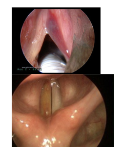
varix/ectasia description:
prominent/enlarged or dilated blood vessel
may have normal voice or fatigue
risk for hemorrhage depending on location
varix/ectasia picture:

sulcus vocalis description:
longitudinal furrow/groove along the medial membranous edge
loss of viscoelasticity
can be congenital or related to a ruptured cyst
deeper types = more dysphonia
sulcus vocalis picture:

vocal fold scar description:
may not be a “mass”
tissue looks/behaves stiff
reduced/absent mucosal wave
permanent microarchitectural change in LP
history of phonotrauma, surgery, radiation, trauma, hemorrage, acid exposure
vocal fold scar picture:

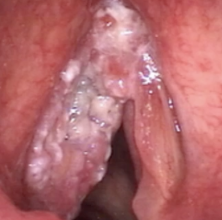
laryngeal cancer/malignant lesion description:
irregular growth
may be opaque/white, with irregular margins, abnormal color, mass, and stiffness
unilateral or bilateral
think irregular invasive-looking lesion rather than smooth benign mass
hoarseness/sore throat
smoking and alcohol are major risks
laryngeal cancer/malignant lesion picture:
granuloma/contact ulcer description:
posterior glottis at vocal process of arytenoid
granuloma = exophytic mass
contact ulcer = superficial ulcer
“cup and saucer” may occur
caused by LPR, intubation, cough/throat clearing, pressed low-pitched voice
granuloma/contact ulcer picture:

recurrent respiratory papillomatosis description:
irregular “grape-like” clusters with punctate/vascular appearance
HPV associated
may recur and require repeated surgery
recurrent respiratory papillomatosis picture:

glottic web description:
tissue bridge most often at the anterior commissure
congenital or acquired after surgery/trauma
reduced vibrating length can produce an abnormally high F0
glottic web picture:

presbylaryngis/presbyphonia description:
bowing/atrophy of the vocal folds with age
a spindle-shaped gap may be seen
loss of tissue bulk
breathy/thin/weak voice, reduced volume, instability
presbylaryngis/presbyphonia picture:

what changes in the phonatory system are brought by aging between infancy and childhood?
larynx descends in the neck
hyoid and thyroid overlap in newborns and separate with descent
the epiglottis becomes less curved/flattens with development
when are changes in the phonatory system brought by aging the most rapid?
the first 3 years, then slows until puberty
describe vocal fold proportion change brought by aging:
at birth: a large portion of the VF length is the arytenoid/vocal process
by 3: the membranous portion becomes dominant
in adults: the membranous portion is about 2/3 in total length
describe the change of the lamina propria brought by aging:
2-layer structure is established by age 5
3-layer fiber composition is established around age 13
prepubertal children common show a _____ _____ ___.
posterior glottal gap
85% of girls, 68% of boys ages 5-11
also common in young adult females
describe changes in the phonatory system brought by puberty:
sexual dimorphism becomes more apparent (roughly 11-16)
male laryngeal/vocal fold growth is greater
boys’ VF grow faster, and F0 drops more dramatically
girls have a smaller degree of voice change
what phonatory system changes are brought by adulthood/older age?
cartilage ossification begins by the end of the second decade and continues with age
aging may involve loss of VF tissue bulk/atrophy, bowing, reduced respiratory muscle strength, and altered neuromuscular control
what is presbyphonia?
voice changes associated with advanced age; may present with vocal fold bowing, breathy/thin or “muffled” voice, inadequate volume, and unstable quality.
what is the clinical relevance of changes in the phonatory system brought by aging?
age changes what is “normal”
pediatric airway size makes even small structural changes clinically important
do not pathologize a normal posterior gap in a child/young woman or normal developmental F0 differences
in older adults, distinguish expected aging from treatable pathology and assess functional impact rather than age alone
what is inflammation?
the immune system’s response to an irritant
what are possible triggers/causes of inflammation in the larynx?
pathogens (bacteria, viruses, fungi)
external injury/phonotrauma
chemical or radiation effects
refluxed gastric contents
allergens/immune reactions
autoimmune disease
other medical conditions
signs of inflammation in the larynx:
redness
heat
swelling
pain
loss of function
what do inflammatory mediators such as histamine and bradykinin do?
dilate/widen small blood vessels
allow immune cells/fluid into tissue
produce swelling
irritate nerves
can increase mucus production
label the 3 stages of wound healing?
inflammation
proliferation
remodeling
when does the inflammation/haemostasis stage take place?
0-2 days
when does the proliferation stage take place?
2-9 days
when does the remodeling stage take place?
~9-14 days onward; may continue up to ~2 years
describe the inflammation stage of wound healing
haemostasis: tissue stops the bleeding
inflammation happens
immune cells start to migrate to the site that needs fixing
describe the proliferation stage of wound healing
number of cells increases and the cells synthesizing proteins needed for extracellular matrix (ECM) reconstruction
granulation tissue starts to form, and it serves as a substitute for the lost tissue
broken blood vessels are repaired by the regeneration of new blood vessels (angiogenesis)
an epithelium that was broken becomes re-epithelialized, meaning a new epithelial layer will start to emerge to cover the wound
describe the remodeling stage of wound healing
the granulation tissue will become remodeled more like the original architecture of the lost tissue
the wound is now closed by the contraction of the epithelium
clinical relevance of the stages of wound healing:
tissue changes over time
vocal loading and treatment decisions should respect the healing phase
repeated trauma can disrupt normal healing and contribute to disorganized/thickened tissue, fibrosis, scar, or recurrent lesions
if tissue is still in a proliferative/healing process and the mass is simply removed without addressing the cause (such as in granuloma/contact ulcer), what happens?
the tissue may re-enter the wound-healing cycle and the lesion can recur
List the systemic conditions that can contribute to voice disorders
endocrine disorders:
hypothyroidism
hyperthyroidism
sexual hormone imbalance
autoimmune disorders:
allergy
HIV and AIDS
Sjogren’s syndrome
Wegener’s disease (GPA)
Describe how the systemic endocrine disorder of hypothyroidism contributes to voice disorders:
prolonged low thyroid hormone may produce vocal fold edema
mild-moderate dysphonia
vocal fatigue
reduced frequency/intensity ranges
Describe how the systemic endocrine disorder of hyperthyroidism contributes to voice disorders:
elevated thyroid hormone
breathy or tremulous quality
reduced loudness
Describe how the systemic endocrine disorder of sex-hormone imbalance contributes to voice disorders:
menstruation, pregnancy, menopause, and exogenous hormones/androgens can alter voice
androgenic effects may lower/masculinize the voice
Describe how the systemic autoimmune disorder of allergies contributes to voice disorders:
histamine-related mucosal edema/inflammation and increased secretions
may cause dysphonia and hyponasality
Describe how the systemic autoimmune disorder of HIV/AIDS contributes to voice disorders:
dysphonia may result secondarily from recurrent upper respiratory infection (URI) or laryngeal candidiasis
Describe how the systemic autoimmune disorder of Sjogren’s syndrome contributes to voice disorders:
dry mucosa/thick mucus
dysphonia, fatigue, compensatory muscle tension dysphonia (MTD), and potentially increased phonotrauma
Describe how the systemic autoimmune disorder of Wegners’s syndrome (granulomatosis with polyangiitis) contributes to voice disorders:
vasculitis may cause subglottic stenosis, nasal obstruction, or neurologic impairment/VF paralysis
voice changes are secondary to structural/neural disease
List the neurogenic conditions that can contribute to voice disorders
peripheral disorders
vocal fold paralysis
unilateral RLN paralysis
bilateral RLN paralysis
central disorders
spasmodic dysphonia/laryngeal dystonia
vocal tremor
Parkinson’s disease
myasthenia gravis
Describe how the neurological peripheral disorder of unilateral RLN paralysis contributes to voice disorders:
one immobile fold
breathy/weak voice if closure is poor
severity depends strongly on resting position and compensation
Describe how the neurological peripheral disorder of bilateral RLN paralysis contributes to voice disorders:
if folds are near the midline, voice may be relatively good but airway can be dangerously restricted with inspiratory stridor
if lateral, airway is better but voice is breathier/rougher
Describe how the neurological central disorder of spasmodic dysphonia/laryngeal dystonia contributes to voice disorders:
task-specific CNS disorder causing involuntary laryngeal spasms during speech
Describe how the neurological central disorder of Parkinson’s disease contributes to voice disorders:
reduced dopamine; basal ganglia dysfunction
weak voice, monoloudness, monopitch, rapid rate, reduced articulatory range
voice can often improve with cueing (“think loud”)