Foundations 1a exam 3
 Call Kai
 Learn
Learn Practice Test
 Spaced Repetition
 Match
Match Flashcards
 Knowt Play
1/100

There's no tags or description
Looks like no tags are added yet.
Name | Mastery | Learn | Test | Matching | Spaced | Call with Kai | Chat |
|---|
No analytics yet
Send a link to your students to track their progress
101 Terms
3 broad components of the innate immune system
physical/physiological barriers
antimicrobial molecules
sentinel/phagocytic cells
2 antimicrobial molecules produced by epithelial cells and leukocytes
defensins
cathelicidins
what do antimicrobial molecules do (4)
direct toxicity to pathogens/tumor cells
activate/recruit leukocytes
bind/neutralize LPS
stim wound healing
3 sentinel cells and thier functions
mast cell - initiate inflammation
macrophages - inflammation and phagocytosis
dendritic cells - process antigens and start adaptive responses
where are innate immune cells produced
in bone marrow via hematopoiesis
what do myeloid cells become (5)
granulocytes
mast cells
monocytes/macrophages
myeloid dendritic cells
platelets
what do lymphoid cells become (3)
T cells
B cells
NK cells
where does hematopoiesis occur in young animals and adults
young - most skeletal bones
adult - plat bones and proximal ends of humerus and femur
myeloblasts diff into what innate immune cells (5)
basophils
neutrophils
eosinophils
monocytes —> macrophages/meyloid dendritic cells
what color will neutrophils stain
none

eosinophil stain color
pink

basophil/mast cell stain color
blue/purple
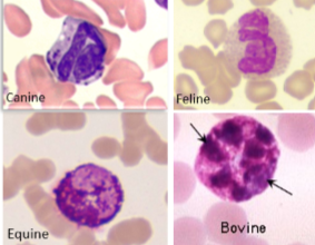
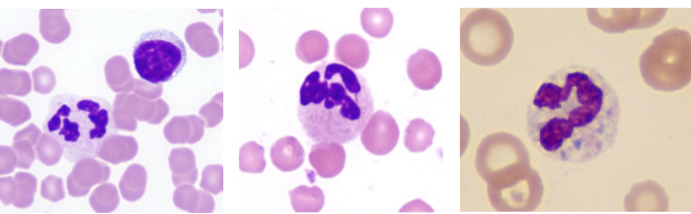
function of neutrophils
kills microbes via
phagocytosis
degranulation
neutrophil extracellular traps
which leukocyte is most abundant
neutrophils
which leukocyte is the “first responder”
neutrophils
how long do neutrophils live in blood
a few days
function of eosinophils
kills parasites,
promotes inflammation
involved in hypersensitivity rxns
func of basophils
similar to eosinophils and mast cells
hypersensitivity rxns
coagulation
parasite rejection

which is the largest leukocyte
monocytes
what color is the cytoplasm of monocytes
blue/grey
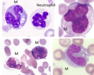
monocytes are the circulating precursors to (2)
macrophages and Antigen presentinc DCs
macrophage func (3)
inflammation response
Phagocytize cell debris, foreign material, and infectious agents
Secretion of inflammatory cytokines to recruit other inflammatory cells
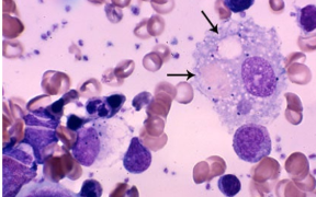
function of dendritic cells (2)
endo/phagocytosis
antigen presentation
mast cell func (4)
vasodilation
vascular permeability
recs other leukocytes
allergic response

do NK cells need prior sensitization
no
where do NK cells mature
2ndary lymphoid organs
NK cell func (2)
1st line of defense agasint viruses and tumors
secrete cytotoxic granules that drive apoptosis
where do NK cells originate and are they part of the innate or adative immune system
from lymphoid tissue and mature in 2ndary lymph tissue but are part of innate
if we see hella neutrophils is the disease acute or chronic
acute
if we see hella macrophages, lymphocytes, and plasma cellsis the disease acute or chronic
chronic
what is the potential pathogen if we see lymphoccytes
viruses or immune mediated tumors
what is the potential pathogen/disease if we see mast cells/basophils/eosinophils
allergies (alos tumors)
what is the potential pathogen/disease if we see eosinophils
parasites or allergy
what is the potential pathogen/disease if we see macrophages
intracellular bacteria/parasites, fungi, foreign bodies
what is the potential pathogen/disease if we see neutrophils
extracellular bacteria
where are PAMPs located
on pathogens
where are DAMPs located
they are released from damages or dying host cells post injury
where are PRRs located
on host sentinel cells to rec PAMP and DAMP
where are signaling PRRs located
can be located on cell surface, cytoplasm or endosomal
toll like receptors are a type of signaling PRR, where is it located and what does it bind
membranous binds various ligands
NOD like receptors are a type of signaling PRRs where are they located
cytoplasm, binds peptidoglycan and muramyl dipeptide (parts of bacteria)
RIG-1 like receptors are a type of signaling PRR where is it located and what does it bind
cytoplasm - viral dsRNA
all 3 signaling PRRs signal through which two transcription factors to make pro-inflammatory cytokines, chemokines, adhesion molecules, or costimulatory molecules
NF-kB or IRF
function of the inflammasome
rapid inflammation to fight a pathogen by producing proinflammaotry proteins (cytokines) IL-1beta and IL-18
how do inflammasomes get activated
1 rec PAMP/DAMP via PRR and activate NF-kB
2.) activation of NOD-like Receptor to activate enzyme to activate precursor IL-1B and IL-18
for signaling PRRs what happens after phagocyte activation (4)
cytokine production (inflammatory cascade)
chemokine production (leukocyte rec)
expression of adhesion molecules on leukocytes (lets them go from blood → tissue)
production of other mediators to help leukocytes (arachidonic acid, platelet-activating factor)
3 types of phagocytic receptors on neutrophils, macrophages, DCs, and what they recognize
complement receptors - microbes coated with complement proteins (C3b)
FcyR - microbes coated with antibodies (IgG)
mannose receptor - sugar residues on microbes

4 steps of phagocytosis
recognition and attachment of microbe
engulfment (formation of phagosome)
phagosome-lysosome fusion
destruction

what cels contain the phagocytic PRRs
neutrophils, macrophages, DCs
4 ways the phagolysosome kills microbe
acidic pH
oxidative/respiratory burst
generation of reactive nitrogen species
granulation
during maturation pf the phagosome all the steps contain what that are harmful to the microbe
free radicals
do birds and reptiles have myeloperozidase
no
function of NET formed by neutrophils in response to cytokines (IL-8) and PAMPs (LPS)
microbicidal
keeps pathogen in place and starts killing it
function of “major basic protein” in eosinophil
helps fight parasites
2 Rs of NK cells and what it recognizes
activation R - viral proteins, glycoproteins, antibody coated cells
inhibitory R - MHC 1
What happens with NK cells when the activation receptor is active but not inhibitory (no MHC 1 seen) (2)
Perforin inserts into membrane (punches hole)
Granzyme enters pore —> apoptosis
where do complement proteins come from (3)
hepatocytes, manocytes/macrophages GI epithelial cells
complement system nomenclature: inactive comlement
simple number: C1, C2, etc
complement system nomenclature: cleavage product
lowercase suffix: C3a, C3b, etc
for the complement system, what does the complement bind for the alternative pathway, classical pathway, and lectin pathway
alt- directly on pathogen
classical - antibody on path
lectin - lectin binds to mannose on pathogen cell wall
3 final outcomes of the CS
phagocytosis via nuetrophils and macrophages
pathogen killing
inflammatory response
all 3 CS pathways lead to what common complement protein being deposited on microbe
C3b
for the complement system, what is involved for the inflammation pathway
C3a - increases vascular permeability, degranulation of mast cells and basophils
C5a - stimulates neutrophil chemotaxis

for CS, what is involved in the opsonization pathway
C3b on bacterium read by a receptor and causing phagocytosis

For CS, what is involved in the lysis of pathogen pathway
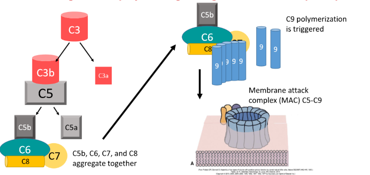
C5-C9 causes polymerization to form MAC that breaks a hole in microbe cell membrane
5 control mechanisms of the complement system
C1- inactivator
CD55, Factor H - prevents complement from attacking host
CD59 - inhibits MAC
complement Rs - ensure removal of antigen-antibody-complement complexes
what are professional APCs
those that present antigen and coordinate response with the adaptive immune system
Dendritic cells, macrophages, B-cells, sometimes neutrophils?
mature vs naive lymphocyte
mature - developed and contains its antigen receptor
naive - lymphocyte that has not encountered specific antigen
what are secondary lymphoid organs and examples
where lymphocytes will activate
-spleen
-lymphatics + nodes
MALT
what part of bone marrow does hematopoiesis occur
red
diferences in TCRs and BCRs
BCRs bind specific protein (antigen) and it doesnt need to be intracellularly processed
TCRs bind processed antigens displayed by MHCs
what happens once B cell and T cells are activated in secondary lymphoid tissue
clonal expansion
does the term humoral relate to B cells or T cells
B cells
does the term cell-mediated relate to B cells or T cells
T cells
function of spleen in terms of adaptive immunity
screens blood

what is occuring at afferent lymph vessels
lymph is being brough in
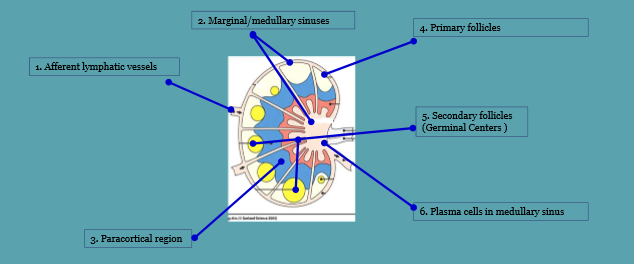
what is occuring at marginal/medullary sinuses
macrophages and denritic cellls can find antigen while filtering thorugh

what is happening at paracortial region
mature T and B communicate with macrophages/dendritic cells

what is happening at primary folicles
T and B cells located and inactive

what happening at secondary follicles
active B and T cells are responding to antigen and differentiating

whats happening at plasma cells in medullary sinus
antibodies are being released
red vs white pulp of spleen
red - where RBCs and macrophages are
white - lymphoid tissue that surrounds arteriols
3 functions of lymphatic system
collection of excess fluid
transport of fats from GI to bloodstream
enable immune response
fluid component of lymph
plasma-like
electrolytes
small and large molecules contained in lymph
small - proteins
large - microbes
right arm/head drains to where, and where does everything else drain to (lymphatics)
R arm and head → Right subclavian v
everything else → left subclavian v
what two structures does lymph move through as it goes from caudal to cranial
cisterna chylii and thoracic duct

2 ways lymph can move
via skeletal muscle increasing external pressure
or SM cells contracting along lymph vessels
what is an immunogen
an antigen that can induce an immune response (binds to BCR and TCR)
antigen def
any substance that can bind to an antigen receptor
hapten def
an antigen that cant induce an immune resposne on its own, needs a more complex molecule to be immonogenic

epitope definition
part of antigen that is recognized
conformational vs linear determinant for BCR epitopes
conformational - determinant is lost by denaturation
linear - determinant can be denatured or native

MHC 1 vs MHC 2 expression and what it binds to
MHC 1 is on all nucleated cells and presents to CD8+ T cells
MHC 2 is on professional APCs and presents to CD4+ T cells
MHC 1 vs MHC 2, which binds a larger seq of AA peptides
MHC class 2
how many MHC genes does 1 have and how is it expressed
several genes
expressed co-dominantly
peptide source for MLH class 1 (intracellular or extracellular)
intracellular
peptide source for MLH class 2 (intracellular or extracellular)
extracellular (endocytosed, lysosome, endosome)
what blocks the binding groove of MHC class 2 proteins until endosomal compartment binds
invariant chain
for MHC class 1 antigen processing how does the peptide transport into the ER where MHC 1 molecule is present
via TAP