SBMS1431 Human Anatomy and Physiology I
 Call Kai
 Learn
 Practice Test
 Spaced Repetition
 Match
 Flashcards
 Knowt Play
1/35

Earn XP
Description and Tags
MECHANICS OF VENTILATION AND LUNG VOLUMES
36 Terms
Respiration
Ventillation (breathing)
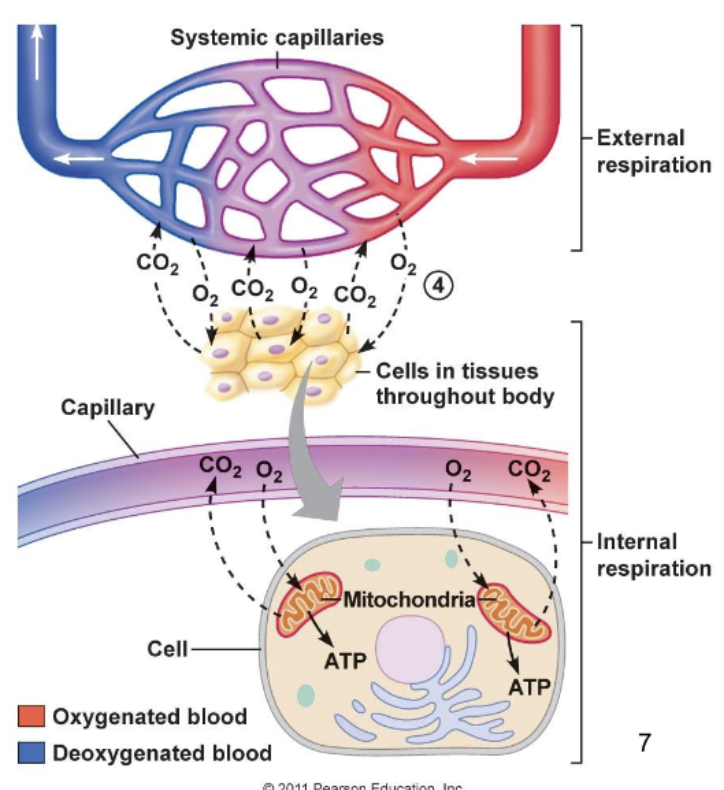
Gas exchange between blood and lungs & between blood and tissues
Oxygen utilization by tissues to produce ATP
External respiration: Ventilation and gas exchange in lungs
Internal respiration: Oxygen utilization and gas exchange in tissues.
External respiration
Pulmonary ventilation: Air moves in and out of the lungs
Gaseous exchange by diffusion (between air and blood): Oxygen and carbon dioxide are exchanged between lung tissue and the blood
Transportation of gases: Oxygen and carbon dioxide are transported in the blood

Internal Respiration
Gaseous exchange by diffusion (between blood and tissues): Oxygen and carbon dioxide are exchanged between systemic tissues and the blood
Use of oxygen and production of ATP and carbon dioxide by cells, primarily within the mitochondria.
Respiratory Zone vs. Conducting Zone
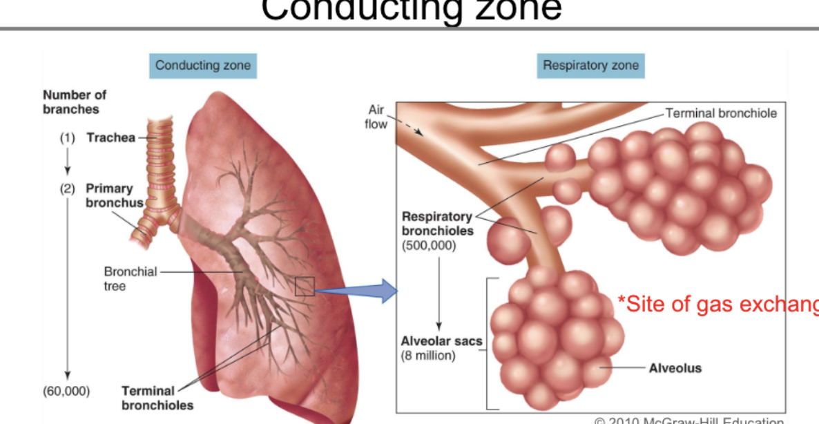
Conducting zone: Air travels from nasal cavity→ pharynx → larynx → trachea → right and left primary bronchi → secondary bronchi → tertiary bronchi → (more branching) → terminal bronchioles
Respiratory zone: Respiratory bronchiles → Terminal alveolar sacs
Functions of Conducting Zone
Transports air to the lungs
Warms, humidifies, filters, and cleans the air
*Mucus (secreted by goblet cells) Traps small particles, and cilia (hair-like projections) move them away from the lungs.
Voice production in the larynx as air passes over the vocal folds.

Respiratory Zone: Respiratory Bronchioles and Alveolar Sacs
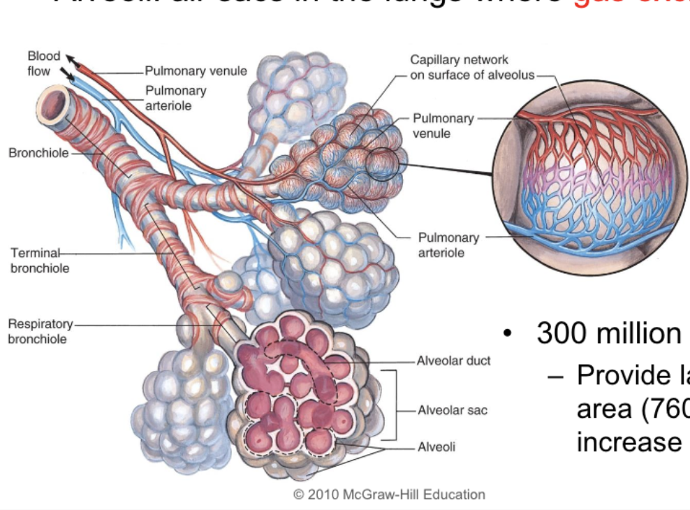
Alveoli: air sacs in the lungs where gas exchange occurs
300 million of them (provide large surface area (760 square feet) to increase diffusion rate
Mechanics of Ventilation: Pulmonary Pressures
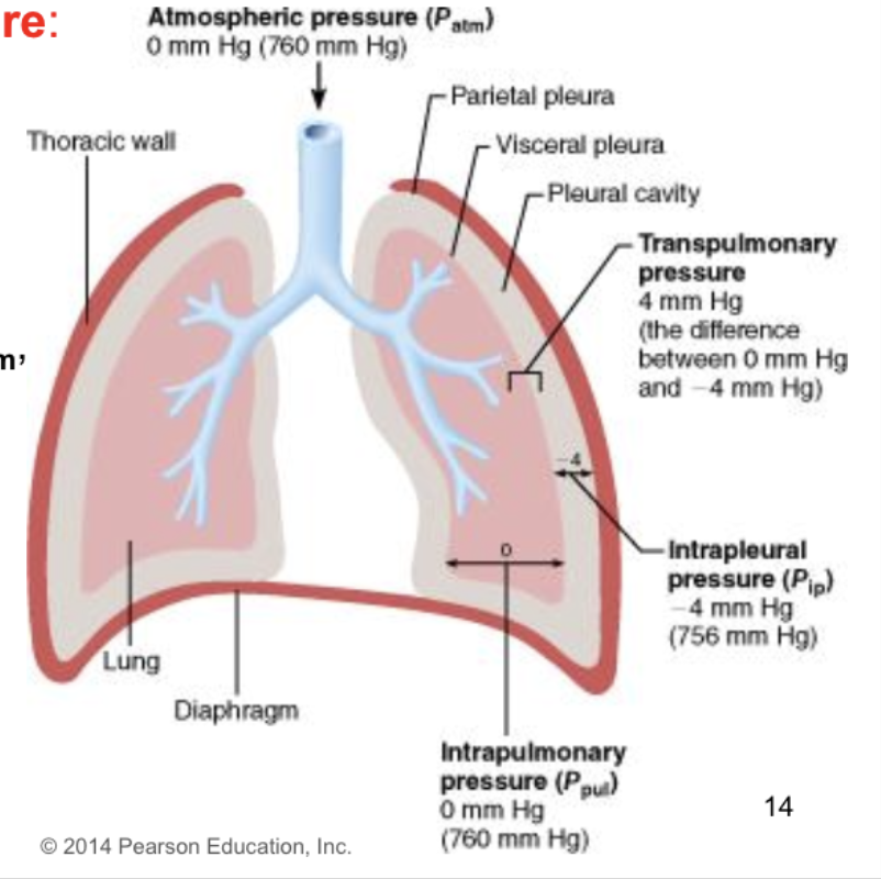
Atmospheric Pressure (Patm): the pressure of the outside air; normal value at sea level is 760 mm Hg
Intra-alveolar pressure (Palv) or Intrapulmonary pressure: pressure of air within the alveoli, which varies with ventilation. At rest it is equal to Patm, and is considered to be 0 mm Hg.
Intrapleural pressure (Pip): pressure within the intrapleural space (between parietal and visceral pleura), which varies with ventilation. At rest it is -4 mm Hg.
Transpulmonary pressure: difference between the intrapleural pressure and the intra-alveolar pressure (Palv- Pip)
Parietal and Visceral Pleura
Thoracic cavity contaisn the heart, trachea, esophagus, and thymus within the central mediastinum while lungs fill the rest of the cavity
Each lung occupies a pleural cavity lined with serous membrane (pleura) and lubricated with intrapleural fluid
The parietal pleura lines the thoracic wall
The viesceral pleura covers the lungs
The parietal and visceral pleura are normally pushed together, with a potential space between called the intrapleural space

Intrapulmonary pressure
During inspiration it is less than Patm
During expiration it is greater than Patm
At rest it is equal to Patm, i.e, 0 mm Hg relative to atmosphere.
Intrapleural Pressure
Varies during ventilation but it is always less than intra-alveolar pressure in both inhalation and exhalation
It is always negative (i.e less than atmospheric pressure) during normal breathing.
This negative pressure results from the elastic forces exerted on the intrapleural space by the chest wall and the lungs.
Significance: The elastic forces are trying to keep the
intrapleural space open, i.e. keep the lungs inflated and
prevent them from collapsing
How to maintain negative intrapleural pressure?
To maintain the negative intrapleural pressure the pleurae
need to be airtight.If conditions such as trauma (gunshot or knife wound) or
disease (pneumonia or emphysema) occur, air enters this
space and the condition is called pneumothorax.The lung collapses and the thoracic wall expands

Transpulmonary Pressure
Equals the difference between the intrapleural pressure
and the intrapulmonary pressure.This force operates across the walls of the lungs and
causes the lungs to expand
Mechanics of Breathing
Air moves down a pressure gradient (from higher to lower pressure)
Pressure differences between the two ends of the conducting zone occur due to changing lung volumes
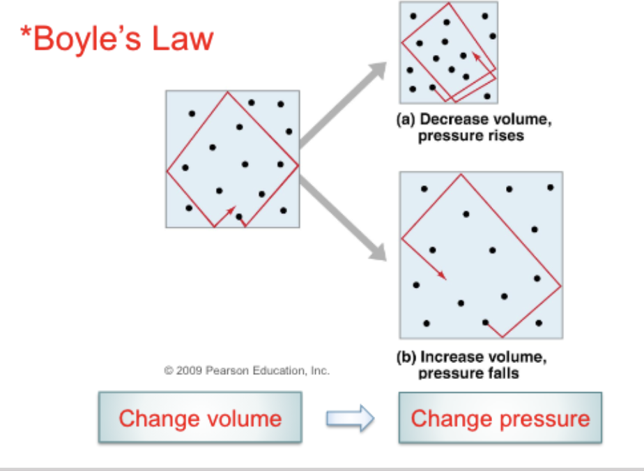
What is the relationship between pressure and volume?
Inversely proportional (stated according to Boyle’s law)
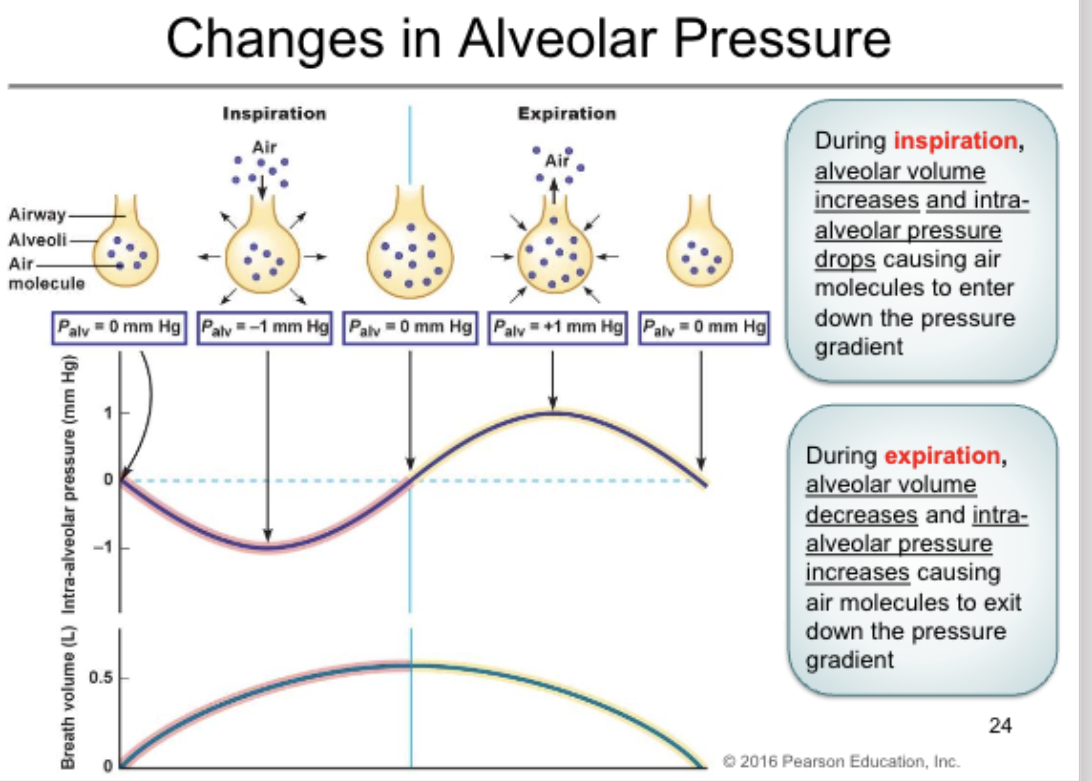
During inspiration, an increase in lung volume decreases intra-alveolar pressure to sub-atmospheric levels, i.e, the pressure in the alveoli is less than that in the atmosphere.
Air moves in
During exhalation, a decrease in lung volume increases intra-alveolar pressure above atmospheric levels, i.e. the pressure in the alveoli exceeds that in the atmosphere.
Air moves out
Pressure Differences during Inhalation and Exhalation
Inhalation: Intra-alveolar pressure is lower than atmospheric pressure.
Pressure below that of the atmosphere is called sub-atmospheric or negative pressure.
ExhalationL Intra-alveolar pressure is greater than atmospheric pressure.
How do changes in volume and pressure drive air in and out of the lungs?
At Rest: Pressure outside equals pressure inside, so no air moves.
Inhalation: The diaphragm contracts and ribs elevate, so volume increases. Pressure inside falls, so air flows in.
Exhalation: Muscles relax, so volume decreases. Pressure inside rises, so air flows out.

How does intra-alveolar pressure change to drive airflow during inspiration and expiration?
Inspiration (Air In): Alveolar volume increases → intra-alveolar pressure drops (to about -1 mm Hg) → air flows in down the pressure gradient.
Expiration (Air Out): Alveolar volume decreases → intra-alveolar pressure increases (to about +1 mm Hg) → air flows out down the pressure gradient.
Key Rule: Air always flows from higher pressure to lower pressure. At the start and end of each breath, alveolar pressure equals atmospheric pressure (0 mm Hg), so no air moves.
Which muscles are primarily involved in breathing, and what do they do during inspiration and normal expiration?
Inspiration: External intercostal muscles contract (moving ribs up and out) and the diaphragm contracts (moving down). This expands the chest cavity.
Normal Expiration: Both the external intercostals and the diaphragm relax, causing the chest cavity to shrink back passively.

What changes during active expiration (like when blowing out candles or exercising)?
The external intercostals relax, but the internal intercostal muscles and abdominal muscles contract.
This forces the ribs and sternum to depress, actively pushing air out faster.

What muscles and structural changes drive Inspiration versus Expiration?
External intercostals contract (raise ribs up and out).
Diaphragm contracts (moves down).
Result: Vertical and A-P (front-to-back) diameters increase.
Expiration:
Internal intercostals contract (pull ribs down).
Abdominal muscles contract (push diaphragm up).
Result: Ribs are depressed and the chest cavity shrinks.
What happens to the diaphragm and rib cage during inspiration vs. expiration?
Inspiration: The diaphragm contracts (moves down) and the rib cage is elevated. This enlarges the thoracic cavity.
Expiration: The diaphragm relaxes (moves up) and the rib cage drops. This reduces the space in the thoracic cavity.
Which muscles are used during forced expiration?
Internal intercostal muscles (pull ribs down).
Abdominal muscles (push diaphragm up).
How does the diaphragm act as a lever during inspiration?
When the diaphragm contracts, it pushes down on the abdominal contents.
This leverage effect pushes the rib cage up and outward, helping to expand the chest cavity.
What is the relationship between barometric (atmospheric) pressure and alveolar pressure at the end of expiration, and what does this mean for airflow?
Barometric air pressure (PB) and alveolar air pressure (Palv) are equal.
Because the pressures are equal, no air moves into or out of the lungs. (This is the brief pause between breaths).
What is Spirometry, and what does it measure?
What it is: A test where the subject breathes into and out of a device to record the volumes of inspired and expired air (creating a spirogram).
What it measures: Lung volumes and lung capacities.
Purpose: It can be used to test for and diagnose respiratory disorders.
Define Tidal Volume (VT) and Inspiratory Reserve Volume (IRV), including their average values.
Tidal Volume (VT): The volume of air that moves in and out in one unforced breath. Average = 500 mL.
Inspiratory Reserve Volume (IRV): The maximum volume of air you can breathe in at the end of a normal inspiration. Average = 3000 mL.

Define Expiratory Reserve Volume (ERV) and Residual Volume (RV), including their average values.
Expiratory Reserve Volume (ERV): The maximum volume of air you can breathe out at the end of a normal expiration. Average = 1000 mL.
Residual Volume (RV): The volume of air remaining in the lungs after a maximum expiration. Average = 1200 mL.

Which lung volumes can be measured directly, and which must be measured indirectly?
Measured Directly: Tidal volume, Inspiratory reserve volume, and Expiratory reserve volume.
Measured Indirectly: Residual volume (because you can't physically breathe it out to measure it).
Define Inspiratory Capacity (IC) and Vital Capacity (VC), including their average values.
Inspiratory Capacity (IC): The maximum volume of air that can be inspired at the end of a resting expiration. Average = 3500 mL.
Vital Capacity (VC): The maximum volume of air that can be forcefully expired after a maximum inspiration. Average = 4500 mL.
Define Functional Residual Capacity (FRC) and Total Lung Capacity (TLC), including their average values.
Functional Residual Capacity (FRC): The volume of air remaining in the lungs at the end of a tidal (normal) expiration. Average = 2200 mL.
Total Lung Capacity (TLC): The total volume of air in the lungs at the end of a maximum inspiration. Average = 5700 mL.
Relationships between Lung Volumes and Capacities
Inspiratory Capacity = tidal volume + inspiratory reserve volume
Vital Capacity = tidal volume + inspiratory reserve volume +
expiratory reserve volume
Functional Residual Capacity = expiratory reserve volume + residual volume
Total Lung Capacity = tidal volume + inspiratory reserve volume +
expiratory reserve volume + residual volume
Pictorial Example

Minute Ventilation (VE)
Minute Ventilation (VE): Total amount of air that flows in or out of respiratory system in a minute
Equals tidal volume x respiration rate or number of breaths per minute
Example:
VE= tidal volume x breaths per minute
= (500 mL/breath) x (12 breaths/min)
= 6000 mL/min
What is Alveolar Ventilation (VA) and Anatomical Dead Space?
Alveolar Ventilation (VA): The volume of fresh air that reaches the alveoli each minute.
Anatomical Dead Space: The combined volume of non-exchanging airways (the conducting zone). Air here does not participate in gas exchange.
How to calculate VA ?
VA =VE - (DSV *breaths per minute)
(Minute Volume minus Dead Space Volume per minute)
(500 mL/breath×12)−(150 mL/breath×12)=4200 mL
What happens to the air in the conducting zone during breathing?
At the end of expiration (a): All air in the airways is "stale" (old air).
During inspiration (b): The stale air in the dead space enters the alveoli first, followed by fresh atmospheric air.
During expiration (c): Atmospheric air in the conducting zone is expired first, followed by stale air from the lungs.
