Morphology — Definitions, Clinical Conditions & Cell Pictures
 Call Kai
 Learn
Learn Practice Test
 Spaced Repetition
 Match
Match Flashcards
 Knowt Play
1/122

Earn XP
Description and Tags
Revised morphology review with focused cards for appearance, causes, and clinical conditions. Objectives 1–11 use the lecture; objective 12 includes matching cell pictures. Missing images: ASH Image Bank (Vicari, Scordino, Schrier, Maslak) and CDC DPDx case 287 (University of Texas HSC).
Name | Mastery | Learn | Test | Matching | Spaced | Call with Kai | Chat |
|---|
No analytics yet
Send a link to your students to track their progress
123 Terms
Anisocytosis — definition
Variation in RBC size.
Poikilocytosis — definition
Variation in RBC shape.
Normochromic — definition
Normal RBC coloration, with central pallor about 2–3 microns across.
Hypochromic — definition
Increased central pallor, greater than 3 microns. MCHC helps assess hypochromia.
Normal RBC — size
About 7–8 microns in diameter. Normal size variation is about 5%.
Normal RBC — membrane proteins
Glycophorin and spectrin.
RBC maturation — main changes
The cell loses its nucleus, becomes smaller, and changes cytoplasmic color as it matures.
RBC morphology — how much of the smear should be reviewed?
At least 10 oil-immersion fields. Check that abnormalities are consistent and not artifacts.
RBC morphology — which indices should be reviewed?
MCV, MCH, MCHC, and RDW.
Anisocytosis and poikilocytosis — slight to 2+ grading
Slight: 5–10% of cells. 1+: more than 10–25%. 2+: more than 25–50%.
Anisocytosis and poikilocytosis — 3+ and 4+ grading
3+: more than 50–75% of cells. 4+: more than 75%.
Normal RBC — appearance
Reddish with Wright’s stain, with a central pale area about 2–3 microns across.

Macrocytic cells — associated conditions and processes
Impaired DNA synthesis; increased erythropoiesis with early reticulocyte release; increased membrane cholesterol and lecithin. Megaloblastic processes may produce macro-ovalocytes or macro-teardrops. Sideroblastic anemia can include macrocytes in a mixed-size population.
Microcytic cells — clinical conditions
Iron deficiency; thalassemic conditions; sideroblastic anemia; anemia of chronic disease; lead poisoning.
Mixed-size RBC population — associated anemia
Sideroblastic anemia can produce a dimorphic blood picture containing macrocytic, normocytic, and microcytic cells.
Macrocytosis — why does impaired DNA synthesis enlarge RBCs?
Fewer cell divisions occur, so the resulting cells are larger.
Macrocytosis — why can increased erythropoiesis cause it?
Reticulocytes or diffusely basophilic erythrocytes enter circulation prematurely.
Microcytosis — why does defective hemoglobin synthesis make cells smaller?
The cells undergo more divisions, producing a microcytic, hypochromic blood picture.
Microcytosis — iron and globin problems
Ineffective iron absorption, release, or utilization; decreased or defective globin synthesis.
Macrocytes — features to assess
Oval or round shape; red or blue color; central pallor; inclusions; and the reason for the size change.
Hypochromia — clinical conditions
Iron-deficiency anemia is specifically associated with hypochromia. Defective hemoglobin synthesis produces a microcytic, hypochromic picture.
Hypochromia — can it occur in different cell sizes?
Yes. Microcytic, normocytic, and macrocytic cells can be hypochromic. Target cells are included in the macrocytic hypochromic category.
Pseudohypochromia — definition and cause
Artificially induced pallor caused by water artifact. The pale area is distinctly outlined.
Hypochromia — 1+ and 2+ grading
1+: central pallor occupies half the cell diameter. 2+: central pallor occupies two-thirds.
Hypochromia — 3+ and 4+ grading
3+: central pallor occupies three-fourths of the cell diameter. 4+: only a thin rim of hemoglobin remains.
Polychromasia — definition and appearance
Immature RBCs enter peripheral blood and appear gray-blue rather than red.
Polychromasia — cause of the blue color
Residual RNA produces basophilia.
Polychromasia — associated process
Increased erythropoiesis with premature release of reticulocytes or diffusely basophilic erythrocytes.
Polychromatophilic macrocytes — cell type and confirming stain
They are reticulocytes. Methylene blue supravital staining demonstrates the reticular material.
Polychromasia — slight to 2+ grading
Slight: 1% of RBCs. 1+: 3%. 2+: 5%.
Polychromasia — 3+ and 4+ grading
3+: 10% of RBCs. 4+: more than 11%.
Target cell — name and appearance
Also called a codocyte. A target-like cell with increased surface membrane; bell shaped in three dimensions and hypochromic.

Target cell — primary defect or cause
Excess membrane cholesterol and phospholipids, or decreased cellular hemoglobin. Osmotic fragility is decreased.
Target cell — clinical conditions
Hgb C disease; Hgb C trait; post-splenectomy; liver disease; iron-deficiency anemia; other Hgb abnormalities.

Spherocyte — name and appearance
Small, round cell with no central pallor and a low surface-area-to-volume ratio.

Spherocyte — primary defect or cause
Membrane loss causes an irreversible shape change. Hereditary spherocytosis involves decreased spectrin quantity and function.
Spherocyte — clinical conditions
Immune hemolytic anemia; hereditary spherocytosis; post-transfusion.

Ovalocyte — name and appearance
Egg-shaped RBC. The picture shows macro-ovalocytes, the enlarged oval form.

Ovalocyte — clinical conditions
Myelodysplastic syndrome; thalassemic syndromes; megaloblastic processes. Ovalocytes may be macrocytic.
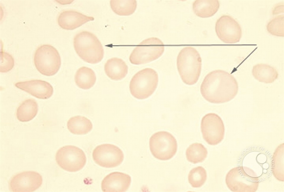
Elliptocyte — name and appearance
Elongated, pencil-shaped RBC.

Elliptocyte — primary defect or cause
Hereditary elliptocytosis involves a membrane defect in spectrin and other membrane protein that may cause hemolysis.
Elliptocyte — clinical conditions
Iron-deficiency anemia; hereditary elliptocytosis; idiopathic myelofibrosis.

Ovalocytes and elliptocytes — terminology
Ovalocytes are egg shaped; elliptocytes are pencil shaped. Some sources use the terms interchangeably.
Stomatocyte — name and appearance
RBC with a slit-like area of central pallor on an air-dried smear.

Stomatocyte — primary defect or cause
The change can be reversible. Chemical agents and artifact can produce the appearance.
Stomatocyte — clinical conditions
Artifact; hereditary spherocytosis; hereditary stomatocytosis; acute alcoholism; Rh null phenotype.

RBC fragmentation — main mechanism
Loss of membrane. Abnormal fluid circulation or an intrinsic RBC defect makes cells less deformable and promotes fragmentation.
RBC fragments — named forms
Schistocytes; burr cells; helmet cells (keratocytes); bite cells (degmacytes).
Schistocyte — name and appearance
A tiny, irregular RBC fragment.

Schistocyte — primary defect or cause
RBC fragmentation due to abnormal circulation or an intrinsic RBC defect, with reduced deformability and membrane loss.
Schistocyte — clinical conditions
Microangiopathic hemolytic anemia; disseminated intravascular coagulation; heart valve surgery; hemolytic uremic syndrome; thrombotic thrombocytopenic purpura; burns.

Echinocyte — name and appearance
Burr cell with multiple short, evenly spaced projections and retained central pallor.

Echinocyte — primary defect or cause
May be an artifact of smear preparation or storage; crenated cells can resemble true burr cells.
Echinocyte — clinical conditions
Uremia; heart disease; stomach cancer; peptic ulcer; post-heparin injection; hypothyroidism; dehydration; azotemia.

Agglutination — name and appearance
RBCs aggregate into irregular clusters.

Agglutination — primary defect or cause
RBC antibodies react with RBC antigens. Cold-related agglutination can occur at room temperature and disappear on warming.
Agglutination — clinical conditions
Cold antibody syndromes, including cold hemagglutination disease and paroxysmal cold hemoglobinuria.

Rouleaux — name and appearance
RBCs arranged in stacks resembling coins.

Rouleaux — primary defect or cause
Increased plasma globulins or fibrinogen, with protein deposition or adsorption on RBC membranes. Associated with a high ESR.
Rouleaux — clinical conditions
Multiple myeloma; Waldenstrom’s macroglobulinemia; chronic inflammatory disorders.

Agglutination — definition and cause
Clustering of RBCs caused by antibody reacting with RBC antigen.
Agglutination — associated conditions and warming response
Cold antibody syndromes. Clumping present at room temperature may disappear when the specimen is warmed.
Rouleaux — definition and cause
Stacks of RBCs resembling coins, associated with increased plasma globulins or fibrinogen.
Rouleaux — clinical conditions
Multiple myeloma; Waldenstrom’s macroglobulinemia; chronic inflammatory disorders.
Rouleaux — effect of saline dilution
The stacks disappear with saline dilution.
Howell-Jolly body — name and appearance
A small, round, dark-purple inclusion. Usually single or double and eccentrically located.

Howell-Jolly body — primary defect or cause
A remnant of nuclear DNA.
Howell-Jolly body — clinical conditions
Splenectomy; thalassemia; hemolytic anemia; megaloblastic anemia; functional hyposplenia.
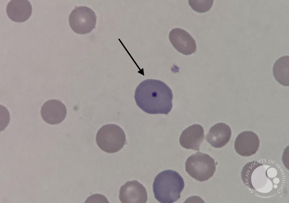
Basophilic stippling — name and appearance
Multiple small blue-purple stipples dispersed within an RBC.

Basophilic stippling — primary defect or cause
Ribosomal material: ribonucleoprotein (RNA) and mitochondrial remnants.
Basophilic stippling — clinical conditions
Lead intoxication; thalassemia; disturbances of heme synthesis.

Pappenheimer bodies — name and appearance
Small, irregular magenta inclusions near the RBC periphery, often in clusters.
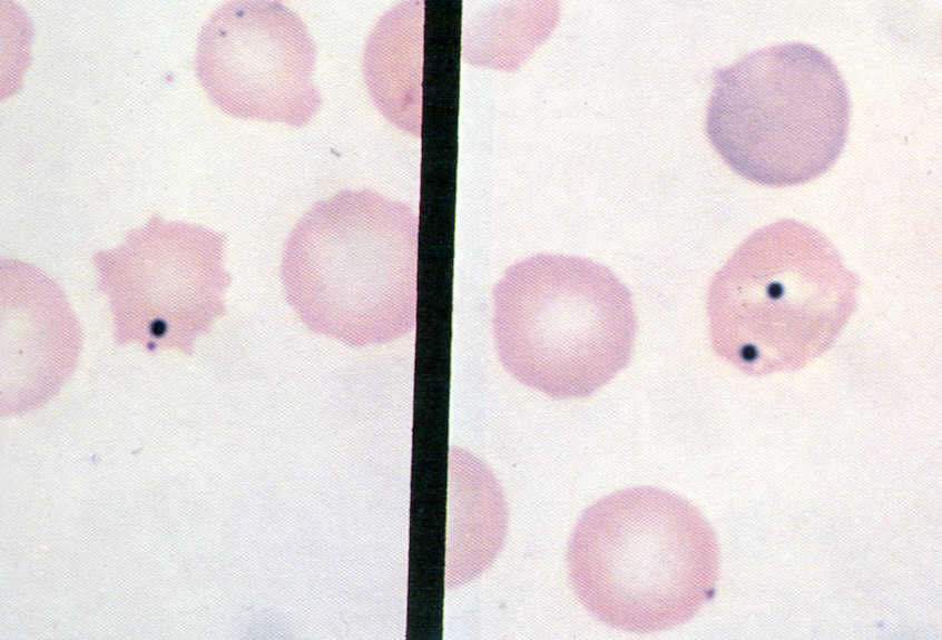
Pappenheimer bodies — primary defect or cause
Iron-containing material. Wright-stained inclusions are called Pappenheimer bodies; Prussian blue confirms iron and demonstrates siderotic granules.
Pappenheimer bodies — clinical conditions
Sideroblastic anemia; hemochromatosis or hemosiderosis; sickle-cell or thalassemic hemoglobinopathies; splenectomy.

Cabot ring — name and appearance
A delicate ring or figure-eight structure, sometimes resembling beads on a necklace, in a stippled RBC.
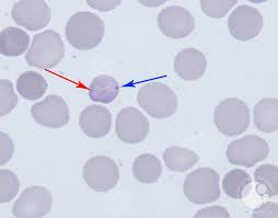
Cabot ring — primary defect or cause
The inclusion contains arginine-rich histone and non-hemoglobin iron.
Cabot ring — clinical conditions
Megaloblastic anemia; homozygous thalassemia; post-splenectomy.

Howell-Jolly bodies — material and location
DNA remnants, present singly or doubly in an eccentric position.
Howell-Jolly bodies — clinical conditions
Splenectomy; thalassemia; hemolytic anemia; megaloblastic anemia; functional hyposplenia.
Heinz bodies — definition and effect
Denatured or precipitated hemoglobin that becomes rigid and distorts the RBC membrane.
Heinz bodies — useful stains
May not be visible with Wright’s stain. Crystal violet and brilliant cresyl blue may demonstrate them.
Heinz bodies — clinical conditions
Alpha-thalassemic syndromes; G6PD deficiency; chemical insult to RBCs.
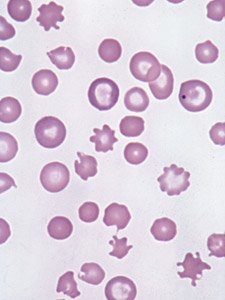
Acanthocyte — name and appearance
A smaller RBC with 3–12 irregular spicules.

Acanthocyte — primary defect or cause
Increased membrane cholesterol and surface area with decreased lecithin. Lecithin-cholesterol acyltransferase deficiency can increase cholesterol and produce acanthocytes.
Acanthocyte — clinical conditions
Severe hepatic disease; alcohol intoxication (Zieve’s syndrome); pyruvate kinase deficiency; congenital abetalipoproteinemia; vitamin E deficiency; post-splenectomy.
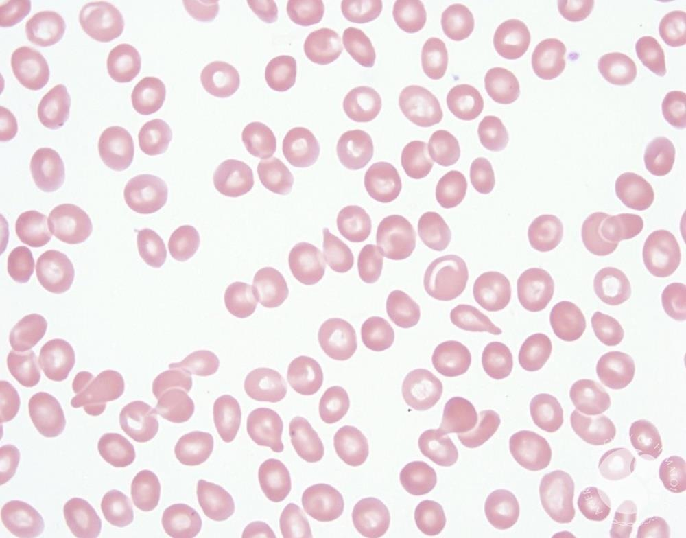
Teardrop cell — name and appearance
A pear-shaped RBC with a tail.

Teardrop cell — primary defect or cause
Distortion into a pear shape with a tail; associated with inclusion-body formation and myelofibrosis.
Teardrop cell — clinical conditions
Idiopathic myelofibrosis with myeloid metaplasia; thalassemia syndromes; iron deficiency; drug-induced Heinz body formation; other inclusion-body conditions; megaloblastic processes with macro-teardrops.
Drepanocyte (sickle cell) — name and appearance
Rigid, inflexible RBC with a pointed projection or sickle shape.

Drepanocyte (sickle cell) — primary defect or cause
Low oxygen causes hemoglobin tubules to form and bundle, deforming the cell. Repeated episodes can make the sickling irreversible.
Drepanocyte (sickle cell) — clinical conditions
Sickle-cell disease. With sickle-cell trait, sickled cells are usually absent from the routine smear.

Sickle cells — can the shape reverse?
Some return to their usual shape with oxygen; repeated sickling episodes can produce irreversibly sickled cells.
Malaria — name and appearance
Plasmodium parasites within RBCs. This Wright-stained example shows delicate ring forms with small chromatin dots in normal-sized RBCs; some are at the cell edge.

Malaria — primary defect or cause
Plasmodium infection of RBCs. The pictured example is Plasmodium falciparum.
Malaria — clinical conditions
Malaria. This example is falciparum malaria; the ring appearance is a parasite finding, not an RBC nuclear remnant.

Normal platelet — morphology
Small and round, with blue granules throughout light-blue cytoplasm.
Abnormal platelet — possible appearances
Agranular, large, tailing, or streaming platelets.
Abnormal platelet morphology — clinical conditions
Variations occur in idiopathic myelofibrosis. Large platelets occur with increased platelet turnover, idiopathic thrombocytopenia purpura, and bleeding.
Toxic leukocyte changes — associated conditions
Severe infection, inflammatory conditions, and leukemoid reactions may show toxic granulation, toxic vacuolization, or Döhle bodies.
Toxic granulation — appearance and contents
Large granules scattered through the cytoplasm of segmented neutrophils. They contain peroxidase and acid hydrolases.