HPL Identification practice
 Call Kai
 Learn
Learn Practice Test
 Spaced Repetition
 Match
Match Flashcards
 Knowt Play
1/176

There's no tags or description
Looks like no tags are added yet.
Name | Mastery | Learn | Test | Matching | Spaced | Call with Kai | Chat |
|---|
No analytics yet
Send a link to your students to track their progress
177 Terms
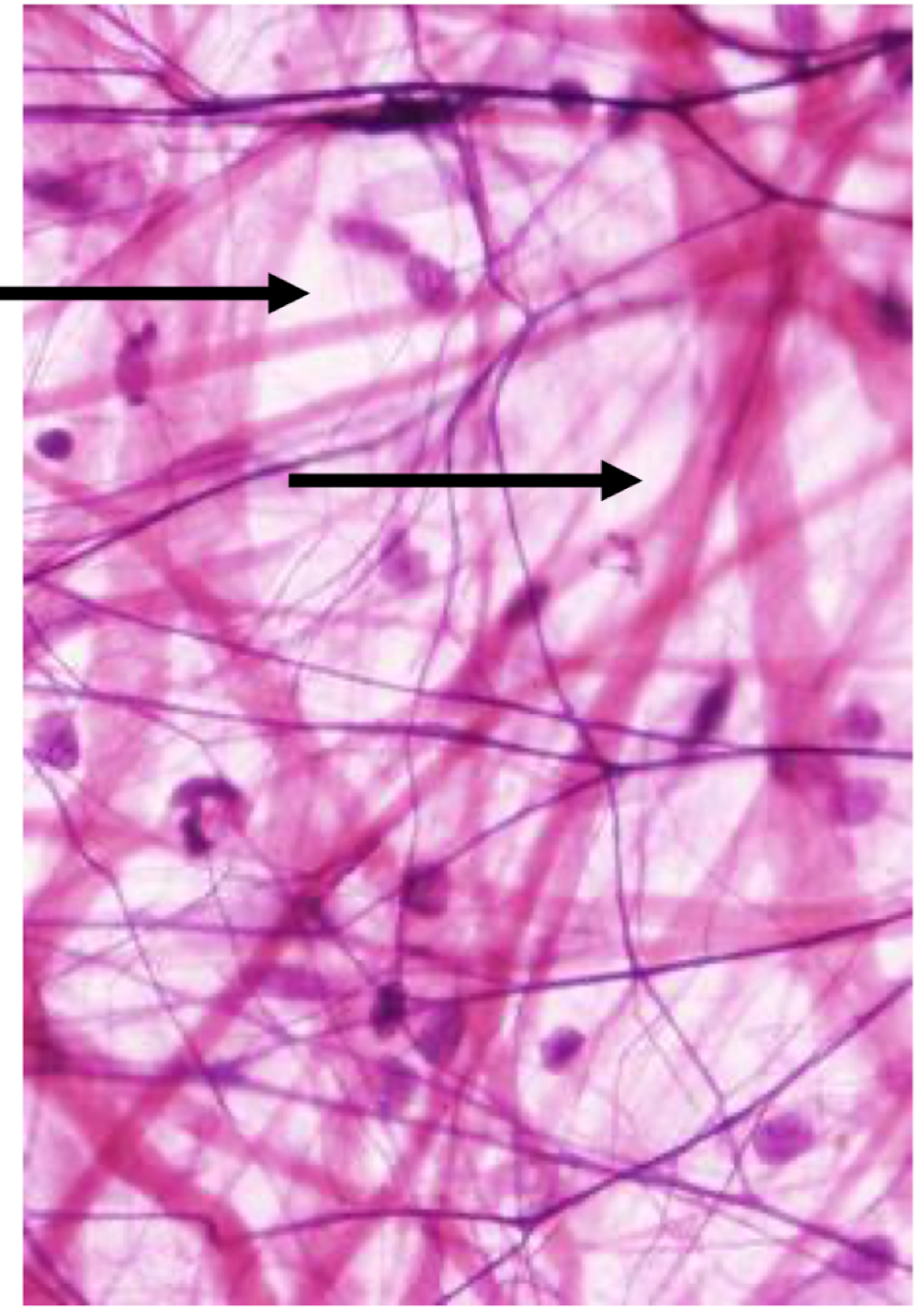
Identify the region of the ECM
ground substance

Identify the body fluid space
extracellular fluid

identify the body fluid space (dark pink)
Intracellular fluid

identify the darkest (black)
heterochromatin

identify the white inside the nucleus
euchromatin

identify what is lining outside the heterochromatin
nuclear membrane

identify the black region in the center of the nucleus
nucleolus

identify the structure seen in the FOV
nucleus

identify the grey circles surrounding the nucleus
mitochondria

identify the black specks inside the cell
glycogen granules

identify the body fluid space
intercellular space

identify the organelle
Rough ER
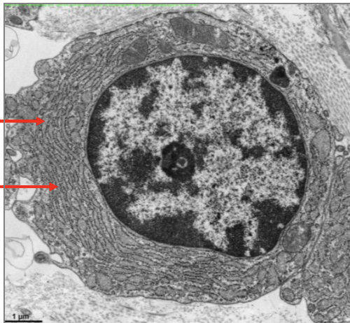
identify the grey space around the RER
RER lumen

identify the top right corner light grey area
protein/protein fibers or type 1 collagen/collagen
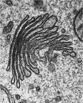
identify this organelle
Golgi Apparatus

identify the lighter purple right outside the nucleus ORGANELLE
golgi apparatus

identify the organelle on the periphery
RER

identify the region of where the Golgi would be
perinuclear clearing region

identify the movement of the intermost space going IN
luminal

indentify the movement of the intermost space going OUT
abluminal

identify the tissue section
cross, oblique, longitudinal
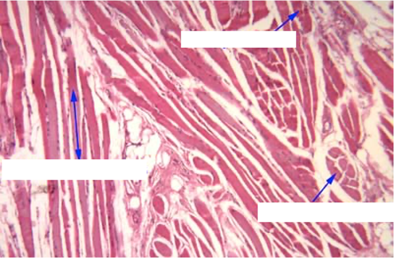
identify the tissue sectioning (left,up, right)
longitudinal, oblique, cross

identify the fluid directly outside the black
plasma
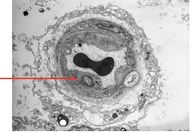
identify the body fluid space outside
interstitial fluid

identify the specific cell type
endothelium

identify the structure
basal lamina/basement membrane

identify the domain of this ell at the tip of the pointer
basal domain

identify this structure
junctional complex

identify the domain
lateral

identify the exocrine secretion modes
merocrine, apocrine, halocrine

identify the epithelial domain in the FoV
apical
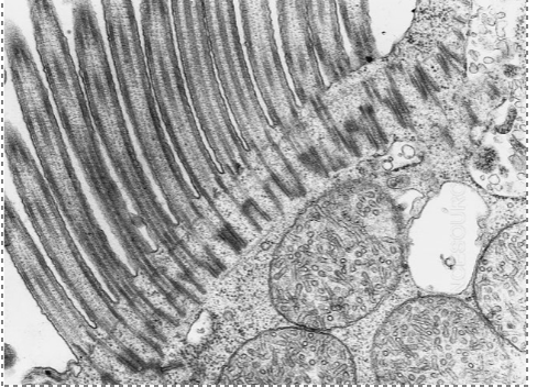
identify the apical specialization in the FoV
cilia

these are (cross/longitudinal) sections of (cilia, microvilli)
cross, cilia

these are (cross/longitudinal) sections of (cilia/microvilli)
cross, microvilli

identify the organelle
cell membrane

identify the body fluid space
ICF/ intracellular fluid

identify the body fluid space
extracellular space

these is/are portions of (1,2, or 3) cells in the FoV
2

the basal domain is in the FoV T or F
false
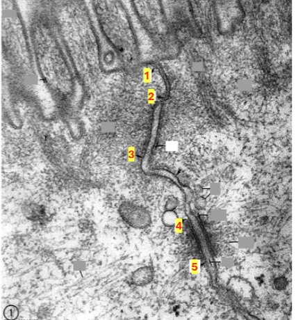
identify the junctional complex component btwn 1 and 2
zona occuldens or tight junctions

identify the junctional complex component btwn 4 and 5
macula adherens or desmosome

identify the junctional complex component btwn 2 and 3
zona adherens

identify the cytoskeletal proteins in the region
actin

identify the cytoskeletal proteins in the region
actin

identify the body fluid space
intraceullar fluid

identify the structure
terminal web

what epithelium
simple squamous

what epithelium
simple cuboidal
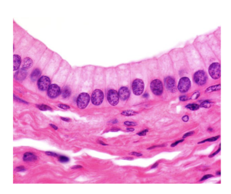
what epithelium
simple columnar

what epithelium
psuedostratified ciliated columnar

identify the structure
basal body
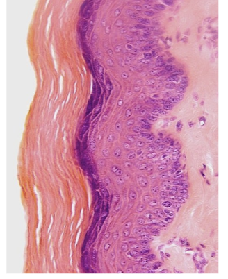
what epithelium
stratified squamous keratinized

what epithelium
stratified squamous nonkeratinized

what epithelium
stratified cuboidal

what epithelium
stratified columnar

what apical specializations (L to R)
microvilli, cilia, sterocilia

cell type
mucous cell or goblet cell

what structure
basement membrane

name the dominant collagen found at the tip of the arrow
type IV

identify structure
hemidesmosome

identify the cell
myoepithelial cell
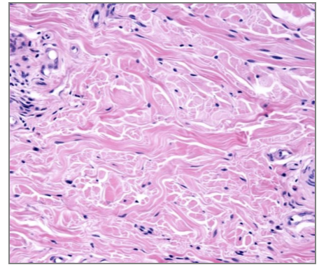
identify tissue
dense irregular connective tissue

identify the tissue
dense regular connective tissue

dense regular CT is in the FoV
false (smooth muscle)

dense regular CT is in the field of view
false (peripheral nerve)
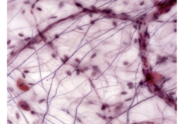
identify the tissue
loose connective tissue OR areolar tissue

what is e
elastin

what is r
reticular fiber
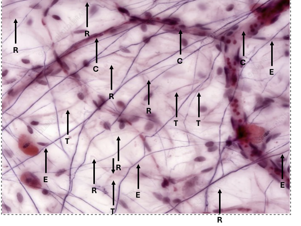
what is t
type 1 collagen

what is c
cappillary

what CT
reticular tissue

what CT
reticular tissue

identify the ECM component
elastic fibers

identify the ECM component
type 1 collagen
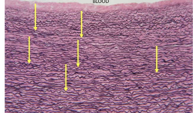
identify the ECM portein
elastin fibers
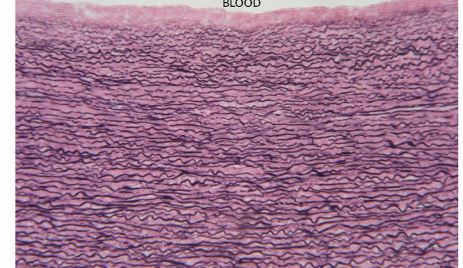
identify the tissue type
elastic tissue

identify the tissue
dense regular CT
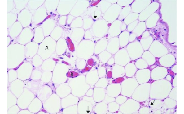
identify the tissue
white adipose tissue

in vivo the letter a is a membrane bound inclusion
false

identify
mast cell

what fibroblasts (a or b) is in its quiescent phase
A (active fibroblasts are larger)

what organ
skin

classify the skin
thin (stratum lucidum not present)

identify this skin
thick

identify the epithelium
stratified squamous keratinized

identify cell type
melanocyte

identify epidermis layer A
stratum corneum

identify B
stratuym granulosum

identify C
stratum spinosum

identify D
stratum basale

identify E
langerhans

letter x identifies a rete ridge (t or f)
false, identifies a papilla

identify the structure
desmosomes

identify the protein
intermediate filaments
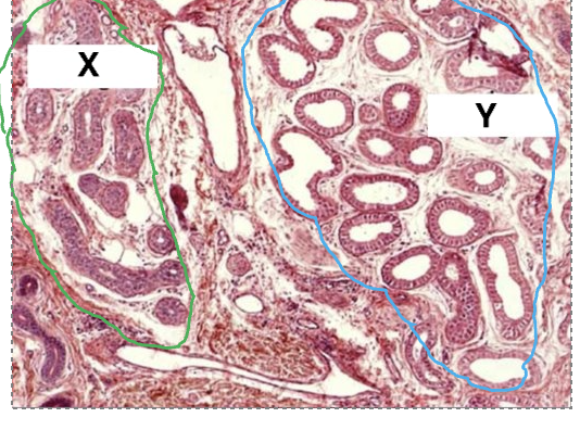
identify x
eccrine sweat glands (smaller lumen, typically two layers of stratified cuboidal, myoepithelial can be seen at higher magnifications)
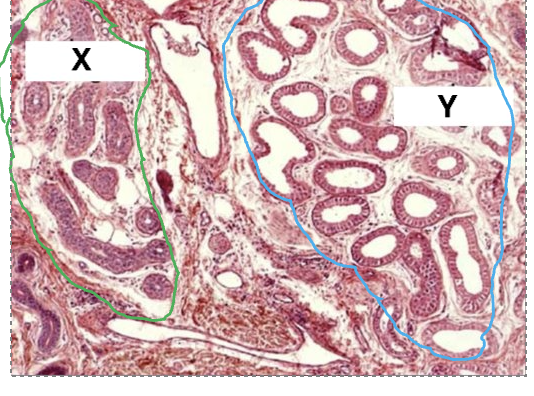
identify y
apocrine gland (larger lumen, simple cuboidal or columnar)

identify x
sebaceous gland

identify the exocrine skin gland
eccrine

what organ’s tissue
bone

what is A
sinusoid, marrow sinusoid, or capillaries