parasites
 Call Kai
 Learn
Learn Practice Test
 Spaced Repetition
 Match
Match Flashcards
 Knowt Play
1/163

There's no tags or description
Looks like no tags are added yet.
Name | Mastery | Learn | Test | Matching | Spaced | Call with Kai | Chat |
|---|
No analytics yet
Send a link to your students to track their progress
164 Terms
key points to remember
Who is affected?
How do we know they are infected?
What happens when infected?
How can we treat?
How can we prevent?
can answer by knowing the life cycle of the parasite
host parasite relationships
symbiosis
organism 1: essential
organism 2: essential
mutualism:
organism 1: benefit
organism 2: benefit
commensalism:
organism 1: benefit
organism 2: no benefit
disease (or parasitic)
organism 1: benefit
organism 2: harm
some can be a commensulate or cause disease. the development od disease depends on the hosts immune system
host-parasite relationships: reproduction
genetics:
asexual: direct replication
sexual: exchange if genetic material
protozoans:
all are asexual, some are sexual
metazoans:
some are asexual, all are sexual
definitive host
SEXUAL; a host that harbours adult or sexual stage
intermediate host
ASEXUAL; a host that harbours juvenile or asexual stages
reservoir host
NON-HUMAN; a non-human host that harbours same stages as humans; serves as reservoir for the parasite
incidental host
DEAD-END; a host that is not the normal part of the life cycle (i.e. transmission will not continue)
host vs vector
must be a living organism
Host:
Where does the parasite live? (as an adult or juvenile)
(Definitive, Intermediate, Reservoir, Incidental)
vector
How does the parasite move from one host to another?
Biological, Mechanical
eg
Biological vector of MALARIA: Female Anopheles mosquito
versus
Mechanical vector of Entamoeba histolytica: housefly
No development or growth of E. histolytica in houseflies, just passively carried.
How do parasites infect (get into the body)
Skin (intact or broken)
Gut
Lungs
Eyes, nose, mouth
Vagina
Any external surface/barrier
how do parasites cause disease?
Overall Pathological Mechanisms:
a. Toxic parasite products
b. Mechanical tissue damage
c. Immunopathology
d. Immune evasion
Remember that they can cause disease through more than one of these mechanisms
Trypanosoma brucei: life cycle
tsete fly take blood meal
injected metacyclic trypomastigites transform into bloodstream trypomastigotes whihc are carried to otehr sites
trypomastigites multiply by binary fission in various bodily fluids
diagnositic stage: circulating trypomastigotes in blood duringa cute phase
usually undetectable in latent stage
tsete fly eats blood from infected person
bloodstream trypomastigotes transform into procyclic trypomastigites in vector midgut. multiply by binary fissin.
procyclic trypomastigites leva emidgyr abd transform into epimastigotes
multiply in salivary gland, transform into metacyclc trypomastigotes, ready to infect
two variants, west and east
trypanosoma brucei: epidemeology
Targeted by WHO for elimination by 2020.
92% of infections are West (T. brucei gambiense).
1998: ~40 000 cases reported but estimated 300,000 undiagnosed.
• 2009: first time below 10,000.
• 2021: 750
• 61% of cases in D.R. of Congo
trypanosoma brucei: symptoms at both stages
Hemolymphatic stage
Nodule or chancre at bite site
Fever
Enlarged lymph nodes
CNS stage
Vacant expression
Sleeping in day
Insomnia
Seizures
Coma
Death
T. brucei gambiense transition takes month (west)
T. brucei rhodesiense transition takes weeks (east)
trypanosoma brucei: diagnosis
Lymph Node Aspiration for Diagnosis of Trypanosomiasis
Lumbar Puncture for Diagnosis of
wrights stain: can see parasite, distinct shape

trypanosoma brucei: disease
African Trypanosomiasis
african sleeping sickness
pathological mechanism of trypanosoma brucei
Antigenic variation
• T. brucei expresses ONE protein on its surface.
• Variable surface glycoprotein (VSG)
• It can switch this protein so that it evades immune recognition.
therefore cannot produce a vaccine for it
trypanosoma brucei treatment
Fexinidazole (oral)
(CNS for T.b.g.)
NECT or fexinidazole
trypanosoma cruzi: life cycle
triatomine bug takes blood meal
metacyclic trypomastogites penetrate various cells at bite wound site. inside cells transfomr into amastigotes
amastigites multiply by binary fission on cells of infected tissues
intracellular amastigites transform into trypomastigotes then burst out of cell and into bloodstream
can infect ourher cells and transform amastigotes in new infection sites. can result in clinical manifestations
triatoamine bug take sblood meal
epimastigites in midgut, multiply
metacyclic trypamastigites in hindgut
metacyclic tryomastigites in hindgut can be transferred when bug takes blood meal as bug defecates into wound when eating
trypanosoma cruzei: disease
chagas disease:
spread by reduviid bugs (kissing bugs) that defecate into wound when eating
trypanosoma cruzei: epidemeology
endemic to south america, present in west europe, north america (current estimate is at 280,000 in the US), nz/aus, japan
8 million infected
transmission of trypanosoma cruzi
vector
• congenital
• blood bank
• oral (rare)
bites at night, comes out of walls of mud huts, can help rpevent by using different building materials, bed nets
trypanosoma cruzi parasitemia
acute phase:
ten days, polyclonal lymphocyte proliferation
indeterminate and chronic phase
month-years
parasitemia detectable by xenodiagnosis or PCR
immunosuppression
trypanosoma cruzi: Romaña’s Sign
Acute stage:
• parasites in blood
• fever
• swelling at site of entrance (Romaña’s sign)
trypanosoma cruzi: chronic manifestation
(20-30% develop 10-20 years after infection)
• mega heart (Chagasic Megacardia, can develop Apical
Aneurysm)
• mega colon (Chagasic Megacolon)
• mega oesophogus
trypanosoma cruzi: diagnosis
acute:
blood smear, distinct C shape, can see kinetoplast and flagellum
can used lymph node aspiration and smear to see Amastigotes and Trypomastigotes, but may also be difficult in chronic phase
chronic
Serology (antibodies)
PCR
xenodiagnosis (feed uninfected bugs on infected persons blood, wait and see if bugs become infected)
Diagnosis of chronic Chagas disease is made after consideration of the patient’s clinical findings, as well as by the likelihood of being infected, such as having lived in a country where Chagas disease is common

Chagas Disease: A Silent Emergency
due to antigenic variation, (can change surface coating/very confusing surface coating) difficult to mount immune response, therefore vaccine doesn't work
Prevention is important, method is bite of bug, therefore if bug come from mud wall, thacthed walls, can provide diffirent housing, pesticides. If come from blood bank, can throw out blood to prevent furtehr infection
Treatments best for acute phase, not so good for chronic as parasite has expanded into tissues, hard to get.
Treatments have such sugnificant side effects that once 50yrs old, have to weigh benefits of treatment side effect and parasite side effects
Treatment does not fix damage, only prevent more damage
trypanosoma cruzi: treatment
Nifurtimox
Benznidazole
no vaccine due to antigenic variation
trypanosomes
t. brucei has less people at risk as less people infected, hopefully eridicated in next 5 years
Both treated by dangerous drugs that must balance risk of drug with risk if parasite before treating, though t. brucei have new oral drug that is better
Trichomonas vaginalis: life cycle
imple life ccycle: doesn't infect into any cells, transmitted from person to person via sexual intercourse
Survives mostly on mucosal surfaces, divides by binary fission, no sexual reproductio
multiplies by longitudinal binary fission
trichomonas vaginalis epidemeology
can be found everywhere in the world, all age groups, women and men: though tehre are preferential places for parasite
is sexually transmitted
Trichomonas vaginalis trophozoites
females:
(10-50% asymptomatic)
vaginitis
discharge
irritation, pain
long term…
mostly diagnosed in female, mostly because while 10-50% are asymptomatic, the rest are symptomatic
Suffer from vaginitis: discharge, irriation, pain
Long term: by having immune response trying to cntrol infection may have long term conseuqence ot fertility and reproduction
males:
mostly
asymptomatic
trichomonas vaginalis: disease
vaginitis
trichomonas vaginalis diagnosis
giemsa stain of vaginal smear

trichomonas vaginalis: treatment
treat with metronidazole,
prevent with protective sex, notfiing partner etc
Giardia lamblia: disease
giardia
Giardia lamblia: lifestyle
primarily faecal-oral, live in faecal are trophozoites but cannot survie in environemnt. Cyst can exits for longer
Both trophozoite and cyst are infective, but since cyst exits for longer, more common infection method
Doe not infect cell, but stays outsid ein gut
Giardia lamblia: scanning electron micrograph
has tear drop shape, multiple flagella and suction disc to stick to villi of small
when tehre is a lot there, immune system has large reaction, chaniges physiology, intestine wall

Giardia lamblia: epidemeology
world wide:
spread:
water-bourne
food less common
person-person in early childhood centres
usually srpead through contanimated water (cysts) but also human to human, normally early childhood centres (creshe)
nz has higher incidence of giardiasis than other developed countries, though to be due to high farming, but actually was person to person spread.
Improving hygeine, handwashing, nappy handling in early chidl hood centres were where coming from
giardia lamblia: clinical picture
infected:
asymptomatic (2/3 cases)
can be commensle, can live and benefit wothout harmt o host (2/3 cases asymtptomatic)
symptomatic (no IgA)
Symptomatic people are so as don’t secreted IgA into gut, tehrefore cannot manage parasite, cause explosion of parasites
giardia lamblia: clinical picture symptoms
malabsorbtion
diarrhea
sudden, explosive, greasy, smelly (1-2 weeks later)
gas & cramping
symptoms can resolve in 1-4 weeks
Giardia lamblia trophozoites diagnosis
usually diagnose thru faecal smaer.
Has flagella, 2 nuclei, ready to undergo cell division
can also see with trichrome stain: teardrop shape
Vets use snap test, to test giardia in faeces, is rapid test

Giardia lamblia: treatment
symptoms will resolve with time but treat to help symptoms
metronidazole
prevention is easy with hygeine measures to ensure no transmission of cysts in faeces, trophozoite
Entamoeba histolytica
does not have flagellum, is amoeba so amorphic
Two life cycle stages: trophozoitic (free living form tjhat can divide) or cyst (when want to exist outside host)
Can be passed thru faeces and reinfect
Entamoeba histolytica trophozoites: blood smear
red dots are red blood cells, as e. histolytica likes ot eat blood

Entamoeba histolytica: epidemeology
world wide spread, but much more common in tropical areas,
Generally spread thru food
causes ~50,000 deaths/year
entamoeba histolytica disease
amoebic colitis
“Shirt button” lesions of amoebic colitis
it doesn’t infect cells but damages intestion linig (shirt button lesions)
Damages cells directly from outside to destroy epithelial layer because wants to cause bleeding to eat
tehrefore symptoms:
If symptomatic, can have bloody diarrhoea, complications incl haemorrhage, or tissue abcesses in liver, brain, lungs(caused by immuen system trying to kill parasite)
can have low level of infection and be asymptomiatic, immune system can overcome it
Flask-like ulcers of amoebic colon disease
entamoeba are in bottom of ulcer causing problems

E. histolytica trophozoites invading colon mucosa (recognition)
can be recognised by eye looking nuclei (one of diagnostic features for this)

entamoeba histolytica: abscess
common in liver, lung, sometime brain
lots of this is immune driven by also release digestive enzymes to eat and create abcess
Entamoeba histolytica diagnosis
can see cyst in faecal smears
see trophozoite in trichrome stain

entamoeba histolytic treatment
treatment also with metronidazole
Prevention is by hygeince, esp with preparation of fruit
Sporozoa (Apicomplexa)
apicomplexa so called for very special organelle that allows them to infect into cells
Go thru sexual phase and asexual in one hsot
sporogony (sexual)
gametocyte—> gamete→ zygote→ oocyst→sporozoite
schizogony (asexual)
sporozoite→ trophozoite → schizont→merozoite
Cryptosporidium lifecycle
asexual and sexual stages in the same host
is ingested then lives in gut, in humans goes thru both asexual and sexual stages
Exits body, gets into water
Usually transmissiion thru contanimated water
Cryptosporidium Sporozoites
(released from cysts by trypsin/bile/37 C)
when oocyst is ingested, brekas open and sphorcyte come out, is ifectius stage can infect epitehlium cells

Cryptosporidium in Colon Biopsy
form ring/butthole shape

Cryptosporidium EM
epithelial cell attachment

Cryptosporidium epidemeology
found world-wide
associated with water, not only drinking but recreational (water reservoirs, swimming pools)
common caue of diarrhoea outbreak
Is zoonotic: aniamls are soruce that infects humans, very little host specificity
nz has higher incidence than other developed coutnries (highly zoonotic: due to animal contanminatin of drinking water)
Cryptosporidium treatment and symptoms
Immunocompetent:
benign, self-limiting diarrhear explosive onset, very watery (5-14 days)
no need to treat
Immunocompromised:
unremitting, persistent diarrhea, abdominal pain, weight loss, mortality
cannot control parasite, must treat
Cryptosporidium diagnosis
Diagnosis is faecal smear
Prevention again is hygiene, more so at organisational level to ensure water sources are clean
Toxoplasma gondii life cycle
cat is definitive host:sexual stage
Cat has oocysts in faeces if ingested by pretty much any other animal, becomes infectied
When infected, forms cysts.
Because infected into cells, can get anywhere (brain, etc) but preferes muscle
Cat get from eating tissue cysts in animal it eats
Cat only gets cysts in gut where forms oocysts
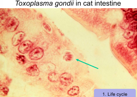
Toxoplasma gondii transmission
transmission can be congenital (mother to child),
blood trnasfusions,
ingestion of oocysts (faecal-oral)
infected meats
toxoplasma gondii: epidemeology
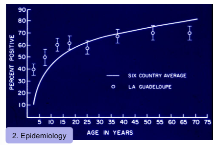
exist world wide prevalence os ~30%, but some areas much higher
Most common protozoan parasite in the wolrd (~30%)
Toxoplasma sporogony in cat intestine
can become infected very ealry in life and remain infected for life
Difficult to show demonstration of parasite for diagnosis, so use serology instead
hard to find parasite itself as live in tissue, so use serology to look to see if antibodies present
In france, most common transmission from raw meat
Central america most common from cats and climate allow oocysts to survive longer
Toxoplasma gondii: immunity
presents differently for imme adn non-immune host.
in non-immune host
After first infection, is continued infection as parasite hidden away, but enough immunity to control parasite, enough so that if reexposed, can kill parasite
immune host
control of infection
extracelkular parasites killed
in immunicomprimised host
If immunocomprise,d cannot control parasite anymore. Issue with controlling parasite already in body
If mother is non-immune host, foetus can become infected and can cause significant damage and cause birth defects
If mother immune, imunitu protects foetus.

HIV+ Person: Toxoplasma Encephalitis
common for cysts to be in brain,
in HIV+ person is immunocomprimised: where cyst become activated in brain, cause encephalitis
cyst may be able to alter nuerology, dopamine levels in brain
Toxoplasma gondii diagnosis
diagnosis:
serology, look for antibodies as evidence of immune response/exsposure
Prevention: cooking meat fully so cysts are killed
Control cats so that cat do not get infecting (keep from eating wild birds, rats etc)
Wash hands esp if dealing with cats
Treatment: if needed
Use pyrimethamine, often reserved for cases when essential to clear parasite because not suffiencent immunological control
Plasmodium spp disease
malaria
Plasmodium spp risk
3 leading cause of death - half of worlds population is at trisk at becoing infected
If put all cancers together, are 3rd
Plasmodium spp life cycle
mozzie: host and vector, responsible for transmitted disease
Life cycle cycles through humans and mozzie
human stage
human stages important for how disease manifest and can be control - vaccine etc
key stages of Plasmodium spp life cycle
1st: starts when mozzie eat blood and inject sphorzoyte into blood, whhc then goes ot target liver, infect liver cells then goes to blood and reproduction
in liver, transforms to trophozoite, then replciates forms schizont, which ruptures and infects blood
One sphrozoite takes six days to replciate enough to go from 1--> 40,000, and go into blood
infects red blood cell, multiplies and becomes schizont, ruptures and infects more blood cells. This cycle can continue indefinetly
Depedning on species, take 36-48 hrs, goes from 1 parasite to 8-24
at some point, once infect blood cel, triggers to become gametocyte, which is triggered to become distinctly female or male
Picked up by mozzie, undergoes sexual reproduction in mozzie
Asexual phase in humans
plasmodium sporozoites
thin, small parasite, infect liver cell

Plasmodium falciparum early trophozoites (rings)
trophozoite, often called ring stage: chromotin dot (with dna), has darker ring around it
Diagnostic for this form
take over red blood cells, multiply inside forming schizont, which later bursts and releases parasites

plasmodium spp. transmission
transmitted only by female anopheles mosquito that eats blood and support development of parasite
plasmodium spp. epidemeology
primarily in tropical regions (must have particular mozzie and support life cycle)
half the world at risk
• ~300 million cases/yr
• 4 species of Plasmodium that cause disease in humans
>400,000 deaths (2018)
• 67% of deaths in children <5 y.o.
Plasmodium species characteristics
P. falciparum
causes the most deaths
high drug resistance;
90% of countries with malaria
P. vivax and ovale
relapses are common (within 2-4 years)
more common in Asia than Africa
p. Vivax and p. ovale act very similar so can be grouped together, but look distinct under microscope
P. malariae
uncommon
Malaria deaths over time (1980-2010)
in this period, rapid development of drug resistance. Had one drug thsat worked for long time, but not anymore
2005 new drug was developed, decreased number of deaths
Endemic versus epidemic areas
when people get and how often different for different areas, not evenly spread
Hyperendemic:
occurs year round
in hyperendemic areas immune system develops partial immunity allows for control to some extentm as get older less likely to die from it. People without partialm immunity are children or people who come from other places who don’t have immunity
Hypoendemic:
get episodes/epidemics, periods of higher incidence (may be relating to seasons, when is standing water so mozzie can breed and trnasmit parasite
In hypoendemic area,s may not develop any immunity, mortality in all age groups
plasmodium spp. presentation
how presents I human depends on stage of life cycle
major symptoms:
fever followed by chills (97%)
headache (94%)
relapsing fever
P. vivax/ovale = 48 hours
P. malariae = 72 hours
P. falciparum = 36-48 hours
uncomplicated vs complicated malaria sysmtoms
uncomplicated
symptoms
eventual recovery
partial immunity
complicated
specially with P. falciparum
serious organ failure
cerebral malaria (coma, loss of consciousness, seizures)
Distinguishing Features of Malaria Species
P/ falciparum
ring stage
multiple rings in red blood cells, “headphones”
female gametocyte
male gametocyte
banana shaped gametocytes

plasmodium falciparum. pathological mechanisms
red blood cell (RBC) becomes “sticky” (Pf)
RBC stick together and form “rosettes”
RBC bind blood vessel wall; sequestration
antigenic variation (Pf esp.)
RBC have P. falciparum erythrocyte membrane protein-1 (PfEMP-1) on surface.
PfEMP-1 has antigenic variation
fever (all species)
When infected RBC rupture to release merozoite, “toxins” are released.
Induces TNF-𝝰 which causes fever.

Distribution of Drug Resistant P. falciparum
worldwide across teh equator
drug resistance big problem 1980s-2000s
Ranges of drugs were used but over time resistantance developed to oral drugs
previous treatments
• chloroquine
• primaquine
• mefloquine
• doxycycline
• atovaquone and proguanii
malaria and coartem
Coartum was dual acting, so parasite couldn't circumbvent - combination therapy
Combines artemether with lumefantrine
Provide orally avaliable dose that very efficent at parasite destruction
6 tablet 2x per day, 3 days, clear parasites
thought to decrease in death by new combination therapys that clear parasite and less likely to induce drug resistence
malarial prevention
preventation also becoming more common.
bed nets
insect cotnrol
More prevalent biting at dusk-night, bet nets for sleep is good
prophylaxis
Prophalayix reserved more for travellers coming in
malaria vaccine
had to find which stage to target (mosquito, RBC, liver)
vaccine look at not prevent infeciton, but reduce severe disease
pilot studies in malawi, ghana, kenya
Reduction in severe disease, prevent further deatsh of children
dracunculus
causes dracunculiasis or Guinea worm infection
from ingestion of infected cyclops (tiny crustacean) in water
trichinella spiralis
causes trichinosis
source: ingestion of uncooked carnivore or pork meat
wucheria bancrofti
causes lymphatic filariasis (elephantiasis)
transmitted by mosquito
Wuchereria bancrofti life cycle
mozzie=both vector and host
When eats blood, injects larvae into blood stream
Larvae mature into adutls that live in lymphatics
Adults produce progeny (live microfilarae worms) into blood
Picked up by mozzie, develop in mozzie into different larval stages, put back into host when eat blood
Human=definitive host
Mozzie=intermediate host
lymphatic filariasis: life cycle diagram
larvae
adults
microfilariae(MF)
microfilarae: very small, during daytime go to ahng out in lung. At nigth, go into peripheral circulation where mozzie can pick it up
Adults always in lymphatics
Important for manifestation and diagnosis of disease

distribution of lymphatic filariasis
only some places, depend s of where mozzie ar einfected and transmitting
Mostly in southeast asis and africa, not much elsewhere
worldwide: 51 million infected (863 million at risk)
W. bancrofti causes 90% of cases
Wucheria bancrofti sympotoms
manifestiations of disease depend on life stage, where is located.
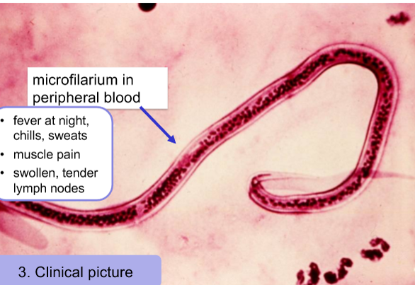
Microfilare in blood at night, cause night fever, chills, sweats fur to immune response
muscle pain, swollen lymph nodes (bc activated immune system)
wucheria bancrofti diagnosis
Diagnosis is looking at microfilarium in blood smear (must do at night, as thats when are found in blood)
distinct sheathed microfilarium in peripheral blood
looks like a worm
Cause adutls in lymphatics, very hard to get
but Can also use serology
lymphatic filariasis: elephantiasis symptoms
adult worm cause lymphatic filariasis: leads to elephantantiasis
Obstruct fluid flow as are located in lymphatics, causes edema, typically in lower extremities
Over longer period of time, elephantiasis occurs
lymphatic filariasis: preventing secondary infections
adults inlymphatics supress immune cells, immuen cells cannot control parasites
As a result, in lower extremities, if have barrier break and bacteria infection, immune cells cannot control, causes significant issue.
Overtime get clearnace of infections, but overtime get thickinging of skin, scarring
wucheria bancrofti treatment
diethyl carbamazine (DEC)
WHO strategy to stop spread (as biting only occurs at night, can sue bed nets to prevent transmission
aim to stop spread
alleviate suffering
over corse of 2000-2018, dramatic reduction in prevalence in central/equatorial africa and south east asia
Dracunculus mediensis life cycle
one of easiest infections to prevent
Two key host: human as definitive host (Also others but focus on huma)
Copepod are intermediate host
Become infected by drinking water cotaining these tin infected crustatceans
When ingested, larvae can escape through gut and spend year migrating until get to lower extremities
For most nematodes, females much larger than males (make fertilise female, then die shortly after)
Female migrate to feet etc, extend through skin and release larvae into water.
Year long cycle
Travel is not nessarily through blood
Dracunculus mediensis transmission
must be contact between human foot and water for release
Tends to occur during rainy period typically aligned with harvest season etc
Large economic burden as people have sores on lower extremitites during thos time
drancunculiasis prevalence
4 cases to July 2024
14 cases in 2021
these cases in very specific areas, likely due to civil unrest that mean has remained
CDC believe will be eradicated very soon