peripheral vascular dysfunction
 Call Kai
 Learn
Learn Practice Test
 Spaced Repetition
 Match
Match Flashcards
 Knowt Play
1/71

There's no tags or description
Looks like no tags are added yet.
Name | Mastery | Learn | Test | Matching | Spaced | Call with Kai | Chat |
|---|
No analytics yet
Send a link to your students to track their progress
72 Terms
peripheral vascular disease
- arterial
- venous
arterial peripheral vascular disease
- stenosis/occlusion
- aneurysm
- dissection
venous peripheral vascular disease
- stasis/insufficiency
- thromboembolism (DVT, PE)
arterial dysfunction
aneurysm
aneurysm
- part of artery wall weakens, allowing it to balloon out or widen abnormally
- named for location and characteristics (aortic aneurysm)
- can occur in any vessel, most notably in the brain, heart, thoracic aorta, and abdominal aorta
- dissection: tear in intima layer allows blood to leak through, separating inner and middle layers
dissecting aneurysm signs and symptoms
- sudden severe chest or upper back pain
- often described as "tearing or ripping" that can spread to the neck or down the back
- sudden severe stomach pain
- syncope
- SOB
- sudden vision problems, difficulty speaking, and weakness or hemiparesis (similar to CVA)
- unilateral weak pulse in one arm or thigh
PAD
- narrowing or occlusion of arteries outside of the heart --> reduced blood flow to extremities, gut, kidneys, head
- primary cause: atherosclerosis
- collateral circulation may develop if atherosclerosis is gradual
risk factors of PAD
- smoking
- HTN
- hyperlipidemia
- obesity
- diabetes
- older age (15-20% over age 70)
- CAD
PAD signs and symptoms: typical presentation
- intermittent claudication
- limb pain or cramping with exercise, resolves with rest (just like angina)
- pain (burning, aching) at rest, lying flat/LE elevated
- ulcers/sores/wounds on toes, feet, or legs that heal slowly, poorly, or not at all
- coldness in affected extremity
- pale or cyanotic, dusky red color
- possible numbness
- weak or absent pulses (dorsals pedis and post tib)
claudication scale
- I: initial discomfort (established, but minimal)
- II: moderate discomfort but attention can be diverted
- III: intense pain (attention CANNOT be diverted)
- IV: excruciating and unbearable pain
claudication (leg pain)
- neurogenic: proximal to distal
- vascular: distal to proximal
claudication (easing)
- neurogenic: sitting, lying
- vascular: standing
claudication (walking)
- neurogenic: uphill easier
- vascular: downhill easier
claudication (cycling)
- neurogenic: no pain
- vascular: painful
claudication (delordosing)
- neurogenic: pain improvement
- vascular: no effect
claudication (lordosing)
- neurogenic: pain exacerbation
- vascular: no effect
claudication (pulse) peripheral
- neurogenic: normal
- vascular: weak
claudication (skin condition)
- neurogenic: normal
- vascular: hair loss, dystrophic
claudication (posture)
- neurogenic: flexed
- vascular: normal
PAD complications
- acute limb ischemia
- critical limb ischemia
- osteomyelitis
- stroke, MI
acute limb ischemia
- sudden decrease in limb perfusion, potentially threatening limb viability
- pain, pallor, paresthesias, poilkilotherma (coldness), pulselessness, paralysis
- due to embolism or thrombosis
critical limb ischemia (chronic)
- pain at rest, with elevation
- arterial insufficiency ulcers
- gangrene
PT management
- maximize quality of life, general health and well-being
- maximize aerobic capacity, O2 transport
- maximize general strength, peripheral O2 extraction
education
- atherosclerosis/heart disease process
- CAD/cardiac risk factors
- disease prevention
- self-management
- good foot and skin care (assess daily for signs of skin breakdown)
chief complaints problems/impairments
- pain: claudication
- poor/non-healing wound or ulcer
- decreased exercise/activity tolerance
- impaired peripheral circulation (ABI, palpation)
- impaired muscle function/strength
- impaired aerobic capacity/endurance
- impaired respiration/gas exchange (PE)
vascular exam - inspection
- color
- ulcers
- edema
- symmetry
vascular exam: palpation
- temp
- pulses
- special tests: ABI, rubor of dependency, capillary refill, pitting edema, venous filling time, calf girth
arterial skin changes
- shiny, tight, dry, hairless
- thickened toenails
- cool/cold to touch
- pallor, pale, or bluish color to skin (peripheral cyanosis) : worse with elevation
- ulcers: may or may not be painful, ischemic, gangrene (tissue necrosis), result of trauma: common in toes and heel, develop rapidly
- rubor of dependence
palpation of pulses
- dorsalis pedis: on dorsum of foot, lateral to extensor hallucis longus
- post tibial: behind medial malleolus
- femoral
- popliteal
- radial
- brachial
- carotid
pulses scale
- 4+ = bounding
- 3+ = increased
- 2+ = normal
- 1+ = diminished
- 0 = absent, unable to palpate
- most important sign of arterial insufficiency
capillary refill
- compress nail beds until blanched
- normal flesh coloration returns in 3-5 sec
- prolonged time = arterial insufficiency
rubor of dependency
- assess for arterial insufficiency
- change in color with position
- pallor with elevation
- deep red color in 20-30 secs in dependent position
- veins must be competent
rubor of dependence
- elevate LE 60 degrees for 1 min
- 0= normal, no pallor
- 1 = definite pallor in 60 secs
- 2 = pallor in 30-60 sec
- 3 = pallor in < 30 sec
- 4 = pallor without elevation
rubor of dependency - LE dependent
- assess time for normal flesh color to return
- normal = 10-15 sec
- moderate occlusive disease = 15-25 sec
- severe ischemia = > 40sec
- deep red color in 20-30 sec indicates arterial insufficiency
venous filing time
- identify superficial veins on dorsum of foot, establish baseline appearance
- elevate LE 60 degrees x 1 min and allow gravity to drain
- place LE dependent : observe amount of time for superficial veins to refill and return to baseline appearance
- normal = 10-15 sec
- > 20 sec = mod to severe arterial insufficiency
- < 5 sec = suspect venous insufficiency
ankle brachial index
- to distinguish LE neurogenic vs vascular claudication pain
- compared ratio of systolic BP of arms and legs
- use doppler on posterior tibial or dorsalis pedis artery
ABI procedure
- 2 ratios/patient
- L leg systolic/highest brachial BP
- R leg systolic/highest brachial BP
- use index plus other signs of vascular impairment to determine risk for PAD
- other signs: color, skin changes, sensation
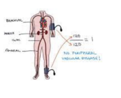
ABI process
- ultrasound device amplifies the sound of arterial blood flow
- systolic pressure recorded in brachial artery of arm
- sound of arterial blood flow located in ankle
- systolic pressure recorded in arteries of the ankle after each arterial flow is located
ABI value: greater than 1.4
- interpretation: calcification/vessel hardening
- recc: refer to vascular specialist
ABI value 1.0-1.4
- interpretation: normal
- recc: none
ABI value 0.9-1.0
- interpretation: acceptable
- recc: none
ABI value 0.5-0.8
- interpretation: moderate arterial disease
- recc: refer to vascular specialist
ABI less than 0.5
- interpretation: severe arterial disease
- recc: refer to vascular specialist
PAD interventions
- at risk for atherosclerotic heart disease
- intermittent claudication: decreased exercise tolerance
PAD - benefit from exercise training
- stimulate development of collateral circulation around stenosed vessel
- improve functional capacity: increased distance walked to pain onset
exercise prescription - PAD
- frequency: 3-5x/week
- intensity: 5-10 min warm up and cool down
- time: start 3-5 min, 1:1 work rest intervals
- type: weight bearing aerobic (walking is mode of choice)
PAD exercise intensity
- moderate 40-60% VO2R
- exercise at level 3/4 claudication pain as long as tolerated (or 3-5/10) (moderate - severe, but tolerable)
- interval training: 8-10 minute bouts of exercise, rest until symptoms subside, repeat
Pad exercise time
- increase by 5 min/session
- goal: accumulate 30-60 min/day of actual exercise
other considerations
- supervised exercise is important for adherence - at least 3 months, > 6 months optimal
- upright positions preferred - maximize LE BP, minimize cardiac workload
- cold may aggravate symptoms of intermittent claudication: need longer warm up
- monitor HR and BP response
- at increased risk of angina or ST segment changes
- resistance exercise (2x/week) for general fitness, not specifically for PAD
- watch foot care
venous insufficiency
- venous dilation, walls weaken, valves unable to close adequately
- venous pump becomes less efficient in returning blood to the heart --> blood pooling (stasis)
- risk factors: age, female, family history DVT, obesity, pregnancy, prolonged sitting or standing, smoking
venous insufficiency signs/symptoms
- dull ache, heaviness or cramping in legs (worse when standing, relieved with elevation of legs)
- flaking, itching skin on legs/feet
- tingling
- skin color changes (redness or hyperpigmentation)
- edema (dependent, worse at end of day)
- ulcers: chronic, slow healing
- often associated with varicose veins
venous skin changes
- warmer than normal
- stasis dermatitis
- ulcers
stasis dermatitis
- itchy, flaky, skin
- edema: red, weepy
- reddish-brown: staining due to burst capillaries
ulcers
- ankle area, most common above medial malleolus
- slowly developing
- shallow base
- unevenly shaped borders
venous dysfunction complications
- cellulitis: bacterial skin infection
- infection of ulcers
- osteomyelitis
- DVT
- PE
cellulitis
- warm, red area of skin, tends to spread/expand
- edema
- pain, tenderness
- fever
pitting edema
- edema pits due to increased tissue fluid: result of high peripheral vascular pressure, movement of fluid into interstitial space
- 1+ barely detectable impression
- 2+ slight indentation, rebounds < 15 sec
- 3+ deeper indentation, rebounds 15-30 sec
- 4+ rebounds > 30 sec
calf girth
- objectively measures LE edema
- circumference around calf at 10 cm below tibial tuberosity
- compare bilaterally
interventions for venous stasis
- prevent DVT: early mobilization, ankle pumps
- compression stockings: promote venous return
venous thromboembolism (VTE)
- deep vein thrombosis (DVT)
- pulmonary embolism (PE)
- blood clot (thrombus) formed in the vein, caused by slowing or changes in blood flow
- can travel to other parts of the body (embolize)
risk factors of venous thromboembolism
- older age
- recent major surgery especially ortho
- LE fractures
- multiple trauma (several serious injuries from something like a fall, attack, or car crash)
- smoking
- cancer
- prior hx of VTE
- oral contraceptives
- pregnancy
- family history
- obesity
- prolonged immobility (bedrest, long distance travel)
DVT signs/symptoms
- changes in skin color and temp: redness and warmth
- leg pain, tenderness especially in calf
- edema
PE signs and symptoms
- sudden, unexplained with SOB
- tachypnea
- tachycardia
- chest pain: worse with deep breath
- cough, hemoptysis
- falling BP
- lightheadedness or syncope
- anxiety
DVT screen
- homan's sign
- wells clinical prediction rule
homan's sign
gentle squeezing of affected calf or slow dorsiflexion of ankle produces calf pain
well's clinical prediction rule
- 0 = low probability
- 1-2 = moderate probability
- > 3 = high probability
well's clinical prediction rule for DVT
-active CA or w/in last 6 months
-paralysis, paresis or recent immobilization
-bedridden for 3+ days
-surgery requiring anesthesia in last 12 wks
-localized tenderness along deep vein
-entire leg swelling
-calf swelling at least 3 cm larger than asymptomatic leg (10 cm below tib tube)
-pitting edema in symptomatic leg
-superficial veins
-prev DVT
-high prob if >3, mod if 1-2, low
anti-coagulation
- used prophylactically to prevent blood clot formation
- may also be used to prevent emboli when thrombus is already formed: PE, DVT, atrial fibrillation
- inhibits thrombin formation and function of clotting factors
- ex: heparin, warfarin (Coumadin), Xarelto
PAD arterial ulcers
- intermittent claudication pain
- no edema
- no pulse or weak pulse no drainage
- round smooth sores
- black eschar
- location of soreness: toes and feet
venous ulcers
- dull, achy pain
- lower leg edema
- pulse present drainage
- sores with irregular borders
- yellow slough or ruddy skin
- location of sores: ankles
clinical practice guidelines action statements
- advocate for a culture of mobility and physical activity
- screen for risk of VTE
- provide preventative measures for LE DVT
- recommend mechanical compression as a preventative measure and for patients with LE DVT
- identify the likelihood of LE DVT when signs and symptoms are present (wells prediction)
- communicate the likelihood of LE DVT and recommend further medical testing
- verify the patient is taking an anticoagulant
- mobilize patients who are at a therapeutic level of anticoagulation
- mobilize patients after IVC filter placement once hemodynamically stable
- consult with the medical team when a patient is not taking anticoagulants and has no IVC filter
- screen for fall risk
- implement management strategies to prevent further VTE
PAD case study: 75 y/o male with CC of increasing difficulty walking over past 6 months, hindering ability to exercise and
socialize in the community.
HPI: Difficulty walking around the block due to
increasing pain and cramping in LEs. Unable to walk >
10 min without 8/10 pain and cramping. Pt reports
stopping and taking a break with stretching tends to
relieve all pain in his legs. Pt has also been suffering
from back pain over the past year.
C:
PMH: smokes 1 pack/day, drinks whiskey 4 glasses/wk,
L4-L5-S1 degeneration
Occupation: retired musician
PLOF: used to walk 2-3 miles with wife every night, has
been cutting back over past 6 months due to increasing
pain in legs. Plays backgammon 1x/wk.
Diagnostic tests: ABI = 0.75
S:
PMH: HTN, hypercholesterolemia, atherosclerosis,
diabetes, overweight, heart disease, kidney disease,
smoking
ROS: any co-morbidities that share risk factors
Neuro, cardiovascular, pulmonary
Pain: onset, timing, aggs/eases, association with activity
Claudication scale
Exercise/activity tolerance
Screen for DVT?