Anatomy Test 4
 Note
 Call Kai
 Learn
 Practice Test
 Spaced Repetition
 Match
 Flashcards
 Knowt Play
 Podcast
1/91

There's no tags or description
Looks like no tags are added yet.
92 Terms
Four major Regions of the Brain
Brain Stem
Diencephalon
Cerebellum
Cerebrum
What structures contain the cardiovascular centers, repiratory centers?
Cardiovascular centers and respiratory rhythmicity centers are located in the medulla oblongata.
What structures contain apneustic and pneumotaxic centers?
Apneustic and pneumotaxic centers (nuclei that coordinate the control of breathing) are located in the pons.
Sections of the brain stem from superior to inferior (top to bottom):
Mesencephalon
Pons
Medulla Oblongata
What sections form the Diencephalon?
Thalamus
Epithalamus (Pineal Gland)
Hypothalamus (Pituitary Gland)
Location and general functions of the Thalamus:
Location: Between the brain stem and the cerebrum as part of the diencephalon.
Functions: Acts as a relay station and information filter for sensory information passing to the cerebrum and brain stem, and coordinates motor activities.
Location and general functions of the Hypothalamus:
Location: Forms the floor of the third ventricle within the diencephalon.
Function: Control autonomic functions, sets appetitive drives and behaviors (thirst, hunger, sexual desire), sets emotional states, integrates with endocrine system.
Know the names of nuclei and centers located in the Thalmus and Hypothalamus:
Paraventricular Nucleus
Supraoptic Nucleus
Suprachiasmatic Nucleus
And more…

Where is the Longitudinal Fissure located?
A deep groove located between the two cerebral hemispheres of the cerebrum.
Where is the Arbor Vitae located?
The white matter of the cerebellum located inside of the cerebellar cortex.
Where is the Vermis located?
The narrow band along the midline separating the two cerebellar hemispheres.
Where is the Primary Fissure located?
On the cerebellum, separating the anterior and posterior lobes.
What structures divide the cerebellum into lobes and hemispheres?
The Vermis (Left and Right)
The Primary Fissure (Anterior and Posterior)
What is the location and function of the Corpora Quadrigemina?
Location: Found in the mesencephalon (midbrain).
Functions: Reflex centers for visual and auditory processing.

Describe the structures that from the white matter of the medulla oblongata:
Formed by ascending somatosensory tracts and descending corticospinal tracts.
Describe the structures that from the gray matter of the medulla oblongata:
Autonomic nuclei (controlling blood pressure, breathing, swallowing, vomiting, the reticular formation), and the gracile, cuneate, and olivary nuclei.
Describe the structures that from the white matter of the cerebrum:
Formed by tracts the allow communication within the cortex, consisting of association fibers, commissural fibers, and projection fibers.
Describe the structures that from the gray matter of the cerebrum:
Formed by the cerebral cortex as well as deep gray matter structures like the basal nuclei and limbic system.
Describe the structures that from the white matter of the cerebellum:
Formed by the branching arbor vitae and connected via the superior, middle, and inferior cerebellar peduncles.
Describe the structures that form the gray matter of the cerebellum:
Formed by the outer cerebellar cortex and the internal deep cerebellar nuclei.
What are the components of the limbic system?
Cingulate Gyrus
Hippocampus
Amygdaloid Body
Connecting Tracts

What is cerebral lateralization?
Each hemisphere has a special function not shared by the other hemisphere.
Functional dominance between hemispheres.
Left hemisphere typically directs analytical tasks, mathematics and language
Right hemisphere specializes in spatial relationships, music, art, and facial recognition.
Location and Function of Warnicke’s Area:
Locations: Posterior part of the left superior temporal gyrus
Function: Language comprehension region; Understanding spoken and written language.
Location and Function of Broca’s Area:
Location: Left frontal lobe.
Function: Responsible for speech production and expressive language.
Location of the Spinal Cord:
Extend from the foramen magnum to the first or second lumbar vertebra.
What forms the dorsal root in the spinal cord?
Formed by afferent axons of sensory neurons (with cell bodies located in the dorsal root ganglion)
What forms ventral roots in the spinal cord?
Formed by efferent motor fibers emerging from the spinal cord (somatic and visceral).
What is the organization of gray and white matter in the spinal cord?
Gray matter is deep, arranged into an “H” or butterfly shape surrounding the central canal.
White matter is superficial, organized into columns or funiculi (dorsal, lateral, and ventral).
What is the organization of gray and white matter in the brain?
Gray matter forms the superficial cortex and deep nuclei (such as the basal nuclei and brainstem nuclei).
White matter is deep.
What is cervical enlargement?
A thickening of the spinal cord containing an abundance of interneurons that supply the pectoral girdle and upper limbs.
What is lumbar enlargement?
A thickening that supplies the pelvis and lower limbs.
What is the Conus Medullaris?
The tapered, cone-shaped terminal tip of the spinal cord.
What is the Cauda Equina?
A tuft of spinal nerve roots extending inferiorly from the conus medullaris through the vertebral canal. (Like a horse’s tail).
What is the Filum Terminale?
Fibrous extensions that connect the spinal cord to the first coccygeal vertebrae.
How many spinal nerves are in the human body?
There are 31 pairs of spinal nerves.
What are dermatomes?
Specific regions of the skin supplied by a single pair of spinal nerves.
What is nervous plexus?
An interwoven network of “braid” of nerve fibers.
What nerve of the cervical plexus has an important role in the process of respiration? Why?
The phrenic nerve.
It innervates the diaphragm muscles.
What are the dorsal and ventral rami? and what are their functions?
Dorsal Ramus: The dorsal branch of spinal nerve; supplies the skin and muscles of the back.
Ventral Ramus: The larger branch; supplies the body wall, limbs, and forms nerve plexuses.
What are rami communicantes?
White ramus (myelinated preganglionic fibers) and gray ramus (unmyelinated postganglionic fibers)
They connect spinal nerves to the sympathetic chain ganglia of the autonomic nervous system.
List the names of the nerves of the lumbosacral plexus:
Lumbar Plexus:
Iliohypogastric Nerve
Ilio-inguinal Nerve
Genitofemoral Nerve
Femoral Nerve
Lateral Femoral Cutaneous Nerve
Obturator Nerve
Sacral Plexus:
Superior Gluteal Nerve
Inferior Gluteal Nerve
Pudendal Nerve
Sciatic Nerve
List the names of the nerves of the cervicle plexus:
Lesser Occipital Nerve
Great Auricular Nerve
Transverse Cervical Nerve
Supraclavicular Nerve
Phrenic Nerve
What nerve innervates the diaphragm?
The phrenic nerve.
What is the longest nerve of the human body and where is it located?
The longest nerve is the Sciatic Nerve, which is located in the Sacral Plexus and running down the posterior lower limb.
List in order the sections that compose the branchial plexus:
Ventral Rami (Roots)
Trunks (Superior, middle and inferior).
Divisions (anterior and posterior).
Cords (posterior, lateral, and medial).
Nerves
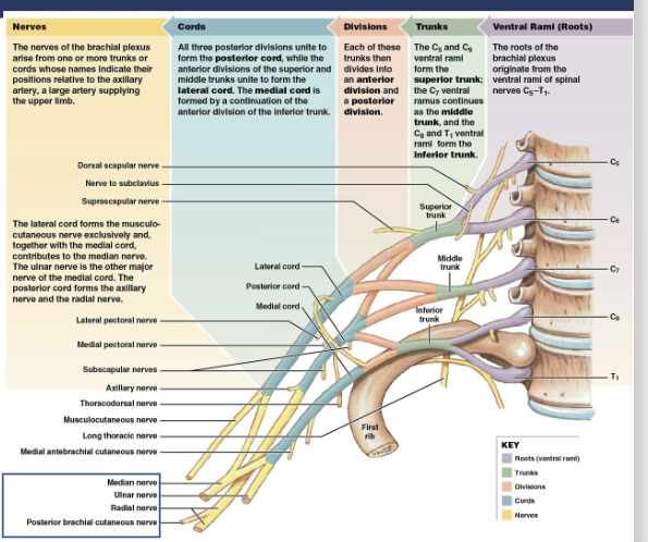
List a nerve that innervates flexors and extensors of the arm:
Musculocutaneous Nerve (Flexors of the arm) or Radial Nerve (Extensors of the arm/forearm).
What is a reflex and what elements for a reflex arc?
A reflex is an immediate involuntary motor response.
A reflex arc begins at a sensory receptor and ends at a peripheral effector.

What is the classification of receptors based on the stimulus they receive?
Thermoreceptors (temperature changes)
Nociceptors (pain and itch)
Mechanoreceptors (touch, pressure, vibration, stretch)
Chemoreceptors (chemical concentration changes; things like respiration, taste, smell, and overall maintaining internal homeostasis).
How are receptors classified by structure?
Free nerve endings vs. Encapsulated nerve endings.
What is the location and function of encapsulated receptors?
Tactile Corpuscles: Located in dermal papillae of hairless skin (eyelids, libs, fingertips nipples, etc.); sense touch and light pressure.
Lamellar Corpuscles: Located in deep dermis, hypodermis, fingers, breast, joint capsules, and walls of urinary bladder; sense pressure and vibration.
Bulbous Corpuscles: Located in the dermis; sense continuous pressure and skin stretch.
What is the location and function of free nerve endings?
Location: Distributed throughout epithelial and connective tissue, including the epidermis and cornea.
Function: Monitor pain, itch, tickle, temperature, and light touch.
What are exteroceptors?
Provide information about the external environment.
What are interoceptors?
Monitor internal organ activity.
What are proprioceptors?
Monitor position and movement. Located in the musculoskeletal organs (tendons and joints).
What is the origin of neurons and ganglia of the sympathetic division?
Origin: Lateral gray horns of spinal cord segments T1-L2.
Ganglia: Trunk Ganglia and Collateral Ganglia.
What is the origin of neurons and ganglia of the parasympathetic division?
Origin: Brain Stem Nuclei and sacral lateral gray matter.
Ganglia: Terminal ganglia and intramural ganglia.
What is the cranial outflow?
Parasympathetic preganglionic fibers originating from the brain stem cranial nerves and innervating structures in the head, neck, thorax, and most abdominal organs.
What is the sacral outflow?
Parasympathetic preganglionic fibers originating from sacral spinal segments and innervating distal portions of the digestive tract and pelvic organs.
What is the trunk ganglia?
Located in chains along both sides of the vertebral column.
Where is the collateral ganglia?
Located anterior to the vertebral column.
What is the Enteric Nerveous system?
A network of neurons embedded in the wall of the digestive tract that governs gastrointestinal behavior.
What is Achlasia?
Progressive inflammation and degeneration of ganglion cells in the myenteric plexus of the esophagus.
What is Gastroparesis?
Delayed gastric emptying in the absence of mechanical obstruction (diabetes being the most common cause).
What are the control areas of the autonomic divisions and what activities do they regulate?
Hypothalamus: Regulates water balance, temperature, hunger, and endocrine functions.
Brian Stem: Regulates visceral functions such as heart rate, blood pressure, pupil size, airflow/respiration, salivation, swallowing.
Spinal Cord: Manages lower-level autonomic reflexes for urination, defecation, erection, and ejaculation.
What is the anatomical arrangement of the autonomic pathways? (sympathetic and parasympathetic)
A two-neuron chain consisting of a preganglionic neuron that synapses onto a ganglionic/postganglionic neuron.
What is the neruoeffector synapse?
The junction between the postganglionic neuron and its effectors.
What is referred pain?
When pain originating in visceral organs is perceived as coming from specific superficial areas of the body.
What type of cells are associated with olfactory epithelium?
Olfactory sensory neurons
Supporting cells
Olfactory stem cells
What substances or ions are involved in taste sensation?
Hydrogen Ions (Sour)
Sodium Ions (Salty)
Sugars (Sweet)
Amino Acids (Umami)
Potential Toxic Components (Bitter)
What are the taste receptors of the tongue?
They are located inside the taste buds, which are found within the elevations of the surface of the tongue called papillae.
What are the different types and locations of the papillae?
Fungiform Papillae: Scattered across the surface of the tongue.
Foliate Papillae: Lateral margins of the tongue.
Vallate Papillae: Arranged in a V-shape on the posterior portion of the tongue.
What structures form the external ear?
Auricle
Helix
Lobule
External Acoustic Meatus
What structures form the middle ear?
Tympanic membrane.
Auditory ossicles.
Pharyngotympanic tube.
What structures form the internal ear?
Bony and Membranous labyrinths
Cochlea
Vestibule
Semicircular canals.
What structures and fluids are associated with the cochlea?
Structures: Scala vestibuli, scala tympani, cochlear duct, vestibular membrane, basilar membrane, tectorial membrane, spiral organ, and hair cells.
Fluids: Perilymph and Endolymph.
Where is the organ of Corti located? What structures form it?
Location: Basilar membrane inside of the cochlear duct of the inner ear.
Structures: Hair cells, supporting cells, and overlying tectorial membrane.
What structures of the inner ear are associated with equilibrium?
Otolith organs (Saccule and Utricle) and Semicircular Canals.
What are semicircular canals?
Three fluid-filled ring-like tubes that detect rotational head movement.
What are otoliths?
Calcium carbonate-calcite crystals embedded in the otolith membrane of maculae.
What are ampullas?
The expanded swelling at the base of each semicircular canal containing snesory receptors.
What are saccules and utricles?
The two otolith sac-like organs of the vestibule that detect gravity and linear acceleration.
What are kinocilium?
A large, specialized hair cell stereocilium that serves as a directional reference point for head movement.
What are macula?
The sensory receptor area in the utricle and saccule containing hair cells and supporting cells.
What structures form the external anatomy of the eye?
Eyebrows
Eyelids
Eyelashes
Conjunctiva
Lacrimal Caruncle
Lacrimal Apparatus
What structures form the internal anatomy of the eye?
Cornea
Sclera
Iris
Pupil
Anterior Chamber
Lens
Posterior Chamber
Vitreous Champer
Choroid
Retina
What structures form the lacrimal apparatus?
Lacrimal gland
Lacrimal puncta
Lacrimal canaliculi
Lacrimal sac
Nasolacrimal duct
Inferior nasal meatus
What type of photoreceptors and photopigments are in the retina?
Photoreceptors: Rods (low-light and night vision) and Cones (color vision during daylight).
Pigments: Red, green, and blue lights.
What structure of the cortex processes visual information?
Visual Cortex (Occipital lobe).
Describe the two components of the blood and their characteristics:
Plasma: (46-63%) mostly water with a little plasma proteins and other solutes.
Formed Elements: (37-54%) 99.9% red blood cells along with white blood cells and platelets.
What are the main characteristics of white blood cells?
What are the main characteristics of red blood cells?
What are the main characteristics of platelets?