Functional Organisation of the Brain
 Call Kai
 Learn
 Practice Test
 Spaced Repetition
 Match
 Flashcards
 Knowt Play
1/28

There's no tags or description
Looks like no tags are added yet.
29 Terms
What is the medulla? What are some of its functions?
Extension of spinal cord
HR, respiration, reticular formation (regulates sleep, wakefulness, levels of arousal)
What is the function of the pons?
Relay between cerebellum + brain
Locked-in syndrome
What is the substantia nigra? Where is it located?
Midbrain
Looks darker because of high levels of dopimanergic neurons (neuromelanin is a byproduct of dopamine synthesis)
Important role in reward + movement
Parkinsons disease
What is the limbic system?
Regulatory functions (e.g. emotion, motivation, memory)
What is the amygdala involved in? What is the evidence of this?
Emotion regulation
E.g. serotonin expression in amygdala is associated with aggression + impulse control in the context of threats
Stimpson (2016)
Lew (2019)

What are the 3 aspects of the cortex and their functions?
Primary motor + somatosensory cortices (M1 + S1)
Topographic organisation
Sensory homunculus
Motor homunculus
Primary visual cortex
Retinotopic organisation of V1
Orientation selective cells
Primary auditory cortex
Changes in air pressure
What are the different types of brain imaging?
CT/MRI = structural imaging
Different tissues show up with different brightness
Diffusion weighted imaging = white matter structure
Functional (metabolic) neuroimaging
Blood flow
Functional activation: neural subtraction method
Functional connectivity: correlated activity in different brain areas

What is computerised axial tomography (CT scan)?
Recombines multiple x-ray photos into a single image
Structural brain imaging
One of the first neuroimaging techniques
Scanner rotates a device around a person’s head → takes a series of X-ray photographs from different angles → computer programs combine these to provide views from any angle
Show different densities of tissue in the brain
Higher density skull looks white on a CT scan
Cortex shows up as grey
Least dense fissures + ventricles in the brain look dark
Used to locate lesions/tumours → appear darker (less dense than cortex)
What is magnetic resonance imaging (MRI scan)?
Powerful magnet causes charged molecules in soft tissue to realign to produce measurable field distortions
Structural brain imaging
Magnetic pulses cause molecules in brain tissue to twist + then relax → releases energy (occurs in short period)
Differently charged molecules respond differently to the magnetic pulses → energy signals reveal brain structures with different molecular compositions
Produces pictures of soft tissue at a better resolution than a CT scan → clearer picture of the structure of the brain + can help localise brain damage
What is functional brain imaging?
Brain in action
Rely on the fact that activated brain areas demand more energy for their neurons to work
Detect changes in blood flow
Can accurately localise changes in the brain
What is positron emission tomography (PET scan)?
Uses radioactive markers to measure blood flow in brain
Harmless radioactive substance injected into bloodstream → brain scanned by radiation detectors whilst PPT performs perceptual/cog tasks
Record the level of radioactivity in each region → computerised image of the activated areas
To adapt to the limitations of PET technology, researchers often have to use experimental designs that are different from ones used in psych lab
What is functional magnetic resonance imaging (fMRI scan)?
Magnet causes haemoglobin molecules to realign, to measure blood flow in the brain
Carries O2 to tissues → concentrated in active areas (blood oxygen level dependent signals = measure of the relative blood flow to locations that are activated in the brain)
Provides a picture of the level of activation in each brain area
No exposure to radioactive substances
Can localise changes in brain activity across briefer periods than PET → more useful for analysing psych processes that occur extremely quickly
Researchers can design experiments that more closely resemble the ones they carry out in the psychological lab
What do functional systems have the capacity for?
Reorganisation → aren’t fixed
E.g.
Ventricles massively enlarged -> reduction in brain tissue
Neurological development + medical history were otherwise normal (lived normal life but had low IQ)
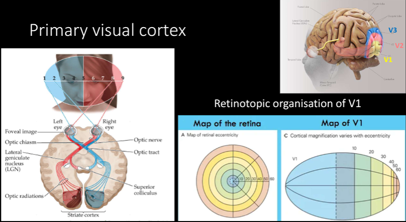
Primary visual cortex (diagrams)

Primary auditory cortex (diagrams)

What is the function of neurons?
Form circuits + pathways → connect with other circuits + pathways
Form nerves = bundles of axons regulated by glial cells that form the insulating myelin sheaths + change in the responsiveness to neuronal activity
What is the nervous system?
Interacting network of neurons that conveys electrochemical info through the body
What is the central nervous system?
Brain + spinal cord
Sensory info from world → processes + coordinates → sends commands to skeletal + muscular systems for action
Brain contains structures supporting complex motor, perceptual, emotional + cog functions of NS
Nerves that process sensory info + relay commands to the body connect to the spinal cord
Damage severs connection from brain to sensory + motor neurons
Describe the process leading to voluntary movement
Brain → sends commands for voluntary movement through spinal cord → motor neurons (axons project out to skeletal muscles) → send the message to contract
What are spinal reflexes?
Simple pathways in the NS that rapidly generate muscle contractions
Describe the process leading to a spinal reflex
Synaptic connections within spinal cord → interneuron relay sensory inputs to motor neurons → connect to muscles
What is the peripheral nervous system?
Connects CNS to organs + muscles through nerves that conduct sensory info into the brain, carry commands our of the brain or both
What are the parts of the peripheral nervous system?
Somatic
Autonomic
Sympathetic
Parasympathetic
What is the somatic nervous system?
Set of nerves that convey info into + out of the CNS
Conscious control
Used to perceive, think + coordinate behaviours
What is the autonomic nervous system?
Set of nerves that carry involuntary + automatic commands that control blood vessels, organs + glands
Largely outside of conscious control
What is the sympathetic nervous system?
Set of nerves that prepare the body for action in threatening situations
Nerves emanates from top + body of spinal cord + connect to different organs (eyes, digestive organs, salivary glands, heart + lungs, sex organs)
What are some actions of the sympathetic nervous system?
Dilates pupils to let in more light
Increases HR + respiration to pump more oxygen to muscles
Diverts blood flow to brain + muscles
Activates sweat glands to cool your body
Energy conservation
Inhibits salivation + bowel movements
Suppresses the body’s immune responses
Suppresses responses to pain + injury
What is the parasympathetic nervous system?
Helps the body return to a normal resting state
Reverses effect of SNS
Generally mirrors the connections of the SNS
What else do the sympathetic and parasympathetic nervous systems do?
Control sexual behaviour
Males
PNS engorges the blood vessels of the penis to produce an erection
BUT SNS is responsible for ejaculation
Females
PNS produces vaginal lubrication
BUT SNS underlies orgasm