PSYC 324: Lecture 11 - Measuring Neural Activity
 Call Kai
 Learn
 Practice Test
 Spaced Repetition
 Match
 Flashcards
 Knowt Play
1/56

Earn XP
Description and Tags
MT 2 Content
57 Terms
4 Steps in Studying Brain Function
Observe and describe:
Examine brain structure and activity.
Correlations between structure/activity and behaviour.
Build theory:
Develop explanations of brain function based on observations.
Develop testable predictions of brain function.
Test theory:
Manipulate brain function or behaviour to test predictions.
Helps establish causal relationships rather than simply correlations.
Refine:
Revise theories based on findings.
Testing Neural Hypotheses with Behavioural Manipulation
Neurons in the primate prefrontal cortex (PFC) appear to maintain information about cues during the delay phase of a working memory task.
These neurons continue firing while information is being held in working memory.
Increasing the delay increases how long these neurons fire.
Prefrontal Cortex (PFC) & Working Memory
Individual PFC neurons fire throughout the delay period when an animal holds information online.
Once the animal makes its response, neuronal activity decreases.
Demonstrates how neural activity can be studied by manipulating behavioural task demands.
4 Dimensions of Brain Observation
Spatial resolution.
Spatial scope.
Temporal resolution.
Temporal scope.
Spatial Resolution vs Spatial Scope
Spatial resolution: How precisely a method can identify the location of brain activity or structures.
Spatial scope: How much of the brain can be observed.
**Generally, greater spatial resolution comes at the expense of spatial scope, and vice versa.
Temporal Resolution vs Temporal Scope
Temporal resolution: How precisely a method can distinguish changes over time.
Temporal scope: How long activity can be monitored.
**Generally, greater temporal resolution can come at the expense of temporal scope, and vice versa.
7 Key Considerations When Choosing a Brain Measurement Method
Direct vs indirect measurement.
Freely moving/naturalistic behaviour.
Invasiveness.
Can it be done pre-mortem?
Signal-to-noise ratio.
Subjects.
Cost.
Direct vs Indirect Measurement
Direct: Measures neural activity itself (e.g., electrical activity using electrophysiology).
Indirect: Measures a byproduct or correlate of neural activity.
Example: fMRI measures changes in blood oxygenation as a proxy for neural activity.
Freely Moving / Naturalistic Behaviour
Whether subjects can move and behave naturally during measurement.
Some methods restrict movement.
Example: fMRI requires subjects to lie inside a scanner, limiting naturalistic behaviour.
Invasiveness & Pre-Mortem Measurement
Invasiveness: Whether a method requires entering or altering the body/brain (e.g., implanting electrodes).
Pre-mortem: Whether measurements can be obtained while the subject is alive.
Post-mortem tissue has limitations because the circumstances surrounding death can substantially affect brain function.
Signal-to-Noise Ratio (SNR)
Measures how robust a signal is relative to background noise.
Higher SNR = clearer, more reliable signal.
Lower SNR = more noise relative to the signal being measured.
Human vs Non-Human Animal Brain Research Methods
Humans:
Behaviour.
Blood/CSF/tissue samples.
Imaging.
Electrophysiology.
Non-human animals:
All of the above, plus cell-type-specific monitoring and neurochemical monitoring.
Behaviour as a Measure of Brain Function
All behaviour is fundamentally derived from brain activity.
Behaviour can therefore be used to infer brain function.
Used in clinical assessment and neuroscience research.
3 Applications of Behavioural Assessment
Psychiatric diagnoses.
Neurocognitive testing.
Neurological examinations.
Psychiatric Diagnoses Through Behaviour
Psychiatric conditions are diagnosed based on behavioural symptoms.
Examples:
Depression.
Addiction.
Anxiety.
Schizophrenia.
Neurocognitive Testing
Assesses specific cognitive processes and abilities.
Can reveal which cognitive functions are impaired.
Example: Identifying cognitive deficits associated with dementia.
Neurological Examinations
Used to assess neurological function.
Can help identify the origin of a stroke.
Can examine dermatomes and myotomes.
Behaviour in Neuroscience Research
Behaviour is often used to assess the causal impact of experimental manipulations.
Researchers manipulate brain function and examine resulting behavioural changes.
Blood / CSF / Saliva Samples
Biological samples used to measure chemicals such as hormones.
Can be collected multiple times.
Allows researchers to examine changes in response to events (e.g., a stressor).
Cerebrospinal Fluid (CSF) Collection
CSF can be collected through a lumbar puncture.
A needle is inserted between vertebrae to access CSF surrounding the spinal cord.
The fluid is withdrawn through the needle for analysis.
Tissue Samples
Localized brain tissue samples can be collected and analyzed for:
Gene transcription.
Protein translation.
Other chemicals.
2 Sources of Brain Tissue Samples
Post-mortem tissue.
Surgical tissue.
Post-Mortem vs Surgical Tissue
Post-mortem: Brain tissue collected after death.
Surgical: Brain tissue collected during a surgical procedure.
Collection is limited by availability and ethical/practical considerations.
Post-mortem tissue has caveats because the circumstances of death can affect brain function and tissue measurements.
Non-Dynamic Imaging
Provides a snapshot of brain structure or molecular characteristics.
Does not continuously measure changes in brain activity over time.
Includes:
Histology/microscopy.
CT/CAT.
MRI.
PET.
Histology / Microscopy
Examines brain tissue using microscopic techniques.
Can identify specific proteins, mRNA sequences, and cellular structures.
Requires tissue sections.
Includes immunohistochemistry and in situ hybridization.
Immunohistochemistry
Uses fluorescently labelled antibodies that bind to specific proteins.
Allows researchers to identify the location of particular proteins in brain tissue.
Fluorescent signals are visualized using a microscope.
In Situ Hybridization
Uses fluorescently labelled nucleotide probes that bind to specific mRNA sequences.
Allows researchers to identify where particular mRNA molecules are expressed.
Detects gene expression rather than directly labelling proteins.
Immunohistochemistry vs In Situ Hybridization
Immunohistochemistry: Fluorescent antibodies bind to specific proteins.
In situ hybridization: Fluorescent nucleotide probes bind to specific mRNA sequences.
Both require tissue sections and allow microscopic visualization.
Computerized Tomography (CT / CAT)
Essentially a 3D X-ray.
Detects differences in tissue density.
Has relatively poor spatial resolution.
Fast and inexpensive.
Particularly useful for detecting brain bleeds.
Can be combined with contrast agents.
CT Angiogram
CT scan combined with a contrast agent.
Improves visualization/labeling of blood vessels.
Helps examine vascular structures.
Magnetic Resonance Imaging (MRI)
Uses an extremely strong magnet and radiofrequency pulses from an electrical coil.
Causes hydrogen protons in water to emit signals.
Signals are processed to produce highly detailed brain images.
Stronger magnets generally produce better image resolution.
MRI – T1 vs T2
Different magnetic field and radiofrequency applications highlight different tissue characteristics.
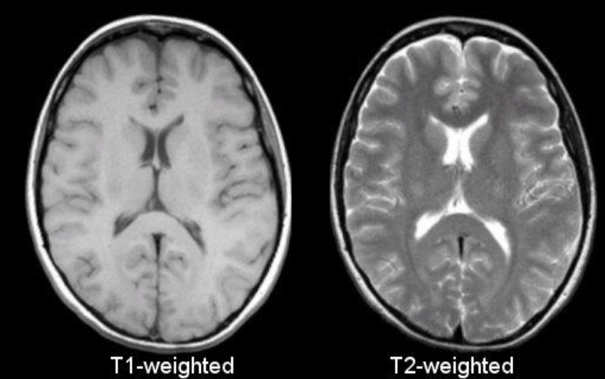
T1 MRI
T1:
White matter = lighter.
Grey matter = darker.
Ventricles = darker.
T2 MRI
T2:
White matter = darker.
Grey matter = lighter.
Ventricles = white/brighter.
Diffusion Tensor Imaging (DTI)
MRI technique used to visualize white matter tracts.
Examines the diffusion of water molecules through brain tissue.
Helps identify the organization and pathways of white matter connections.

Positron Emission Tomography (PET)
Detects radioactive isotopes.
An isotope is often attached to a drug or tracer that binds to a specific brain target (protein or other molecule).
The signal indicates where and how much of the target molecule is present.
Also frequently used in cancer detection.
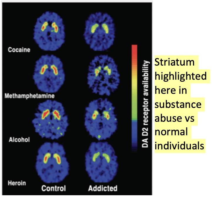
PET – Radiotracers
Radioactive isotopes are attached to molecules that bind to specific targets.
The tracer travels through the body and binds to its target.
PET detects the radioactive signal to determine the location and amount of the target molecule.
CT vs MRI vs PET
CT: Detects tissue density; useful for identifying bleeds.
MRI: Produces highly detailed structural images of brain tissue.
PET: Detects radioactive tracers to examine the distribution of specific target molecules.
Dynamic Imaging
Examines changes in brain activity over time.
Includes:
Functional MRI (fMRI).
Electrophysiological techniques.
Allows researchers to monitor activity rather than simply obtain a structural snapshot.
Functional MRI (fMRI)
MRI specialized to detect changes in blood oxygenation.
More active brain regions require increased blood supply.
Measures changes in oxygenated blood as an indirect indicator of neural activity.
Can examine changes in brain activity over time.

fMRI – Limitations
Measures a proxy for neuronal activity, not neuronal firing directly.
Cannot distinguish the activity of individual neurons.
Does not identify specific cell types.
Requires subjects to remain in a scanner, limiting naturalistic behaviour.
fMRI & Fear Generalization
Researchers can pair one stimulus with an aversive event and present another, different stimulus.
Examine how much fear generalizes from one stimulus to another.
Individuals with PTSD may show greater fear generalization.
This can be associated with particular patterns of brain activity.
Electrophysiology
Measures electrical potentials in the brain.
Can measure the activity of individual neurons or populations of neurons.
Includes intracellular, juxtacellular, local field potential (LFP), and EEG recordings.
Intracellular vs Juxtacellular Recordings
Intracellular: Measures electrical activity from inside a neuron; typically performed ex vivo.
Juxtacellular: Measures activity from very close to an individual neuron; typically performed in vivo.
Local Field Potentials (LFPs)
Measure collective electrical activity from populations of nearby neurons.
Recorded using electrodes inside the brain.
Reflect the combined activity of multiple neurons rather than one individual neuron.
Electroencephalography (EEG)
Measures collective electrical activity from outside the brain.
Uses multiple electrodes attached to the scalp.
Records electrical signals generated by populations of neurons.
Can measure oscillatory brain states.
EEG – Temporal vs Spatial Precision
Very high temporal precision (millisecond timescale).
Poor spatial precision.
Much more flexible than MRI.
Can monitor rapid changes in electrical brain activity.
3 Applications of EEG
Seizure diagnosis and monitoring.
Sleep staging.
Measuring oscillatory brain states.
Single-Unit Recordings
Measure electrical activity from individual neurons.
Electrodes are implanted in specific brain regions.
Can be used to examine the activity of individual neurons.
Often performed in clinical contexts involving neurological disease.
Single-Unit vs LFP Recordings
Single-unit: Measures electrical activity of individual neurons.
LFP: Measures collective electrical activity of nearby neuronal populations.
Both can be recorded using electrodes implanted in the brain.
Often used in seizure monitoring.

Clinical Electrophysiology
Electrodes may be implanted in different brain regions to monitor neural activity.
Commonly performed in the context of neurological disease, particularly seizure monitoring.
Often involves monitoring activity in the hippocampus.
Neural activity can be monitored and, in some cases, targeted with electrical stimulation or current.
Pros vs Cons of Non-Human Animal Research
PROS:
Simpler brains make them easier to study.
High anatomical, genetic, and behavioural homology with humans.
Greater control over environment and past experiences.
Can record genetically defined cells.
Can manipulate genes, molecules, and neural circuits.
CONS:
Findings are not always generalizable to humans.
Certain psychological phenomena may not be present (e.g., gender).
Simpler brains do not fully represent human brain complexity.
Anatomical, Genetic & Behavioural Homology
Homology refers to shared characteristics due to common evolutionary ancestry.
Non-human animals share many anatomical, genetic, and behavioural similarities with humans.
Makes animal models useful for investigating brain function.
Genetically Defined Cells
Animal models allow researchers to identify and monitor specific populations of neurons based on their genetic characteristics.
Allows more precise investigation of particular cell types.
Researchers can also manipulate specific genes, molecules, and neural circuits.
GCaMP (Genetically Encoded Calcium Indicator)
Genetically encoded indicator used to monitor neural activity.
Fusion of modified green fluorescent protein (GFP) and calmodulin.
Fluorescence increases when calcium binds to GCaMP.
Allows researchers to monitor activity in genetically defined neural populations.
GCaMP – Bound vs Unbound States
Unbound state (low calcium): Little/no fluorescence.
Bound state (calcium present): Calcium binds to GCaMP, producing fluorescence.
Neuronal activity/action potentials lead to calcium influx.
Changes in fluorescence can be used as an indicator of neural activity.
Miniature Microscopes (Miniscopes)
Small microscopes used to record GCaMP fluorescence in the brain.
Allow researchers to monitor neural activity in behaving animals.
Can record activity from many neurons and multiple neural cell types.
Provide a way to study brain activity during more naturalistic behaviour.