Anatomy Unit 3 - Muscular and Nervous Systems
 Call Kai
 Learn
Learn Practice Test
 Spaced Repetition
 Match
Match Flashcards
 Knowt Play
1/103

Earn XP
Description and Tags
Omg it feels like we justttt haddd a tessttt, anywhooo, l o c k ! i n ! 9/30-10/14
Name | Mastery | Learn | Test | Matching | Spaced | Call with Kai | Chat |
|---|
No analytics yet
Send a link to your students to track their progress
104 Terms
What are the functions of the muscular system?
Movement, heat generation, protection, and posture

What are the types of muscle
Skeletal muscle
smooth muscle
cardiac muscle

What is the muscle organ?
The muscle belly, the largest unit of the muscle, basically just the muscle

What is the muscle fascicle?
Bundles of muscle fibers that make up the muscle belly. Run from end to end of muscle belly.

Muscle fibers
The muscle cell unit, make up muscle fascicles. Made of myofibrils

What are myofibrils?
They make up the muscle fibers

What are sarcomeres
The contracting units of the muscle

What is the sarcoplasmic reticulum?
A specialized organelle that wraps around sarcomeres. It…
Stores Calcium ions
Surrounds myofibrils

What are the steps to a muscle contraction?
An electrical charge
Sarcoplasmic reticulum releases calcium ions
A bunch of chemical reactions take place :/
Myosin and actin fivers move along each other

Where are the myosin fibers?
branching out from the m line, they’re thicker, have grabby thingies

Where are the actin filaments?
branching out from the z line, they’re thinner

Energy is needed for…
Both Contraction and Relaxation
What is rigor mortis?
Body stiffening after death. All the sarcoplasmic reticulum release all their Ca+ ions, so reactions occur and all the muscles contract.
Timeline of rigor mortis:
It starts 2-3 hours after death because all the Ca+ ions are released
It ends after 12-18 hours, the body is very rigid at this point b/c of all the ions
It fades after 2 days because the cells start to disintegrate, so u can move the body easily now.
Hypertrophy
More stress on the muscles, so there are more myofibrils (muscles grow in size.)
This is caused by exercise (demand)

Atrophy
Less stress on the muscles, so there are less myofibrils (muscles lose their size.)
This is caused by laziness, so muscles are lost for efficiency’s sake

What are the two muscle fiber types
Red/dark/slow
White/light/fast
Where are red muscle fibers found?
Your trunk and legs
Where are white muscle fibers found?
Your eyes and hands
Properties of red muscle fibers
best for endurance
have more adipose (fat) for longer energy storage
have more capillaries to provide oxygen
have myoglobin for oxygen storage
Properties of white muscle fibers
best for quick strength
have a lot of glycogen (glucose storage for shorter, quick energy storage)
What is an origin
the stationary attachment point for a muscle

What is an insertion
the moving attachment point for a muscle

What is an antagonist muscle
Muscles that work against one another (eg- biceps and triceps)
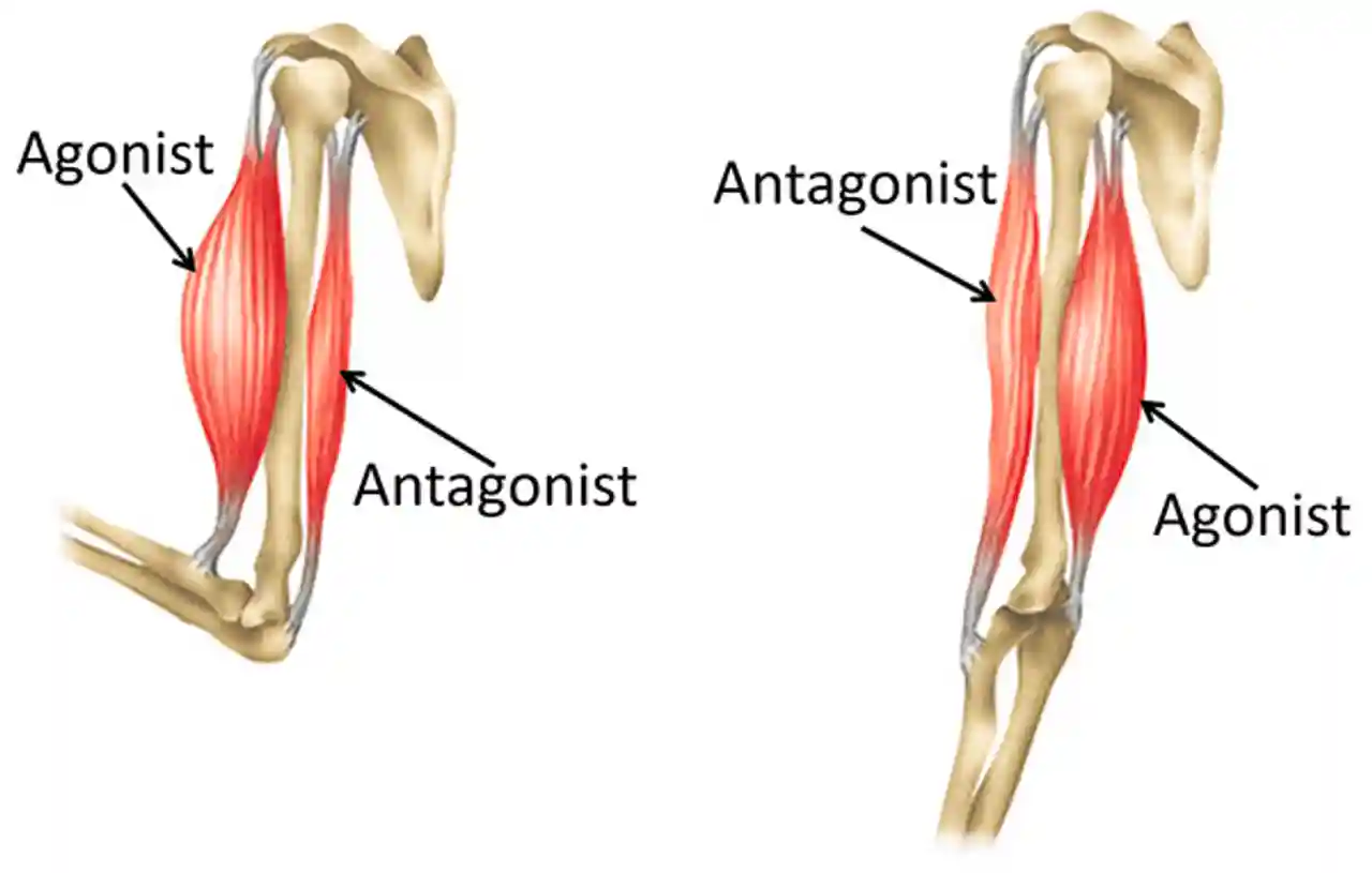
What is a muscle action? har har harr.
what it does duuuuuuhhh boioioi why did even a queshtoun
What is a synergist muscle?
Muscles that work together (eg- brachialis and biceps brachii)

How is the strength of a muscle determined?
It’s cross-sectional area

What happens in an injury
Damage is done to the muscle tissue
What is a sprain
when a ligament is torn
what is a strain/pull/tear
when a tendon or muscle is torn
What is a parallel muscle type
A muscle with muscle fibers that run parallel from end to end

what is a pennate muscle type
several muscle bellies pull on a tendon for more strength

what is a circular/sphincter/orbicularis muscle type?
A circular area of muscle, for openings

Masseter action
elevate the mandible (molars)
Masseter origin and insertion
Origin- Zygomatic arch
Insertion- Angle of the mandible

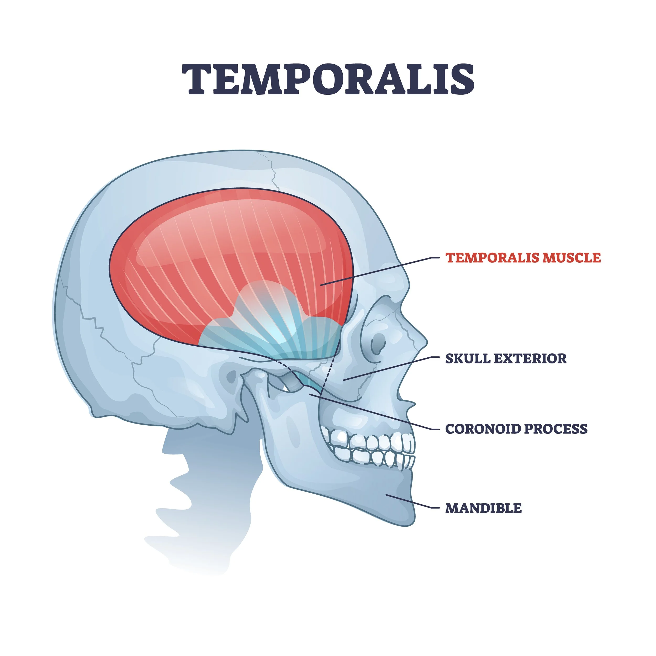
Temporalis action
elevate the mandible (front teeth)
Temporalis origin and insertion
Origin- Lateral skull
Insertion- Coronoid process of the mandible
Lateral Pterygoid action
Depress the mandible, lateral mandibular movement

Medial pterygoid action
elevates the mandible, lateral movement of the mandible

Genioglossus aciton
depress and protract (stick out) tongue
Genioglossus origin and insertion
Origin- The genio region of the mandible (below the mental)
Insertion- Tongue

Hyoglossus action
depress and retract the tongue
Hyoglossus origin and insertion
Origin- Hyoid bone
Insertion- Tongue

Palatoglossus action
elevate tongue
Palatoglossus origin and insertion
Origin- Hard palate bonde
Insertion- tongue

Styloglossus action
Retract the tongue and elevate the lateral sides to make a hawt dawwwggg (yermmmy)
Styloglossus origin and insertion
Origin- Styloid process of the temporal bone
Insertion- Tongue

Medial Rectus action
Moves eye medically
Medial rectus orig and insert
Origin- posterior orbit
Insertion- medial eye

Lateral rectus action
Moves eye laterally
Lateral rectus origin and insertion
Origin- posterior orbit
Insertion- lateral eye

Superior Rectus action
Moves the eye superiorly
Superior rectus origin and insertion
Origin- posterior orbit
Insertion- superior eye

Inferior Rectus action
Move eye inferiorly
Inferior rectus origin and insertion
Origin- posterior orbit
Insertion- inferior eye

Superior oblique action
Move eye inferiorly and laterally
Superior oblique origin and insertion
Origin- posterior orbit
Insertion- posterior lateral eye

Inferior oblique action
Move eye superiorly and laterally
Inferior oblique origin and insertion
Origin- medial orbit
Insertion- posterior lateral eye

Diaphragm action
Move the central tendon inferiorly, this increases volume and decreases pressure in lungs to rush air in to breathe
Diaphragm orig insert
Origin- costal margin
Insertion- central tendon of the diaphragm

Deltoid action
Abduct, flex, and extend the arm (not actually a part of the rotator cuff)
Deltoid origin and insertion
Origin- spine of the scapula and clavicle
Insertion- deltoid tuberosity on the humerus

Supraspinatus action (rotator cuff)
Arm abduction
Supraspinatus origin and insertion
Origin- Supraspinous fossa
Insertion- greater tubercle of the humerus

Infraspinatus action (rotator cuff)
Lateral arm rotation
Infraspinatus origin and insertion
Origin- Infraspinous fossa
Insertion- greater tubercle of the humerus

Teres minor action (rotator cuff)
Lateral arm rotation and arm adduction
Teres minor origin and insertion
Origin- lateral border of the scapula
Insertion- greater tubercle of the humerus

Subscapularis action (rotator cuff)
Medial arm rotation
Subscapularis origin and insertion
Origin- Subscapular fossa
Insertion- lesser tubercle of the humerus

What do the rotator cuff muscles do?
Their tendons actually hold the humerus in the AC joint
What is the labrum
the cartilage cuff in the shoulder joint

Flexor retinaculum
Holds the digit tendons except the palmaris longus

Carpal tunnel
the area the tendons to the digits go through, under flexor retinaculum:
When digits constantly move, tendons fray and rub, swelling begins and pressure is put on the median nerve

Flexor digitorum superficialis action
Flexes the middle phalanx of each digit
Flexor digitorum superficialis origin and insertion
Origin- humeroulnar and radial head
Insertion- middle phalanx of digits 2-5

Flexor digitorum profundus action
Flexes distal phalanges for digits 2-5

Flexor digitorum profundus origin and insertion (deep)
Origin- proximal ulna
Insertion- distal phalanges digits 2-5
Lumbrical action
Flexes the proximal phalanges of digits 2-5
Lumbrical origin and insertion
Origin- flexor digitorum profundus tendons
Insertion- proximal phalanges of digits 2-5
Extensor digitorum action
Extends digits 2-5
Extensor digitorum origin and insertion
Origin- lateral epicondyle
Insertion- both middle and distal phalanges of digits 2-5
Interossei (between the digits) action
Abduction and adduction of digits 2-5
Why is it easier to move digits 2 & 5 than 3 & 4?
digit 2 has the indicies muscles
Digit 5 has the digiti minimi
Digits 3 & 4 don’t have their own muscles
Pronator Quadratus action
Pronation
Pronator quadratus origin and insertion
Origin- distal ulna
Insertion- distal radius
Pronator Teres action
Pronation
Pronator Teres origin and insertion
Origin- medial epicondyle of humerus
Insertion- Coronoid process of ulna
Supinator action
Supination
Supinator origin and insertion
Origin- lateral epicondyle
Insertion- proximal radius
What is the function of the nervous system?
To transmit ELECTRICAL signals
What are the 2 divisions of the nervous system?
central nervous system (CNS)
Peripheral nervous system (PNS)
What is the CNS
made up of the brain and spinal cord (reflexes)
It’s where processing takes place
It is isolated from the rest of the body by layers of tissue called meninges
What is the PNS
Made of the nerves throughout the body (peripheral)
Incoming and outgoing electrical signals
Not isolated from the body
Have myelinated axons for more speeeeed!!!
What are neurons
The cells that actually send the electrical signals throughout the body
Neuron cell body
Is the living part of the neuron, has the ER, mitochondria, and other organelles etc.
Dendrites
Connect to cell body and take in incoming electrical signals
Axon
Long part that conducts the nerve impulse towards the synaptic terminals
Axon hillock
Between the axon and cell body, where the electrical signal is generated, packed with mitochondria