Anatomy II
 Call Kai
 Learn
 Practice Test
 Spaced Repetition
 Match
 Flashcards
 Knowt Play
1/46

There's no tags or description
Looks like no tags are added yet.
47 Terms
kidney functions
excretion of waste products
regulation of:
blood ionic composition
blood pH
blood volume
blood osmolarity
glucose levels
production of hormones:
calcitriol (active form of vitamin D, regulates Ca homeostasis)
erythropoietin
enzymatic blood pressure regulation by secreting renin
kidneys
retroperitoneal organs
located on posterior abdominal wall between T12 and L3
renal hilum on concave border
surrounded by 3 layers of tissue: renal capsule, adipose capsule, renal fascia
contains the adrenal (suprarenal) gland on the top
kidney tissue surrounding
renal capsule (dense irregular connective tissue), closest to kidney
adipose capsule
renal fascia (dense irregular connective tissue)
kidney internal structure
renal cortex is the outer layer
renal medullar is the inner portion making the renal pyramids, base pf pyramids face cortex
apex of pyramids, final papilla, points towards hilum
renal columns are areas of cortex between pyramids
renal lobe: pyramid and overlying cortex with half-columns on each side
renal calyces
minar calyx receives urine from papillary ducts of one renal papilla
each kidney has 8-18 minor and 2-3 major
renal pelvis is single large cavity within kidney connected to ureter
renal sinus: renal pelvis, calyces, renal blood vessels and nerves
renal blood supply
renal arteries are branches of abdominal aorta
divide into segmental, interlobar, arcuate, interlobular aa, afferent arterioles, glomerular, efferent arterioles, peritubular capillaries, vasa recta
renal veins are tributaries of inferior vena cava
divide into interlobular veins, arcuate veins, interlobar veins, renal vein
ureters
muscular ducts that carry urine from kidneys to urinary bladder
retroperitoneal
constrictions at:
pelviureteric junction
pelvic brim where it crosses iliac vessels
ureterovesical junction
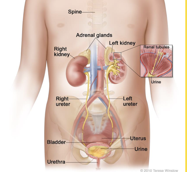
urinary bladder
hollow organ with strong muscular walls, characterized by distensibility
situated in pelvic cavity, posterior to the pubic symphysis. superior surface covered by peritoneum
when empty is tetrahedral in shape with:
trigon (floor): ureteral openings and internal urethral orifice (internal urethral sphincter, more inferiorly external)
apex points to pubic symphysis
3-layered wall: mucosa, muscular, adventitia, serosa

urinary bladder walls
mucosa (transitional epithelium, lamina propria, forms rugae
muscularis
adventitia (posterior and inferior aspects)
serosa (superior)
female urethra
ends in external urethral orifice
wall: mucosa and muscular (circular layer)
mucosa: transitional → stratified columnar/pseudostratified columnar → non-keratinized simple squamous
shorter than male

male urethra
longer, double passage (urine, semen)
prostatic; openings: ducts from prostrate, seminal vesicles and ductus (vas) deferens
membranous
spongy (penile); bulbourethral (Cowper’s) glands, urethral glands

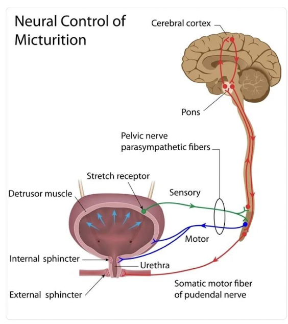
micturition
discharge of the urine from the urinary bladder occurs as a combination of involuntary and voluntary muscle contraction
urinary system function
maintaining optimal blood quality
balancing:
water vs electrolytes → pH balance
excretion:
metabolic waste
water
electrolytes
bioactive (drugs)
secretion:
renin
erythropoietin
conversion:
Vit D → calcitriol
glucogenesis
kidney structures
renal pyramids
regions of urine collection and transport
renal papilla
regions of renal pyramid apex and funnel
medulla
contains renal pyramids
cortex
contains renal filtration apparatus
renal columns
extensions of the cortex into the medullary region
renal lobe
renal pyramid + cortical vase + cortical sides
renal lobule
multiple nephrons draining into a shared collecting duct and associated cortical tissue
nephrons
function units of kidney
each kidney contains 1.25 million nephrons
each nephron comprises a corpuscle
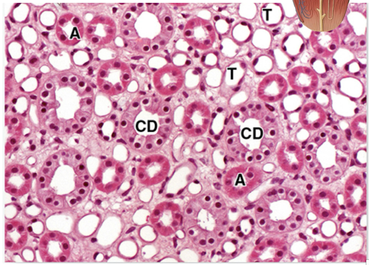
simple epithelial tubule consists of three regions
proximal convoluted tubule
loop of henle
distal convoluted tubule

loop of henle
has four regions and is the loop into the medulla of the nephron
proximal straight tubule
largely continuous with PCT
thin descending limb
simple squamous epithelium
passive resorption of Na+ and Cl-
thin ascending limb
simple cuboidal epithelium (no brush border/many mitochondria)
active resorption of electrolytes
active excretion of Na+
thick ascending limb
renal corpuscle
glomerulus + bowman’s capsule
bowman’s capsule (visceral layer):
podocytes with interdigitated pedicels
podocyte basal laminae + endothelium = Glomerular Basement Membrane
bowman’s capsule (parietal layer)
simple squamous epithelium → cuboidal at tubular pole

proximal convoluted tubule
comes directly after the corpuscle
histological features
simple cuboidal epithelium
central nuclei
brush border - microvilli
functions
reabsorption of 2/3 total water, all organic nutrients, all proteins and electrolytes

distal convoluted tubule
histological features
simple cuboidal epithelium
smaller cells
no brush border (short microvilli)
functions
reabsorption of electrolyes
regulation of Na+ by adrenal glands

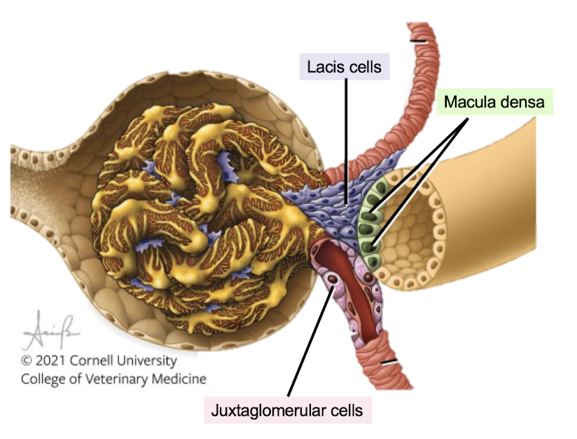
juxtaglomerular apparatus
maintains glomerular filtration rate
macula densa
simple columnar epithelium
densely packed
apical nuclei
NaCl sensor
juxtaglomerular cells
tunica media of afferent arteriole
modified smooth muscle cells
round nuclei
blood pressure sensor
lacis cells
support cells
contractile capacity
physiological connection

collecting ducts kidney
histological features
in cortex: simple cuboidal epithelium
in medulla: simple columnar
functions:
reabsorption of water if needed
carrying filtrate to calyces

ureters
3 layers:
mucosal layer
transitional epithelium
basal cells
cuboidal/low columnar cells
umbrella cells
large elliptical surface cells
sometimes binucleated
laminate propria
muscular layer
inner longitudinal
outer/middle circular
outer longitudinal
adventitial layer
fibroeslastic connective tissue

urinary bladder hist
3 layers
mucosal layer:
transitional epithelium
laminate propria
submucosa
muscular layer
inner longitudinal
middle circular
outer longitudinal
adventitial layer
upper bladder only covered by serous peritoneum
umbrella cells
unique thick apical membrane
lipids assembled into plaque
numerous tight junctions on lateral membranes
provides protection from hypertonic urine

urethra hist
female: 3-5 cm
transitional → stratified squamous
male:
prostatic urethra
transitional epithelium
membranous urethra
stratified/pseudostratified columnar epithelium
spongy urethra
stratified/pseudostratified columnar epithelium
stratified squamous distally
primary reproductive organs
gonads: testes and ovaries produce gametes (sperm and ova) and secrete sex hormones
primary sex characteristics are directly involved in reproduction of the species
accessory reproductive organs
ducts, glands and supporting structures
secondary sex characteristics are features not directly concerned with reproduction, such as voice quality, facial hair, and breast size
male reproductive organs
grouped by function:
gonads (testes)
ducts - epididymis, ductus deferent, ejaculatory ducts and urethra
accessory sex glands - seminal vesicles, prostate and bulbourethral glands
supporting structures - copulatory organ (penis, scrotum)
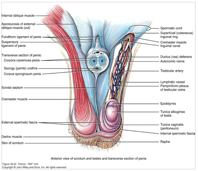
scrotum
cutaneous sack:
internally divided by the scrotal septum into two sacks and externally by the scrotal raphe
musculature of the scrotum:
dartos muscle (smooth muscle, wrinkles and relaxes the skin of the scrotum)
cremaster muscle (skeletal muscle, elevates and lowers the testes)

testes
oval paired glands
produce sperm and hormones
testes are covered by several layers of connective tissue
connective tissue extends inwards and divide testis into lobules
lobules contain 2-3 seminiferous tubules (site of sperm production) → rete testis → efferent ducts → ductus epididymis

epididymis
located along the posterior border of each testis; head, body, tail
single highly coiled tube enveloped by smooth muscle cells
site of sperm maturation, storage; sperm is propelled into the ductus (vas) deferens

ductus (vas) deferens
continuation of the duct of epididymis, conveys sperm to the urethra

spermatic cord
supporting structure of the male reproductive system that ascends from the scrotum and consists of
ductus deferens
testicular artery
veins (venous plexus)
nerves and lymphatics
seminal vesicles
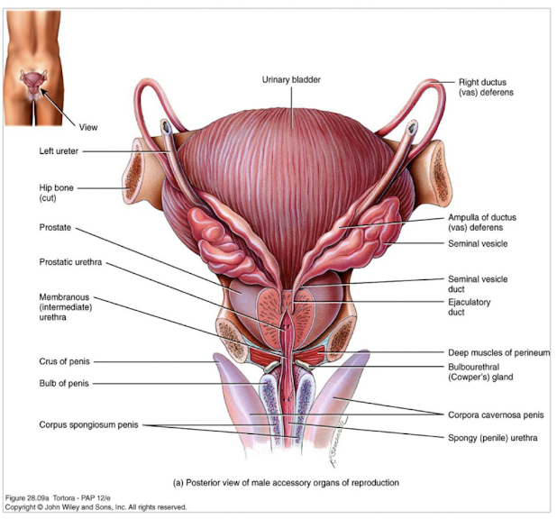
paired seminal vesicles lying posterior on the base of the urinary bladder and secrete alkaline fluid rich in fructose, clotting agents
duct of seminal vesicle unite with the ductus deferens and form ejaculatory duct which opens into the prostatic urethra
prostate
inferior to the urinary bladder
single accessory gland - prostate, surrounds prostatic part of the urethra
prostate produce slightly acidic secretion, citric acid, proteolytic enzymes (prostate specific antigen, amylase, protease)
enlargement of the prostate after the age of 45 common

bulbourethral glands
inferior to prostate
lateral to membranous urethra
alkaline secretions, mucus
semen
sperm (50-150 million per 1 mL) + seminal fluid (produced by seminal vesicles, prostate, bulbourethral glands); coagulates after ejaculation
penis
copulatory organ contains: body, glans, root
body has three cylindrical masses of tissue covered by tunica albuginea
corpora cavernosa: two dorsolateral masses; comprised of erectile cavernous tissue
corpus spongiosum: ventral mass: contains covernous erectile tissue and spongy (penile) urethra, expanded region — glans (prepuce)
root of penis is attached position consisting of:
bulb - extended portion of the base of the corpus spongiosum
crura - extended portions of the corpora cavernosa
remale reproductive organs
internal
ovaries
uterine (fallopian) tubes
uterus
vagina
external
vulva
mammary glands
ovaries
almond shaped glands, located on the lateral walls of the lesser pelvis
functions:
production of gametes
production of hormones (estrogen, progesterone, relaxin)
covered by:
germinal epithelium (simple cuboidal/squamous)
tunica albuginea (dense irregular connective tissue)
two regions:
cortex (contains ovarian follicles)
medulla (contains blood vessels, nerves and lymphatic vessels

uterine (fallopian) tubes
extend from the uterus, between the fold of broad ligaments and open near the ovaries; transport the secondary oocyte
divided into:
infundibulum - ends in fimbriae
ampulla - longest, widest portion, usual place of fertilization
isthmus

uterus
thick-walled, pear shaped muscular organ, located in the lesser pelvis with its body on the top of urinary bladder and its neck (cervix) between the urinary bladder and rectum; projects anterosuperiorly
site of implantation, fetal development, menstrual flow
attached by “ligaments” including broad and round
divided into:
fundus - superior to the isthmus of the uterine tube
body (between the layers of broad ligament), uterine cavity
cervix - cervical canal, internal and external os


vagina
fibromuscular canal between uterine cervix and the exterior of the body (vaginal orifice)
located posterior to the urethra and urinary bladder and anterior to rectum
receptacle for penis, menstrual flow, childbirth
vaginal fornix - recess around the cervix
hymen vascularized mucous membrane, partially closes the vaginal orifice

vulva
external genitals of the female:
mons pubis - adipose tissue covered with the skin and pubic hair
labia majora - two longitudinal folds of skin covered with pubic hair, contains adipose tissue and sweat and oil glands
labia minora - skin folds, devoid of pubic hair and fat
clitoris - cylindrical mass of erectile tissue and nerves
vestibule - the region between the lava minora

vestibule
hymen
vaginal origice
external urethral orifice
ducts of the greater vestibular glands (bartholin’s)
bulb of vestibule - consists of two elongated erectile bodies deep tot he labia on either side of the vaginal orifice
perineum
external surface area located inferior to the pelvic diaphragm (formed by muscles)
osseous boundaries (pubic symphysis, ischial tuberosisties and coccyx)
transverse line joining the anterior ends of the ischial tuberosities divides perineum into two triangles: urogenital and anal
midpoint of the line joining ischial tuberosities is the central point of the perineum overlying the perineal body (fibromuscular mass)

mammary gland
suspensory ligaments firmly attach breast to the dermis and fascia
each lobule (glandular tissue) is drained into mammary ducts
