Histology and Tissues Vocabulary Flashcards
 Call Kai
 Learn
Learn Practice Test
 Spaced Repetition
 Match
Match Flashcards
 Knowt Play
1/108

Earn XP
Description and Tags
Vocabulary flashcards based on lecture notes covering tissue categories, epithelial types, muscle types, nervous tissue, and epidermal structures.
Name | Mastery | Learn | Test | Matching | Spaced | Call with Kai | Chat |
|---|
No analytics yet
Send a link to your students to track their progress
109 Terms
Tissue
A group of associated cells and their intercellular substance working together to perform a specific function or functions.
Epithelium
it covers body surfaces, lines body cavities, and makes up many glands.
Muscle Tissue
An excitable and contractible tissue category consisting of elongate, tube-shaped cells (fibers) that generate electrical signals and shorten to produce movement.
Nervous Tissue
excitable tissue category responsible for generating impulses and electrical signals for communication within the body, located in the brain, spinal cord, and peripheral nerves.
Connective Tissue
A primary tissue category that connects and supports body parts and fills in spaces.
Simple Epithelium
Found in the air sacs of the lungs (alveoli) and the lining of heart, blood vessels, and lymphatic vessels.
Stratified Epithelium
An epithelial tissue classification consisting of more than 1 layer of cells.
Squamous Epithelial Cells
Epithelial cells that are very thin and scale-like in shape.
Cuboidal Epithelial Cells
Epithelial cells that are cube-like in shape.
Columnar Epithelial Cells
Epithelial cells that are longer than they are wide (like a column).

Classes of Epithelium and Cell Shapes
Diagram illustrating simple, pseudostratified columnar, and stratified epithelium classes, along with squamous, cuboidal, and columnar cell shapes.
Simple Squamous Epithelium
An epithelial tissue composed of a single layer of flattened, scale-like cells that functions in absorption; located in the alveoli or lungs, kidney glomerulus, and walls of capillaries.

Cheek Cell Smear (400×)
A slide preparation of simple squamous epithelial cells from the oral cavity, displaying the cell membrane, nucleus, and cytoplasm.
Simple Cuboidal Epithelium
An epithelial tissue consisting of cube-shaped cells that lines cavities, ducts, and tubules, such as kidney tubules.
Simple Columnar Epithelium
An epithelial tissue composed of column-shaped cells longer than they are wide, containing Goblet cells to produce mucus, located in the respiratory tract.
Goblet Cell
A specialized epithelial cell that produces mucus, found in simple columnar epithelium and pseudostratified columnar epithelium.
Pseudostratified Columnar Epithelium
A columnar tissue (often ciliated, abbreviated PCCE) containing Goblet cells, located in the respiratory system where it sweeps debris from air and traps it in mucus.
Stratified Squamous Epithelium
An epithelial tissue made of multiple layers of scale-like cells that functions in protection, located in the skin and vagina, featuring stratum corneum, stratum basale, and keratin.
Stratum Corneum
The surface layer of the epidermis consisting of flattened, dead cornified cells impregnated with dense amounts of fibrous keratin.
Stratum Basale
The deepest layer of the epidermis where cells originate and constantly undergo mitosis.
Melanocytes
Cells located in the stratum basale of the epidermis that produce a pigment called melanin for UV protection.
Merkel's Discs
Sensory structures present in the stratum basale of the epidermis.
Keratinocytes
Cells that make up most of the cells of the epidermis.
Langerhan's Cells
Immune cells located in the skin.

Skeletal Muscle
Voluntary muscle tissue made of long, cylindrical, peripherally multinucleate cells with distinct striations, located in arms and legs to provide voluntary movement.

Cardiac Muscle
Involuntary muscle tissue of the heart consisting of short-branched cells with 1-2 nuclei, less distinct striations, and intercalated discs.
Intercalated Discs
Specialized structures connecting adjacent branching fibers of cardiac muscle cells.

Smooth Muscle
Involuntary muscle tissue found in visceral organs (stomach, intestines, uterus) composed of spindle-shaped cells with a large, central, football-shaped nucleus and no striations.

Neurons
The functional cells of the nervous system responsible for producing impulses and electrical signals, characterized by a soma and long processes (axons and dendrites).
Neuroglia
Supporting cells of the nervous system that protect and support neurons.
Soma
The cell body of a neuron.
What are the general characteristics for the Epithelium?
a. small cells, closely packed together
b. Apical or luminal surface
c. Avascular
d. Basement Membrane
What are the 4 examples of Epithelium?
a. simple squamous epithelium
b. simple cuboidal epithelium
c. simple columnar epithelium
d. stratified squamous epithelium

Simple Squamous Epithelium

Simple Squamous Epithelium

Simple Squamous Epithelium

Cuboidal Epithelial Cells

Simple Cuboidal Epithelium

Identify Basement membrane and Simple Cuboidal Epithelium

determine the lumen and luminal surface
Cuboidal Epithelium and a thyroid follicle.
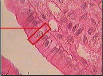
Simple Columnar Epithelium
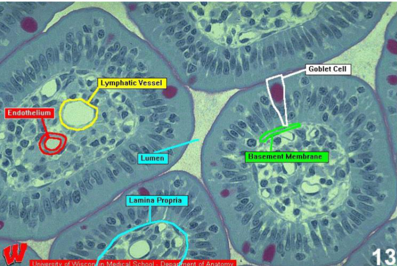
Identify Goblet, basement membrane, lumen, and luminal surface

Simple Columnar Epithelium
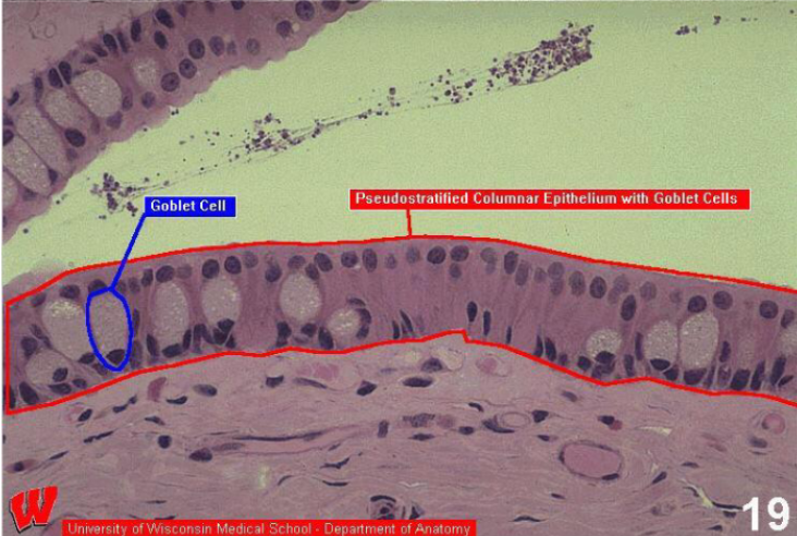
Pseudostratified Epithelium
Simple Squamous Epithelium
Function: Absorption, single layer,
Location: alveoli or lungs kidney glomerulus walls of capillaries
The flat, scale-like form and single-layer arrangement of simple squamous epithelium create an extremely thin barrier that maximizes the speed of passive diffusion and filtration
Simple Cuboidal Epithelium
Function: line cavities, ducts and tubules
a single layer of cube-shaped cells that feature a large, spherical, and centrally located nucleus
Locations: ducts and tubules kidney tubules
provides a balanced trade-off between a short diffusion distance and sufficient internal volume to house the organelles required for active secretion, absorption, and excretion
Simple Columnar Epithelium
Function: Produces Mucus
Goblet Cell Produces mucus
Respiratory tract
provides extra cytoplasmic volume and surface area required for high-capacity absorption, extensive secretion, and robust physical protection
Stratified Squamous Epithelium
Function: Protection
-stratum corneum -stratum basale -keratin
Location: Skin Vagina
creates a thick, resilient barrier that protects underlying tissues from mechanical wear, friction, and pathogen invasion

Stratified Squamous Epidermis and identify stratum basale.

Stratified Squamous epithelium

Identify stratum corneum and stratum basale
Muscle
a. Elongate, tube-shaped cells (fibers)
b. Specialized for Contraction
c. Excitable: Can propagate and electrical charge
d. When contract produce movement
Skeletal Muscle
peripherally
multinucleate, distinct striations, provides voluntary movement
Location: arms and legs.
Cardiac Muscle
-intercalated discs, short-branched, 1-2 nuclei, striations (less distinct)
Location: Heart
Smooth Muscle
-Large, central nucleus (football
shaped), no striations
Location: stomach, intestines, uterus
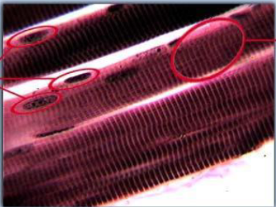
Skeletal Muscle
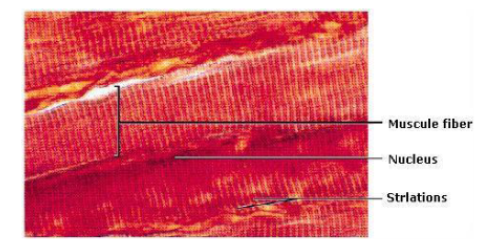
Muscle Fiber, Striations
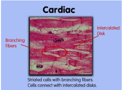
Cardiac Muscle

Identify Intercalated discs and weak striations.
Nervous Tissue
a. Highly Specialized Tissue
b. Responsible for generating impulses
c. Extremely long processes
d. Two main cell categories
i. Neurons: Functional Cells of the Nervous System
1. Soma = cell body
2. Processes=extensions (axons and dendrites)
ii. Neuroglia: Protects and supports neurons
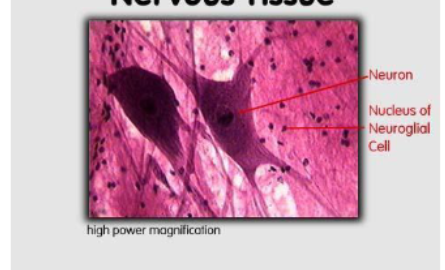
Identify nucleus and neuroglial of the cell
Nervous Tissue

Identify neuron and neuroglial
Nervous Tissue
Connective Tissue
(Grout) tissue with cells that are
widely spaced and separated by matrix.
What are the (Connective Tissue) Matrix 2 components?
Ground Substance and Protein Fibers
Matrix
Intercellular substance that both surrounds and is secreted by the cells of the connective tissue.
What is the exception of the Matrix?
Blood
Ground Substance (Matrix)
background material in which the cells embedded
i. amorphous: No specific shape, liquid or semiliquid
ii. structural: Very definite shape
Protein Fibers (Matrix)
Lie within or embedded in ground substance (Table 5.4 in your e-
lab manual and page 150 in e-textbook describes these fibers)
i. collagen bundles: Look like pink, thick smeared lines
ii. elastic fibers: appear as distinct, dark lines
iii. reticular fibers: small
Cells (Connective Tissue)
the type of cell in Connective Tissue depends on the type of tissue. Often, the
name of the cell ends in “cyte”. For example, a chondrocyte is a cartilage cell. An
adipocyte is an adipose cell
Examples of Connective Tissue
A. Areolar Connective Tissue
B. Adipose Tissue
C. Blood
D. Bone
E. Hyaline Cartilage
F. Fibrocartilage
G. Elastic Cartilage
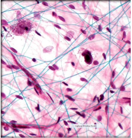
Areolar Connective Tissue
these cells
include: macrophages, fibroblasts, monocytes and mast cells. This tissue has a lot of collagen fibers and elastic fibers
Areolar Connective Tissue
You find this tissue underneath the skin attaching skin to muscles
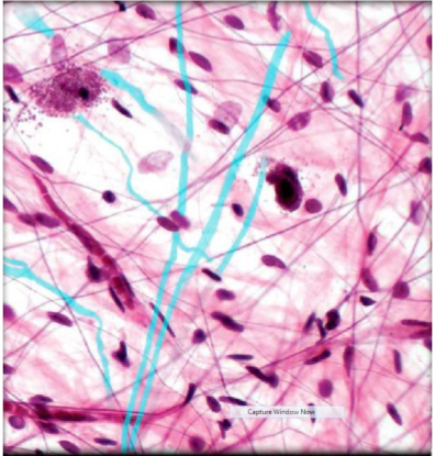
Areolar Connective Tissue
Collagen Bundles
Areolar Connective Tissue
Elastic Fibers
Areolar Connective Tissue
acts as a flexible packing material that binds skin to underlying muscles, cushions organs, and supports blood vessels and nerves
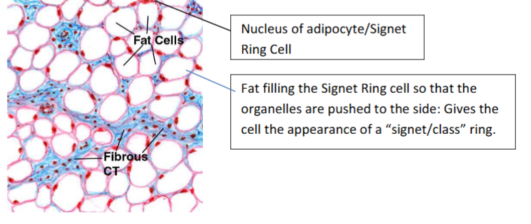
Adipocyte (Connective Tissue)
It has a fat storage vacuole where fat is stored
Signet Ring Cell
When it is full, the organelles of the cell, including the nucleus, are pushed to the side giving the
cell the appearance of a class ring. So, we call a mature adipocyte (one filled with fat)
Adipocyte
Identify the nucleus and the signet ring cell
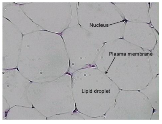
Signet Ring cell and Plasma Membrane

Adipose cell

Adipose (Chicken Wire)
Blood (Connective Tissue)
Blood is unusual. It is a liquid tissue. It is responsible for circulating oxygen and carbon dioxide in the body and for protecting the body against invaders.
What are the 2 (main) cell types for Blood?
erythrocytes and leukocytes
Erythrocytes
red blood cells
a. small, but great in number
b. anucleate
leukocytes
a. larger, but few in number
b. have a nucleus
Platelets (Thrombocytes) (Blood Tissue)
Cell fragments involved in clotting. Note that platelets are not true cells. They are only cell fragments. They look like dust bunnies in a sea of red and
white blood cells.

Identify red blood cells (erythrocytes) and platelets
Osseous Bone (Connective Tissue) (Compact)
Used for structural support for the muscles.
What is the basic unit of compact bone?
Osteon
What does an osteon look like?
Cross-section of a tree.

Identify central canal, canaliculi, osteocytes, lucunae, lamelle
What are parts of the bone?
Osteon: Basic unit of a compact bone
• Central canal: Region where blood vessels pass through the bone
• Lacunae: Fluid-filled cavity containing bone cells
• Osteocyte: Bone cell
• Lamellae: It is the matrix
• Canaliculi: Hair-like canals from the lacunae to the central canal; allow osteocytes to get
nutrients.
Where are the osteocytes?
In the lacune
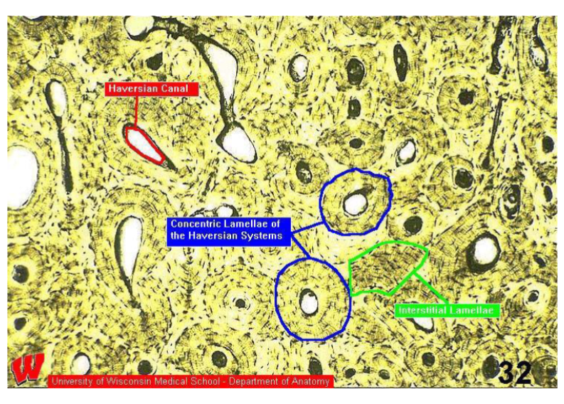
Label Lamellae
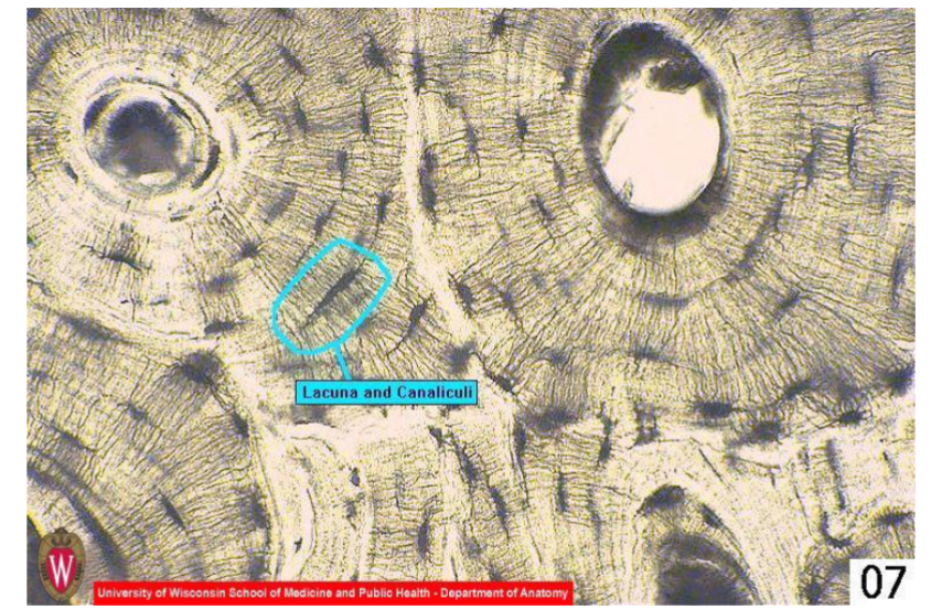
Lacuna & Canaliculi
Cartilage (Connective Tissue)
categorized based on flexibility and support. Some cartilage has more flexibility, some offers more support. Some cartilage has more of certain types of fibers than others giving it more strength of flexibility.
Hyaline Cartilage
offers equal flexibility and support. The matrix does not have any fibers and so looks smooth. The cell is called a chondrocyte and is found in a lacunae (a space or sack in the matrix).
Where is a Hyaline Cartilage located?
It is found in the ribs and nose.

Chondrocytes within the lacune
Hyaline Cartilage
Fibrocartilage
has more support and less flexibility. You find it in between the vertebrae in the back. There are many collagen fibers in fibrocartilage, so it has a streaky look.