54- Normal Resp
 Call Kai
 Learn
 Practice Test
 Spaced Repetition
 Match
 Flashcards
 Knowt Play
1/82

There's no tags or description
Looks like no tags are added yet.
83 Terms
what are the components of the resp system
nostrils, sinus, trachea, nasopharynx, etc
pulm vascular system
diaphragm, intercostal m
what is the function of the lung
facilitate o2 and co2 exchange
nostrils, nasal cav, paranasal sinuses, nasopharynx, larynx, trachea, and bronchi are part of what component
conducting
describe the conducting component histo
lined by pseudostratified, CILIATED columnar cells
lined by sensory olfactory epithelium
squamous epithelium
variable portion of goblet mucous cells

what is this image
conducting component
what serves as the transition zone bt conducting comp (ciliated) and air exchange (alveolar)?
bronchioles that branch off bronchi
describe the transition component cilia
the ciliar become scarce in the prox bronchiolar region until they disappear
what cells can be found in the trans component
club cells, neuroendocrine cells
NO goblet cells
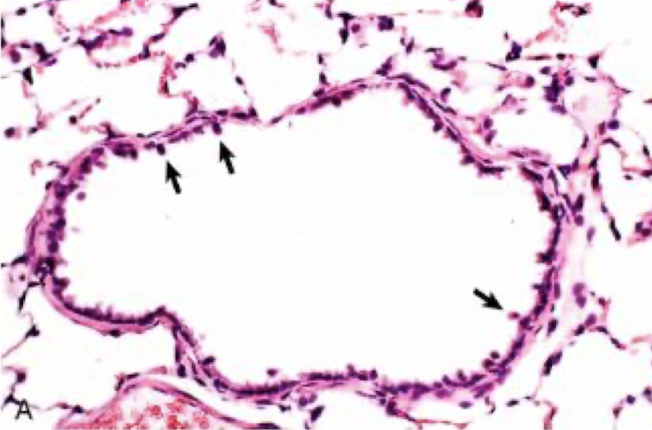
what helps detox xenobiotics, repairs resp stem with stem cells, and contributes to the INNATE imm of lungs by secreting protective proteins and surfactant?
club cells of trans component
what is the air exchange component formed by
alveoli
what does the interstitium of the air exchange component support
alveolar epithelium and alveolar capillary endothelium
in the air exchange component, whats the blood air barrier formed from
vascular endothelium, basement mem, alveolar epithelium lining cells
whats a thin delicate network
alveolar walls
describe type I alveolar surface endothelial cells
membranous pneumocytes
cover majority of alvolar wall
func in gas exchange
describe type II alveolar surface endothelial cells
granular pneumocytes
larger cuboidal cells
produce surfactant

label the pneumocytes
purple- type 2
blue- type 1
whats the degree of lobulation determine?
the degree of air movement between lobules
collateral ventilation
movement of air between lobules and between adj alveoli
is collareral ventilation good in dogs or pigs
DOGS
pigs have thick CT of the interlobar septa seperating lung lobes

are these normal or pathological lungs
normal
what is pleura
single layer of mesothelial cells overlying mult layers of connective tissue
parietal pleura
lines thoracic walls, diaphragm, mediastinum
visceral pleura
lines lung surface
pleural space
between parietal and visceral pleura, sep by mediastinum
what animals have complete mediastinum
cattle goats and pigs
what animals have fenestrated mediastinum
horses and sheep
where is the resp system microbiota most abundant
nasal cavity, pharynx, larynx, trachea, bronchi, alveoli
what is complicated about mannheimia haemolytica in bovines
its part of their nasal flora. but can also cause pneumonia
aerogenous routes of entry
virus, bacteria, toxic gases, pneumotoxicants
hematogenous route of entry
virus, bacteria, fungi, parasites, emboli, IV inj, pneumotox
direct extension route of entry
penetrating wounds, migrating awns, bites, hardware
t or f: inflamm can extend locally regionally or systemically
t
what can laryngeal exudate from laryngitis cause
aspiration
what can a large vascular bed present in the lung lead to
sepsis and septic shock
why does hematogenous portal of entry occur commonly
bc pulm system is well vascularized
physical unidirectional movement and removal of deposited particles and gases disolved in the mucus
mucociliary clearance of the conducting/transitional component
what helps the main defense mechanism of the conducting component, mucociliary clearance
mucociliary escalator with mucus
why is cilia imp in the mucociliary clearance
the rapid movement of cilia causes synchronized waves that propels mucus and particles to the pharynx where its terminated and coughed up
what is the main defense mechanism of alveoli
phagocytosis by pulm alveolar macrophages
the # of macrophages in alveolar space is related to…
the # of inhaled particles reaching the lungs
describe the life span of alveolar macrophages
few days long
replaced by newly migrated monocytes
migrate to bronchiolar region to mucocil elevator
exits to pharynx
pneumoconiosis
lung dz caused by dust deposition
anthracosis
lung dz caused by carbon deposition
can can occur in animals living in pollution/smoking/fireplaces
anthracosis
whats the defense mech for the conducting and transitional components
mucociliary clearance
whats the defense mech for the air exchange component
phagocytosis from pulm alveolar macrophages
What are the portals of entry for injury into the lungs?
Inhalation
•Blood
•Direct extension
What are the portals of entry for injury into the thoracic cavity, mediastinum, or pleura?
•Hematogenous (common)
•Direct extension

Grossly, we see tiny black pinpoint foci on the lung that is associated with black pigment within macrophages on histology. What condition causes these lesions?
Alveolar phagocytosis when deposition of inhaled material/dust exceeds removal, leading to accumulation in the lung

what are these cells
pulm alveolar macrophages
Name the defense mechanisms against spec types of injury `
aerogenous
hematogenous
oxidants and free radicals
what is the underlying pathogenetic mechanism in many pulm diseases
abnormal retention of particles (aerogenous)
deposition
process of particles being trapped within resp sys
clearance
process which particles are destroyed from mucosal surface like sneezing, coughing, mucociliary transport, phagocytosis
if a particle is 10 um or greater…
lodges in nasal passage
if a particle is smaller than 10um,,,
progresses to tracheal and bronchial bifurcations
if a particle is 2um or smaller…
can progress to bronchiole and alveoli
what are intravascular macrophages responsible for in terms of hematogenous defense
removing circulating particles, bacteria, endotoxin
in systemic conditions like sepsis, ____________ by macrophages can cause acute lung injury
excessive release of cytokines
in terms of oxidant/free radical defense, scavengers can be found
in alveolar and bronchiolar epithelial cells and interstitium
what types of oxidant/free radicals cause damage
inhaled oxidant gases, xenobiotic toxin metabolites, free radicals released by phagocytic cells during inflamm (these can impair repair ability)
inhaled bacteria can colonize in bronchi can cause /…
infection leading to fatal pneumonia
what occurs 5-7 days after a viral infection
the phagocytic func of pulm alveolar macrophages and mucociliary clearance are impaired
what are known viruses to predispose to secondary BACTERIAL INFECTIONS
influenza in pigs and horses
BRSV in cattle
canine distemper
felid herpes and calicivirus
what are ex of immunodef that can increase susceptibility to resp conditions
SCID in arabian foals that succumb to adenoviral pneumonia and corticosteroids/chemo drugs
name some of the impairment defenses leading to pneumonia
stress, hypoxia, dehydration, anesthesia, starvation
what are 2 main conducting components
epithelium and bronchi
seq of events of injured pseudostratified ciliated epithelium
irreversible injury
ciliated cells swell
lose their attachment to BM
rapidly exfoliate (ulcerate)
viral infections replicate in ____. examples include:
resp epithelium
bovine herpes, felid herpes, canine adenovirus 2, canine parvovirus 2
if epithelial injury is chronic how does mucus production increase
goblet cell hyperplasia
what does chronic bronchial irritation cause
metaplasia of highly functional ciliated epithelium to nonfunctional squamous epithelium
bronchioles are part of the ___ component
transition
what is bronchiectasis
when an injury results in perm dilation of a bronchus with rupture of the bronchial wall as a result of obstruction or chronic inflammation

what does bronchiectasis form
prominent nodules from bronchi distention that contain exudate
describe alveolar injury
epithelial side lined w type 1 pneumocytes that are suseptible to noxious agents
when damaged, they detach and leads to edema and fibrin leakage
type 2 pneumocytes proliferate to compensate
describe bronchiolar injury
leads to goblet cell production turning the secretions more mucousy
this means the secretions arent removed as easily and can block airway
what happens with alveolar repair if BM stays intact?
cuboidal type II pneumocytes, club cells, and stem cells undergo mitosis and create large pool of new cells
what if alveolar injury persists
fibroblasts and myofibroblasts proliferate in alveolar walls and cause alveolar septal fibrosis
this alters lung function bc loss of type 1 pneumocytes = loss of gas exchange
what are the most numerous cells in pleural space and what do they do
mesothelial cells
capable of secreting pro and anti inflam mediators
can help repair and fibrosis

what occurs with chronic stimuation and mesothelial cells
mesothelial hyperplasia (plaque like proliferations diff to distinguish from neoplasia)

what is this
mesothelioma

what is this
mesothelial cell hyperplasia

What is shown in this histology?
Septal fibrosis and club cells that protruding into the alveolar space