Pulm - Blueprint
 Call Kai
 Learn
 Practice Test
 Spaced Repetition
 Match
 Flashcards
 Knowt Play
1/45

There's no tags or description
Looks like no tags are added yet.
46 Terms
pneumocystis jivorecii pneumonia sx
HIV or CD4 < 200
bilateral ground-glass opacity
caused by a fungus and can present with PTX
pneumocystis jivorecii pneumonia dx
confirm with staining or PCR of respiratory specimen
pneumocystis jivorecii pneumonia tx**
bactrim or steroids
which TB med causes optic neuritis
ethambutol
→ discontinue immediately
isoniazid SE
hepatitis
peripheral neuropathy
pyrazinamide SE
hepatotoxicity
polyarthralgia
rifampin SE
orange discoloration to body fluid (urine, tears, sweat)
Idiopathic pulmonary fibrosis (IPF)
progressive dry cough
inspiratory fine crackles
fingernail clubbing
ct = honeycombing (small cystic spaces enveloped with thick defined walls at the bases)
croup scoring
moderate → stridor at rest and retractions
= oral steroids, nebulized epinephrine, observe in ed for 3 hours, discharge if sx go away
you were bit by a bird. what med do you get
doxy (psittacosis)
macrolide abx MOA
irreversibly binding to the 50S subunit of bacterial ribosomes, thereby blocking translocation during protein synthesis
tetracyclines MOA
binds to the 30s ribosomal subunit
FLQ MOA
inhibiting DNA gyrase
sulfonamides MOA
inhibit folic acid synthesis by blocking dihydropteroate synthase
normal d dimer level
< 500
COPD + seasonal flu ( muscle aches, headache, and malaise)
→ oseltamivir

Löfgren syndrome
sarcoidosis - hilar adenopathy, erythema nodosum (tender reddish-violet subcutaneous nodules on the anterior aspects of the lower extremities)
women
for pulm embolism, when do you give V/Q scan instead of CT?
HIGH creatinine
pregnancy
abdnormal chest radiograph results
central = sputum cytology
peripheral = CT-guided transthoracic biopsy
(if one is negative, test the other)
when does asthma become moderate persistent?
waking up at night more than 1 time (but not everyday)
acute chest syndrome
fever, chest pain, wheezing, cough, hypoxia, and a new lung infiltrate in someone with sickle cell disease (vaso-occlusive cause)
tall and thin, breathlessness (but PTX does NOT have fever)
common complications in sickle cell disease
vaso-occlusion
acute chest syndrome
splenic crisis
aplastic crisis
stroke
leg ulcers
sepsis
cholelithiasis
osteonecrosis
priapism
what kind of bronchoscopy for foreign body aspiration?
rigid
(flexible is for PNA)
first step for foreign body aspiration
bronchoscopy
(heimlich only if they can’t breathe)
croup tx
support
corticosteroids (dexamethasone)
mod-severe = racemic epi
first line med for epiglottitis*
ceftriaxone
you find a well-defined solid lesion in the lung but no pulmonary symptoms at all. what’s your next step?
low dose chest CT w/o contrast (low risk)
if nodule >8 mm → PET
coughing → syncope =
pertussis

lung abscess
cough with putrid sputum, pleuritic chest pain, weight loss, and night sweats, crackles
cavitry lesion and air fluid level
lung abscess tx
augmentin or carbapenem (imipenem, meropenem)

atypical PNA
gradual onset, cough, fever
course breath sounds
Chest film is likely to reveal perihilar and interstitial infiltrates

atypical PNA tx*
azithromycin
tension PTX tx*
Thoracostomy with chest tube placement**
if not → needle decompression
thoracentesis is for what condition?
pleural effusion

what is this
bilar hilar adenopathy → sarcoidosis
(granulomatous disease that can have nodules/erythema nodosum)
( lupus pernio and erythema nodosum)

you see ferruginous bodies =
asbestosis

Mucocutaneous ulcers that are recurrent and usually painful =
behcets disease

Cryptococcus meningitis
Cryptococcus neoformans (dried animal droppings - pidgeons)
cough with scant sputum and pleuritic pain
molluscum contagiosum
tachycardia
high diastolic BP and encapsulated yeast organisms upon India ink staining
tx: intravenous amphotericin B and oral flucytosin → fluconazole

ARDS
accumulation of fluid in the alveolar wall
(happens FAST, like 1 day)
latent TB tx
just rifampin and isoniazid
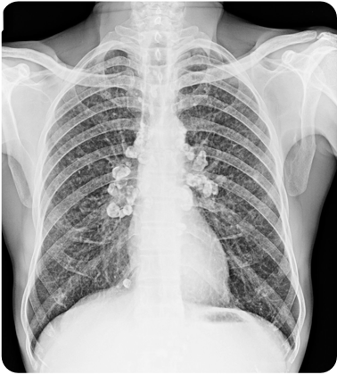
silicosis
chronic, fibrotic lung diseases
inhalation of free silica particles
small (< 1 cm), diffuse, round opacities “eggshell calcifications” on periphery
tx = stop exposure and support
coal miners lung is located primarily where
upper lobes
shipyard construction
asbestosis
lower lobes on lungs
nodular interstitial fibrosis, primarily at the lung bases, and plaques on the parietal and diaphragmatic pleura
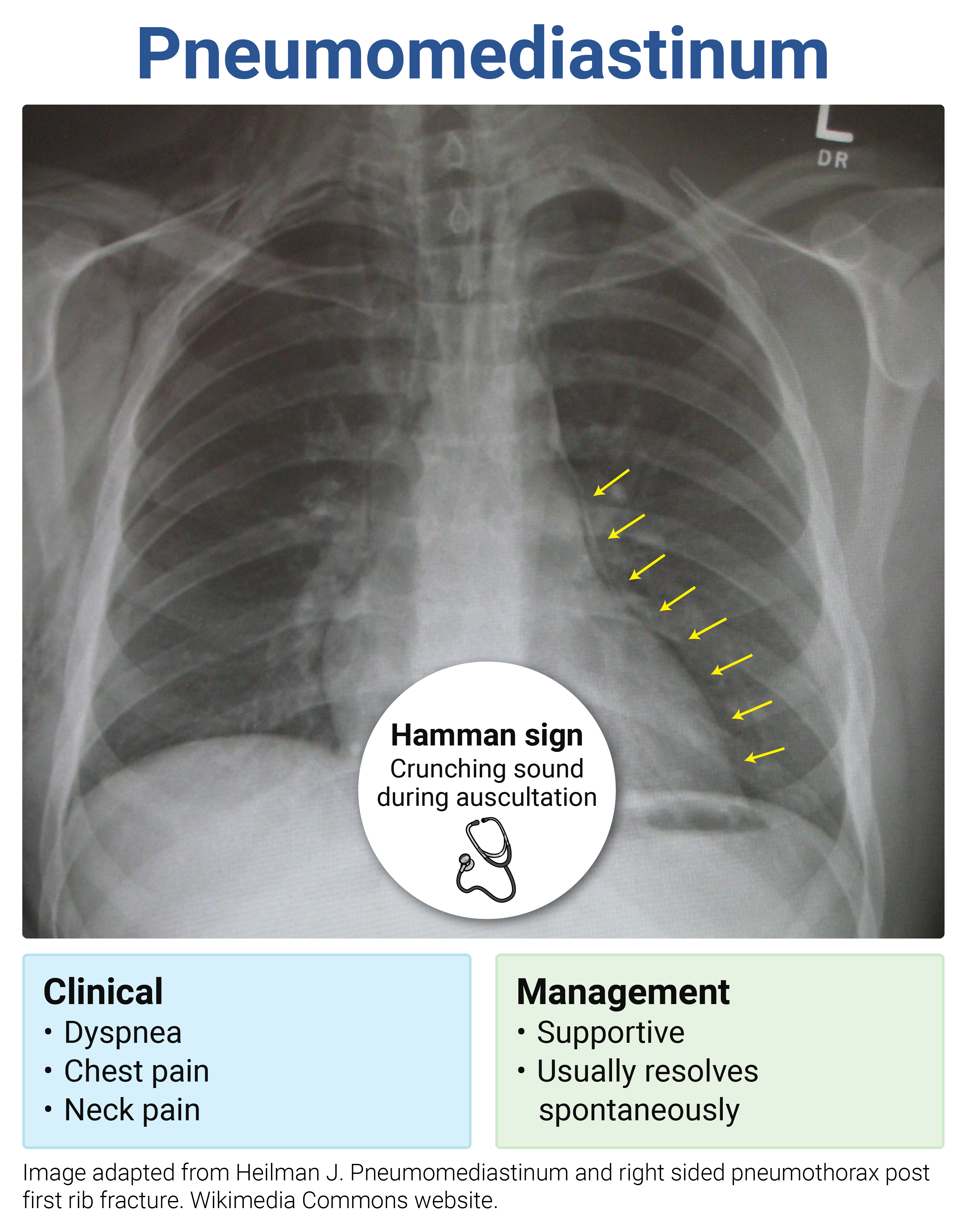
pneumomediastinum
“hamman sign = crunching sound on auscultation”
dx = CT scan
self limited
Mesothelioma cause
exposure to asbestos (pleura)
latent tuberculosis (TB) tx
rifampin daily for 4 months
rifampin and isoniazid for 3 months