NEUR30080 L5 SQ synaptic transmission at NMJ
 Call Kai
 Learn
 Practice Test
 Spaced Repetition
 Match
 Flashcards
 Knowt Play
1/40

There's no tags or description
Looks like no tags are added yet.
41 Terms
learning goals

chemical vs electrical synapses
chemical: use a NT intermediary, imposing a delay of up to 2 ms
electrical synapses (gap junction): pass ionic current directly from cell to cell (NOT released into cleft), pre- and post-synaptic cells fused together, almost instantaneous
which synapse is FASTER?
electrical
which synapse releases neurotransmitters (NTs)?
chemical
chemical vs electrical synapse image

electrical synapses - 0.1 ms delay
current flows virtually instantaneously (< 0.1 ms delay)
allows tightly coupled networks to fire in perfect synchrony
most pass current bidirectionally, though some (e.g. crayfish motor synapse) act as one-way-rectifiers
electrical synapse locations
retinal cells
neocortex
cerebellum
inferior olive
thalamus
brainstem
electrical synapse: pre- and post-synaptic neuron graph

gap junction channel structure
gap junction channel is two docked connexon hexamers that bridge a 2-4 nm gap and pass solutes up to about 1kDa
pore is large enough to pass ions, ATP and secondary messengers (e.g. IP3 and cAMP)
massive influx of Ca2+ physically closes the channel, instantly uncoupling a damaged or dying cell from the healthy network (so toxin doesnt spread from one cell to the next via gap junction)
gap junction image

connexon
made of connexins
form continuous pore
calcium and gap junction
Ca2+ control chemical gating at gap junctions
chemical transmission - cause of 0.5-2 ms delay
chemical transmission adds gain, sign inversion and plasticity (resulting in delay)
electrical vs chemical synapse table

roles of pre- and post-synaptic cells
pre-synaptic: has an AP, or not. Can release NT
post-synaptic cell: activated by this signal, amplifies signal
LT depression
weakening of synapses
LT potentiation
strengthening of synapses
EPSC
the tap
current, excitatory flow of ions
the actual flow of ions through open receptors
the current ends exactly when the channels close
EPSP
the bucket
depolarisation, change in voltage
the resulting voltage change, outlasts the current because the memb capacitor (tau) takes time to drain

EPSP decay formula
tau
governs the time it takes for the lipid bilayer to release charge
‘time constant’
EPSP and EPSC graph

unitary cortical EPSP of 0.1-1 mV is 1 to 2 orders of magnitude smaller than the 10 mV separating rest from threshold
many central synapses release zero or one quantum per impulse
rest: -65 mV, threshold: -55 mV
a single cortical synapse produces a tiny EPSP (0.1-1 mV), falling far short of the 10 mV required to reach threshold
to fire an AP - a central neuron must structurally summate 50-100 coincident inputs (to summate to threshold)
summation image

synaptic current is the prod. of synaptic conductance and driving force (formula)
(Vm - Erev) is how far away the ion is from its equilib. potential

when is a synapse inhibitory?
when Erev lies below spike threshold, whether or not it hyperpolarises the cell
since Erev sits between Ek and Erest, depolarisation occurs

the same Cl- conductance hyperpolarises a cell at -65 mV, and depolarises it at -80 mV
synaptic current always drags Vm directly towards the receptor’s Erev
the magnitude of the current scales exactly with the distance between Vm and Erev
the voltage will never overshoot Erev: once the membrane reaches Erev, net synaptic current drops to zero

an inhib conductance with Erev at the resting potential reduces EPSP amplitude without changing baseline Vm
opening Cl- channels near the resting potential acts as a massive local leak that drastically drops the input resistance (Rin)
the punctured hose: like a massive puncture in a garden hose (or hole in the bucket), this leak bleeds out the presence of advancing EPSPs, preventing them from reaching soma
shunting inhibition acts divisively, it scales down the amplitude of the EPSP rather than simply subtracting from the baseline voltage
if there was IPSP and then EPSP: would bring back to baseline. Distance is larger when hyperpol current occurs instead of shunting current
‘punctured hose’ image

EPSP amplitude at the soma declines exponentially with the distance of the synapse, with length constant lambda
dendritic voltage decays exponentially with distance because current constantly escapes through resting K+ leak channels (lambda)
because distal inputs lose most of their amplitude before reaching the soma, multiple synapses must fire simultaneously across the dendritic tree to successfully push the hillock to threshold = SUMMATION
image: EPSP amplitude at the soma declines exponentially with the distance of the synapse, with length constant lambda

spatial summation
multiple pre-syn cells have to signal on one post-syn cell for syn current to actually reach soma of post-syn cell.
Has to sum all E- / IPSPs. If there is sufficient depol across them, then AP can fire.
a longer membrane time constant (tau) widens the interval over which successive EPSPs sum
a long tau = voltage from a first input does not drain away before the second arrives
the memb capacitance allows rapidly firing inputs from the exact same synapse to stack on top of one another to cross threshold
spike threshold is lowest at axon initial segment (AIS) - so summed potential is converted to firing rate there
all graded spatial and temporal summation physically converges at the AIS
because Nav channel density is highest here, the AIS has the lowest threshold in the cell, acting as the solo site where the integrated analogue signal is converted into a firing rate
frequency that AP fires = how strong the signal is (strength)
one AP = happens or not, frequency of firing matters
summation at AIS image

AMPA (aka kainate) vs NMDA receptors
both open in response to Glut
conduct both K+ and Na+, therefore Erev is close to zero (bc Na and K equilibriums almost cancel out)
NMDA:
Mg2+ blocks NMDA channels and needs to be removed first
Ca2+ involved in syn. plasticity

the EPSC at a Glut synapse has a fast AMPA component and slow NMDA component
AMPA: rapid ligand-gated inward Na current, decays in few ms
NMDA: ligand-voltage-gated, decays over 10s-100s ms, blocked by D-AP5 - must be depolarised to activate
often act hand-in-hand: AMPA activates and depol. memb. slightly, Glut then binds to NMDA and NMDA already depol enough and therefore NMDA can open
at -70 mV the EPSC carried by AMPA, at +40 mV, both contrib

AMPA receptors provide rapid initial depol
AMPA receptors are non-selective cation channels (K+ and Na+) - Erev is therefore near zero
at resting potential (-65 mV), the strong inward DF creates rapid Na+ current
this fast, voltage-dependent EPSC provides crucial initial voltage change required to depol the local memb for NMDA receptors
(IA is vertical axis current)

NMDA receptor is coincidence detector
cog-agonist: glycine or D-serine required
depol. supplied by AMPA receptor current at the same or neighbouring sites
Ca2+ entry couples the conductances to CaMKII and other EC signalling
coincidence detector: because it required both pre-syn Glut and post-syn depol., NMDA Ca2+ entry strictly reports the simultaneous activation of both cells

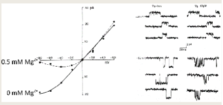
at -65 mV EC Mg2+ blocks NMDA receptor pore
depol relieves the block
when Mg2+ block freed, NMDA looks more like AMPA
Mg2+-free: linear I-V reversing near 0 mV
physiological Mg2+ (about 1 mM): inward current suppressed negative to -40 mV
block is voltage-dependent because Mg2+ binds withing the membrane field
*NMDA receptor both voltage- and ligand-gated
learning outcomes
