Body Log: Session 3 + 4
 Call Kai
 Learn
 Practice Test
 Spaced Repetition
 Match
 Flashcards
 Knowt Play
1/25

There's no tags or description
Looks like no tags are added yet.
26 Terms
Describe the ‘anatomical position‘: (what is their scientific name)
In the anatomical position, a person is:
• Standing upright
• Facing forward
• Head level, with eyes looking straight ahead
• Arms at the sides of the body
• Palms facing forward
• Legs together
• Feet flat on the floor and facing forward
Define the limits of body regions and major parts of the body:
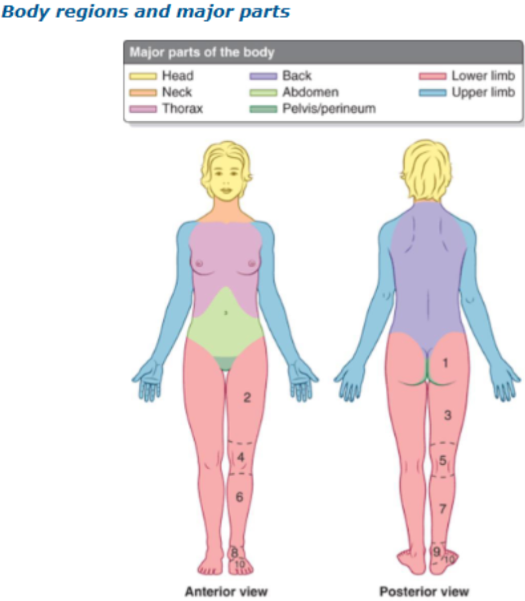

Define terms ‘anterior, posterior, medial, lateral, inferior, superior, transverse, axial, Coronal, Sagittal‘:

Superior closer to the top half of the body
Inferior closer to the bottom half of body
Anterior closer to the front half
Posterior is closer to the back

Medial: Nearer to the Median plane
Lateral: Further from the median plane
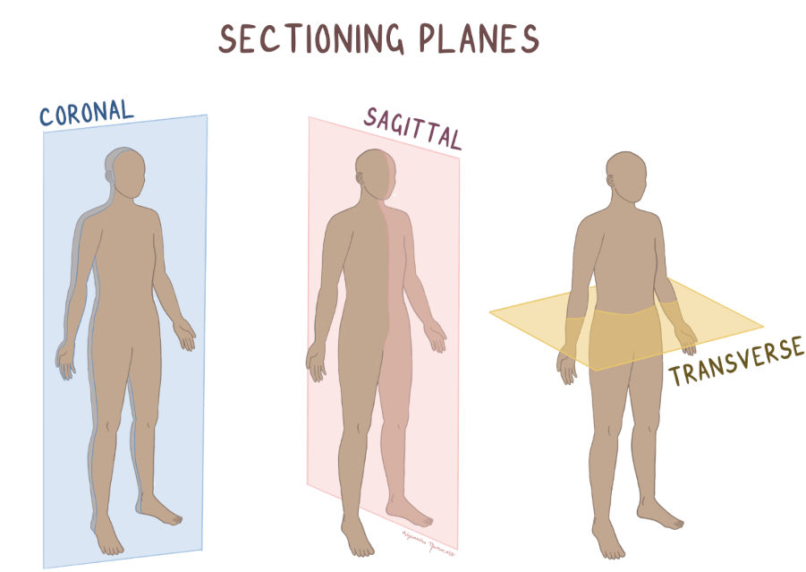
Coronal: Separates the front and the back sides of the body
Sagittal: Longitudinal (that separated the body right down in middle)
Transverse: Axial, separates the top half from the bottom half
Compare and contrast to the terms ‘superficial, intermediate, deep, proximal, distal’ with clinical term usage, such as ipsilateral and contralateral.
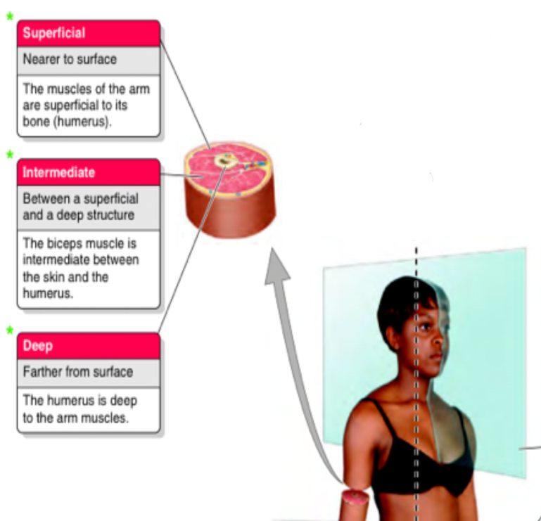

Structures on the same side: Ipsilateral
Structures on the opposite side: Contralateral
Define and recognise common anatomical terms, such as ‘flexion and
extension, abduction and adduction, lateral and medial rotation,
circumduction, supination and pronation’
Flexion: Decreasing the angle between body parts, (Bending of elbow) occurs in the sagittal plane
Extension: Increase the angle between body parts (Straightening the knee) Sagittal
Abduction: Move limb away from midline (Lifting arm to the side) Coronal
Adduction: Move limb towards the midline (Bringing arm back to the body) Coronal
Medial Rotation: Rotates limb towards midline (Turning leg inward) Transverse
Lateral Rotation: Rotates limb away from midline (Turning arm outward) Transverse

Circumduction: Circular movement combining, flexion, extension, abduction, adduction occurring in multiple planes (Drawing circle with an arm)
Supination: Rotating forearm so palm faces up/ anterior (receiving change in hand) Transverse
Pronation: Rotating forearm so palm faces down/ posterior (turning doorknob inward)
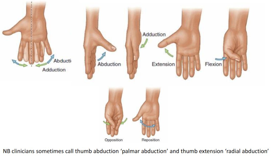
Define the specialised terms associated with movements in the hand and
foot


Define a gland and secretion:
A gland = An epithelial cell or aggregate of cells that are specialised for the secretion of a substance
Secretion = The production and release of materials by cells
Secretion: Proteins (enzymes), lipids (sebum), complex carbohydrates (Mucus)
Describe the role of secretions in cell functions
Communication: e.g. Hormones, neurotransmitters
Protection: e.g. Mucus, tears, antimicrobial proteins (antibodies)
Digestion: e.g. Enzymes in saliva and pancreas
Describe and discriminate between the simple mechanisms of secretion control
Hormonal: Where Hormones control the secretion of further hormones
Hypothalamus (TRH) → Anterior pituitary gland (TSH) → Thyroid gland (T3 and T4) → target cells → Action

Neural: Hormone release is controlled by nerve stimulation e.g. Salivary secretions
Humoral: Hormone release is controlled by substances in the blood e.g. High blood glucose (insulin)
Classify different types of glandular tissue

Exocrine: Secrete onto an epithelial surface
Endocrine: Secrete into bloodstream
Describe the different mechanisms for secretion

Explain the mechanism and importance of glycosylation of newly synthesised proteins in the Golgi apparatus
What is Glycosylation: Enzymes that covalently attach carbohydrate groups to protein/ lipids
Where does it happen: Proteins are synthesised in the RER => transport vesicle enters the cis face, and as it travels through the Golgi enzymes remove particular sugars/ add different sugars/ modify carbon chain length => vesicle leaves the trans face in vesicle transported to destination
Why is it important:
Carbohydrate groups on the protein enable the protein to achieve more stability (structure and folding)
Carbohydrates are hydrophilic, so glycosylation can increase the solubility of proteins (Solubility)
Carbohydrate chains can help protect from being broken down via proteases (Protection from degradation)
The glycoproteins are on the surface so can act as identification markers (cell recognition)
There is two types of secretion:
(Constitutive) Continuous and unregulated, where the transport vesicles once being formed fuse with the plasma membranes, No external trigger or chemical signal required, so rate of release of products is continuous and steady
Regulatory: Controlled process, proteins stored in vesicles and when there is a particular signal the vesicles with fuse with plasma membrane and contents released
Describe the mechanisms of endocytosis and exocytosis:
Exocytosis: Secretion of molecules outside the cell via a vesicle fusing to a membrane
Endocytosis: Engulfing of molecules inside the cell via vesicle formation
Describe how endocytosis and secretion combine to give transepithelial transport
Transcytosis:
Endocytosis → travels through the cell → Exocytosis
Discuss the differences between exocrine and endocrine glands

Describe the molecular and cellular interactions that lead to gland formation and branching:
Growth factor (FGF) = Fibroblast Growth Factor, which signal cells to grow and divide
Proliferation of daughter cells and extracellular protein degradation enzymes: Epithelial cells continue to grow, they release enzymes that breakdown the extracellular proteins (this creates space for more growing epithelial cells)
Then the epithelial cells grow into the newly created space, extending deeper into the underlying tissue
Angiogenic factors: blood vessel (Angio) and genic (forming/ producing)

Branching of Exocrine glands:
FGF10 is released by immature fibroblasts (stem cells), which is a chemical attraction signal for nearby epithelial cells
Either keep getting longer: GF1 active // GF2 inactive
Branch off: GF1 inactive // GF2 active
Then, when Shh a signalling protein is detected both elongation and branching stop
Simple terms:
Exocrine: Develop from epithelial buds that grow down into underlying mesenchyme// maintains connection to the surface epithelium// connection becomes a duct// repeated branching often occurs forming duct system// cells differentiate into secretory units that release products onto an epithelial surface
Endocrine: Develop from epithelial buds growing into mesenchyme// loose their connection to surface epithelium// duct disappears// cells organise into clusters or cords// develop rich capillary network// secretion directly into bloodstream
Describe the classification of glands based upon the following terms:
o Simple or compound
o Acinar or tubular
o Serous or mucous
Serous: Rich in proteins (enzymes) and watery (thin and clear)
Mucous: Rich in glycoproteins (mucins) and viscous (thick and slimy)
Acinus: Is a small ball of secretory epithelial cells containing a tiny central lumen, usually formed by serous cells
Tubules: Elongated tube shaped
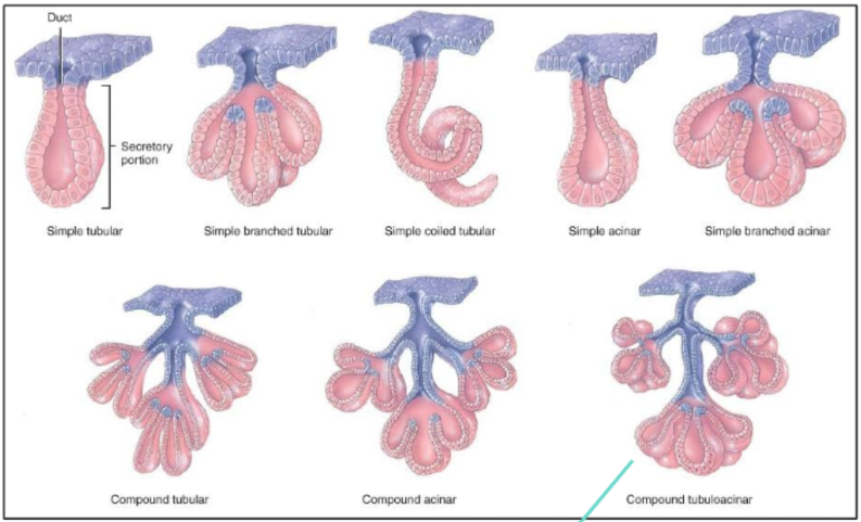
Example of compound tubuloacinar = Salivary glands, where the end of the gland has myoepithelial cells, that contract to release serous substances.
Explain the location and function of myoepithelial cells in relation to
exocrine gland secretion
Contractile cells located between the secretory epithelial cells and the basement membrane of exocrine glands
Their contraction helps move secretions from the secretory units into the duct system.
Recognise exocrine glands in photomicrographs, discuss their anatomical
locations and salient histological features in relation to their function (of salivary glands):
o Unicellular glands (goblet cells) in jejunum and colon
o Parotid gland
o Submandibular & sublingual glands

The function of goblet cells in jejunum: Lubricates the intestinal contents so they move smoothly, protects the epithelium from digestive enzymes and mechanical damage, contributes to barrier against microorganisms.
The function of goblet cells in colon: Contents become more drier and more solid as water is absorbed, therefore lubricates faeces// protects the colonic epithelium from abrasion by solid contents, protective barrier between gut bacteria and epithelial surface
Salivary glands and their function:
Lubrication: keep mouth moist // Digestion: containing enzymes that begin the breakdown of food // chewing and swallowing: Making it easier to eat // Speech: Assisting with clarity of speech // Dental health: Protecting teeth from cavities (also sodium carbonate released which is alkaline, so neutralises any acidity from foods// stomach acid when vomiting)

Parotid gland: Compound acinar, serous exocrine gland (it is similar to pancreas exocrine, so to distinguish pancreas has islets of Langerhans, while parotid has fat cells (adipocytes))


Recognise the following (mixed) glands from photomicrographs, discuss
their approximate anatomical locations and salient histological features in
relation to their function:
o Pancreas
Pancreas contains 95% exocrine tissue and 5% endocrine tissue. Where most of the exocrine is serous gland, producing digestive enzymes released into duodenum. The duct cells secrete bicarbonate to neutralize acid from the stomach.
Compound tubuloacinar = Pancreas
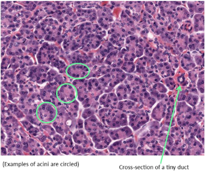
You can recognise the duct, because surrounded by capillary network
Identify the exocrine and endocrine histological components of the pancreas
Contains: Compound tubular acini => acini can be identified by the circular imagining // duct can be identified by close nuclei together and rich blood supply
Contains: Islets of Langerhans, which have spherical nuclei, with multiple capillary spaces showing a rich blood supply

Name the secretions of the exocrine and endocrine pancreas
Zymogen: Is an enzyme that can be activated when part of the enzyme is cleaved


Briefly discuss the structure of endocrine glands and their products
(hormones) and the signalling pathways that regulate their growth and
function
Key structural features of Endocrine glands:
No ducts, since they secrete hormones directly into bloodstream
Cells arranged in cords, clusters or follicles


Surrounded by dense network of capillaries
Supported by delicate connective tissue stroma
Their products:
Peptide/ protein hormones
Water-soluble
Stored in secretory vesicles
Bind to cell surface receptors
Example: Insulin
Steroid hormones:
Lipid-soluble, so can pass through cell membranes
Synthesised from cholesterol
Not stored in large amounts
Diffuse through cell membranes
Bind intracellular receptors (within cell nuclei)
Example: Cortisol
Amine hormones:
Derived from amino acids
Some bind to cell surface receptors// some intracellularly
Regulation of Growth and Function:
Hormonal (Trophic) control:
One hormone stimulates another gland => Operates via negative feedback, where the final hormone can inhibit eariler steps
Humoral control => Changes in blood composition (glucose/ calcium levels) can stimulate hormones to release
Neural control => regulated by autonomic nervous input (sympathetic/ parasympatheitc nervous system stimulation)
Recognise the following endocrine (ductless) glands from
photomicrographs, discuss their approximate anatomical locations and
salient histological features in relation to their function:
o Thyroid gland
o Parathyroid glands
o Adrenal (Suprarenal) glands
Thyroid gland: It is a butterfly shaped gland, located in front of trachea, made up of follicles that produce and store thyroid hormones.
T3 and T4 = Thyroid hormones:
Control metabolic rate (how fast the body uses energy)
Influences growth, development mostly of brain and skeleton
Help regulate temperature, heart rate and weight
Calcitonin: Lowers blood calcium levels by inhibiting bone breakdown
It has simple cuboidal epithelium, filled with colloidal thyroglobulin.

Parathyroid glands: 4 small glands (pea-sized) on the posterior surface of gland. They release parathyroid hormone, increases calcium release from bone // increase calcium absorption in gut (via vitamin D activation) // Increase calcium reabsorption in kidneys.
Mainly has chief cells, which release PTH
Oxyphil cells not completely know, but thought to sense calcium levels and regulate PTH
PTH and calcitonin work together

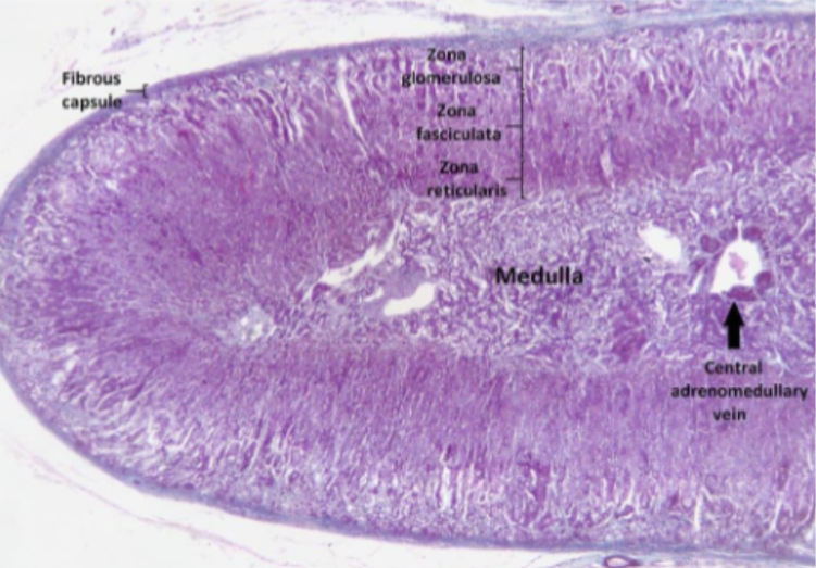
Adrenal glands: One located on top of each kidney
Cortex (outer layer): produced steroid hormones:
Aldosterone (mineralocorticoid) - regulates sodium and water balance which influences blood pressure
Cortisol - (glucocorticoid) - regulates carbohydrate metabolism, stress response and inflammation
Androgens (sex chromosomes) - Contribute to secondary sexual characteristics
Medulla (inner layer): produce catecholamines (hormones for fight or flight response)
Chromaffin cells secrete adrenaline and noradrenaline => increasing heart rate // blood pressure and blood glucose
Recognise the following (mixed) glands from photomicrographs, discuss
their approximate anatomical locations and salient histological features in
relation to their function:
o Liver
Roles of the liver: Exocrine gland and secretes bile into intestine, endocrine gland and a blood filter, storage of glycogen, detoxifying drugs and poisons, cleansing bacteria from blood, catabolism of haemoglobin from worn-out RBC
Blood travels into the liver via hepatic portal vein (deoxygenated blood from spleen and GI tract), and the hepatic artery which brings fresh oxygenated blood from aorta. The blood mixes together in the hepatic sinusoid before leaving the liver in the hepatic vein.

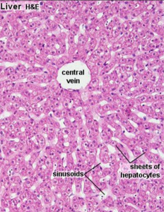


Kupffer cells, special macrophages that form part of the sinusoidal lining// exposed to gut-derived bacteria, microbial debris => trap and phagocyte any damage or aged erythrocytes (RBC) that was missed by spleen

Ito cells: Contain vitamin A // in liver cirrhosis loose vitamin A and differentiate into myofibroblasts
Provide a detailed description of a portal circulatory route, and give examples
A portal circulatory system is the arrangement of blood vessels where the blood flows from one capillary bed to another to a second capillary bed, before bypassing to the heart.
Pituitary gland: Contains a anterior and posterior gland
Hypothalamus releases TRH which enters the first capillary bed
Instead of blood returning to the heart, it travels to the hypophyseal portal vein, where it makes a portal system
Second capillary bed, where the hypophyseal portal vein enters anterior pituitary
TRH stimulates anterior pituitary cells to release TSH
Thyroid gland:

We have main blood supply travelling into the liver via hepatic artery from the aorta
We have another blood supply which travelled through the spleen and small intestine and enters into the liver via portal vein.
Then when both of these blood mixes form blood with oxygen supply (aorta) and high in soluble food components (portal vein) => Hepatic sinusoid