Micro Exam 4
 Call Kai
 Learn
 Practice Test
 Spaced Repetition
 Match
 Flashcards
 Knowt Play
1/363

There's no tags or description
Looks like no tags are added yet.
364 Terms
Taxonomy
science of organizing organisms into two hierarchal groups based on shared characteristics
Microbial genomics
study of structure, function and evolution of microbial genomes
clinical microbiology
branch of microbiology focused on identifying and understanding microbes that cause human disease
immunology
study of immune system components and how they detect and respond to microbes
Taxa
hierarchal groups of organisms formed through classification
Identification
process of characterizing an organism using morphology, physiology and genotype
morphology
observable physical features of an organism -
physiology
functional traits of an organism, such as metabolism or biochemical capabilities
genotype
genetic makeup of an organism
classification
organization of organisms inot groups based on identified similiarities
nomenclature
formal system used to assign standardized names to organisms
species (prokaryotic)
group of closely related strains that share high genetic similarity and differ distinctly from other groups
strain
population of cells derived from a single ancestor that accumulates minor genetic differences over time
prokaryotic taxonomic hierarchy
ordered system of ranks used to classify prokaryotes from species up to domain
16S rRNA gene sequence similarity
molecular comparison used to assess relatedness in prokaryotes
genotpic characteristics
identification features based on an organism’s genetic sequence, often used to determine evolutionary relationships
Phenotype-dependent characteristics
observable traits such as morphology or metabolism used for practical identification
Algorithm/Dichotomous key
step-by-step series of yes/no questions used to narrow down microbial identity
microscopic morphology
size, shape, arrangement, and staining traits observed under a microscope

culture characteristics
observable colony traits such as color, texture and growth patterns that appear on culture media
req. pure culture

metabolic capabilities
biochemical traits that reveal how a microbe uses specific nutrients or produces metabolic by-products
req. pure culture
serology
identification based on antigen-antibody interactions involving microbial surface structures
protein profile
characteristic pattern of cellular proteins, often measured by MALDI-TOF MS, used for rapid microbial identification
req. pure culture
acid-fast stain
describes bacteria such as mycobacterium species, that retain certain dyes due to waxy mycolic acids in their cell walls
mycolic acid
long, waxy lipids found in the cell walls of acid-fast bacteria, responsible for their staining behavior and resilience
Auramine O
fluorescent dye used in modern acid-fast staining to detect organisms like mycobacterium tuberculosis
colony morphology
visible structure and appearance of a microbial colony, including shape, elevation and pigmentation
selective media
culture media that inhibit some microbes while allowing others to grow
differential media
media containing indicators that reveal metabolic differences between microbes through color or pattern changes
MacConkey agar
selective and differential medium used to isolate G- bacteria and detect lactose fermentation
blood agar
differential medium used to detect hemolysis patterns produced by certain bacteira
B-hemolysis
complete lysis of RBCs around a colony, producing a clear zone
biochemical tests
lab tests that detect specific metabolic processes, often through visible color or gas changes
catalase test
test that detects the enzyme catalase by observing the breakdown of H2O2 into O2 and water
sugar fermentation
metabolic process in which bacteria breaks down sugars to produce acidic end products, often altering the pH of the medium

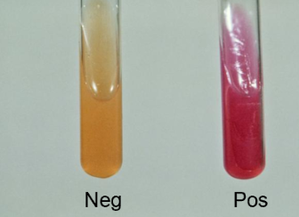
Phenol Red
pH indicator commonly used in biochemical tests that turns yellow in acidic conditions and remains red or pink when no acid is produced

Urease
enzyme that hydrolyzes urea into ammonia and CO2, raising the pH of the surrounding medium
urea breath test
diagnostic method for H. pylori that detects carbon-labeled CO2 produced when urease breaks down labeled urea
commercial kits
preassembled biochemical test systems that allow multiple metabolic reactions to be assessed at once
ELISA (enzyme-linked Immunosorbent Assay)
biochemical test that uses antibodies and enzyme-linked color changes to detect antigens or antibodies
Sandwich ELISA
an ELISA format that detects microbial antigens using antibody-coated wells to capture the pathogen’s surface structures
traps them b/w 2 specifc antibodies
detects antigen → current infection

Indirect ELISA
an ELISA format that detects host antibodies produced during infection rather than microbial antigens themselves
detects antibody → current/recent past infection

antigen
molecule, often found on microbial surfaces, that triggers antibody binding
antibody
protein produced by the immune system that specifically recognizes and binds antigens
anti-pathogen antibody
antibody designed to recognize and bind a specific microbial antigen
used as the ‘capture’ antibody in Sandwich ELISA
Enzyme-linked detection
method in which an antibody-associated enzyme produces a color change to indicate a pos reaction
pathogen-specific antibody
antibody produced by the immune system that binds to a distinct microbial antigen
Point-Of-Care (POC) test
diagnostic test performed at or near the site of patient care, providing rapid, actionable results
lateral flow assay
rapid immunochromatographic test that detects antigens or antibodies using capillary movement across a membrane
Immunochromatographic assay
detection method in which antigen-antibody binding produces a visible signal as the sample flows through the test device

sensitivity
ability of a test to correctly identify true positives
often lower in rapid POC tests
IgM detection
identifying early-stage antibodies that indicate a recent infection
IgG detection
identifying long-lasting antibodies that signal past exposure or late stage infection
False-negatives
test result that incorrectly indicates no infection when the pathogen is actually present
false-positive
test result that incorrectly indicates infection when the pathogen is absent
Co-infection
presence of more that one infectious agent in a patient, which POC tests may overlook
Serovar/Serotype
strain defined by its unique antigenic profile
O antigen
variable polysaccharide portion of LPS used to differentiate G- strains
H antigen
flagellar protein antigen used to distinguish motile bacterial strains
K antigen
capsular antigen used to classify strains with distince protective surface capsules
E. coli O157:H7
pathogenic strain identified by its O157 and H7 flagellar antigens
E. Coli K12
non-pathogenic lab strain ID’d by its capsule (K) antigen
Immunomagnetic bead (IMB) technology
method that uses antibody-coated magnetic beads to selectively capture and isolate specific microbes from complex samples

Immunofluorescence
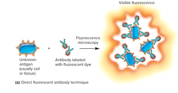
technique using fluorescently labeled antibodies to visualize specific microbes or antibodies under a fluorescence microscope
Direct immunofluorescence
detection method in which fluorescent antibodies bind directly to microbial antigens on a specimen
Indirect immunofluorescence
method in which patient antibodies bind to fixed antigens, followed by a fluorescent secondary antibody to reveal the reaction

fluorochrome-labeled antibody
antibody linked to a fluorescent dye used in immunofluorescence assays
MALDI-TOF MS
rapid, mass-spec technique that identifies microorganisms by measuring the masses of their cellular proteins

mass spectrum
unique distribution of protein mass signals produced during MALDI-TOF, used as a molecular fingerprint for Identification
specific nucleotide sequences
DNA regions unique to particular microbial groups, used as genetic targets for ID
NAATs (Nucleic Acid Amplification Test)
tests that amplify target DNA sequences to detect microbes with high sensitivity
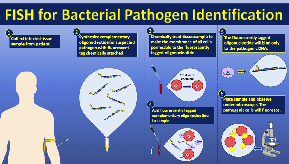
FISH (Fluorescence in situ hybridization)
method that uses fluorescent DNA probes to identify and visualize microbes based on rRNA or other genetic sequences
rRNA (ribosomal RNA)
highly abundant RNA molecule often targeted in FISH due to its high copy number in bacterial cells
PCR (polymerase chain reaction)
technique that amplifies DNA and visualizes the result on an agarose gel to confirm the presence of a target organism
qPCR (Quantitative PCR)
fluorescent version of PCR that measures DNA amplification in real time, allowing quantification of microbial load
primers (F&R primers)
short DNA sequences that bind to the target region and guide DNA synthesis
Taq polymerase
heat-stable DNA polymerase from Thermus aquaticus used to synthesize new DNA strands during PCR
dNTPs
nucleotide building blocks used by polymerase to make new DNA
Denaturation
high-temperature step that separates double-stranded DNA into single strands
Annealing
lower-temperature step where primers bind to their complementary sequences, controlling PCR specificity
Extension
step where Taq polymerase synthesizes new DNA strands, enabling exponential amplification
exponential amplificiation
repeated doubling of target DNA during PCR cycles, producing very large quantities of DNA
Positive control (PCR)
sample known to contain the target DNA, ensuring the PCR rxn worked as expected
Negative control (PCR)
rxn lacking template DNA, used to confirm that no contamination or unintended amplification occurred
DNA ladder
set of DNA fragments of known sizes used as reference markers during gel electrophoresis
SYBR Green
fluorescent dye that binds to double-stranded DNA and increases in fluorescence as amplification proceeds
TaqMan probe
fluorescent reporter-quencher probe cleaved during DNA synthesis, releasing fluorescence only when the target sequence is amplified
Ct value (cycle threshold)
PCR cycle at which fluorescence first rises above background, inversely related to starting DNA quantity
reporter dye
fluorescent molecule that emits light once separated from the quencher
Quencher
molecule that suppresses reporter fluorescence until cleavage occurs
5’ → 3’ exonuclease activity
ability of Taq polymerase to degrade DNA ahead of it during extension, enabling probe cleavage
background fluorescence
baseline signal before amplification produces detectable fluorescence
standard curve
reference graph created by amplifying known quantities of DNA or cells to relate Ct values to actual abundance
Standards
samples containing known number of cells or DNA copies used to build the standard curve
Quantification
process of estimating how much microbial DNA is present in an unknown sample comparing it to the standard curve
fluorescent tag
molecule attached to a probe that emits light under fluorescence microscopy
hybridization
binding of a probe to its complementary nucleotide sequence within intact cells
In situ
“in the original place”
refers to detecting microbes directly in the sample without culturing
conserved regions
Stretches of DNA that change very little over evolutionary time, allowing universal primer binding
variable regions
segments of the 16S gene that differ among microbial groups and help distinguish taxa